. 2009 Jan 1;18(1):164-77.
doi: 10.1093/hmg/ddn326. Epub 2008 Oct 17.

Dissociation of tau toxicity and phosphorylation: role of GSK-3beta, MARK and Cdk5 in a Drosophila model

Affiliations

Dissociation of tau toxicity and phosphorylation: role of GSK-3beta, MARK and Cdk5 in a Drosophila model

Shreyasi Chatterjee et al. Hum Mol Genet. .

Abstract

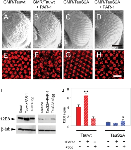
Hyperphosphorylation of tau at multiple sites has been implicated in the formation of neurofibrillary tangles in Alzheimer's disease; however, the relationship between toxicity and phosphorylation of tau has not been clearly elucidated. Putative tau kinases that play a role in such phosphorylation events include the proline-directed kinases glycogen synthase kinase-3beta (GSK-3beta) and cyclin-dependent kinase 5 (Cdk5), as well as nonproline-directed kinases such as microtubule affinity-regulating kinase (MARK)/PAR-1; however, whether the cascade of events linking tau phosphorylation and neurodegeneration involves sequential action of kinases as opposed to parallel pathways is still a matter of controversy. Here, we employed a well-characterized Drosophila model of tauopathy to investigate the interdependence of tau kinases in regulating the phosphorylation and toxicity of tau in vivo. We found that tau mutants resistant to phosphorylation by MARK/PAR-1 were indeed less toxic than wild-type tau; however, this was not due to their resistance to phosphorylation by GSK-3beta/Shaggy. On the contrary, a tau mutant resistant to phosphorylation by GSK-3beta/Shaggy retained substantial toxicity and was found to have increased affinity for microtubules compared with wild-type tau. The fly homologs of Cdk5/p35 did not have major effects on tau toxicity or phosphorylation in this model. These data suggest that, in addition to tau phosphorylation, microtubule binding plays a crucial role in the regulation of tau toxicity when misexpressed. These data have important implications for the understanding and interpretation of animal models of tauopathy.


Figures

Figure 1.
Figure 1.
Misexpression of PAR-1 (68) using GMR-GAL4 produces a rough eye with the disruption of retinal architecture, whereas Sgg (63) misexpression produces a relatively normal eye. (A–C) SEM images. The normal-eye phenotype observed using the GMR-GAL4 driver (A) is disrupted in eyes misexpressing PAR-1 (B) but not in eyes misexpressing Shaggy (C). (DF) Retinal whole mounts stained with phalloidin-TRITC to identify rhabdomeres of photoreceptor neurons (single tangential confocal sections). At this apical section, normally, clusters of seven photoreceptors within each ommatidium form a characteristic chevron-shaped structure (D). Full genotypes: (i) w1118, GMR-GAL4/+ (A and D); (ii) w1118, GMR-GAL4/+, UAS-PAR-1/+ (B and E); (iii) w1118, GMR-GAL4/+, UAS-Sgg/+ (C and F). Scale bars: 100 µm (A–C); 10 µM (D–F).
Figure 2.
Figure 2.
Schematic representation of the tau constructs used. TauS2A was constructed by mutating two serine residues (S262 and S356) to alanine; these are the phosphoepitopes recognized by the 12E8 antibody. TauS11A has 11 serine and threonine residues mutated to alanine. The epitopes recognized by phosphorylation-dependent antibodies, e.g. AT8, AT180, PHF1 and AT100, are shown. GMR-GAL4 driver was used to drive combinations of UAS-Tauwt, UAS-TauS2A and UAS-TauS11A along with PAR1 and Shaggy. gl drivers were also used for the same purpose.
Figure 3.
Figure 3.
The retinal phenotype of wild-type tau (Tauwt) under the control of GMR-GAL4 is more severe than that of S2A tau. (A and B) SEM images. The mild rough-eye phenotype produced by Tauwt (A) is not observed with TauS2A (B). Scale bars: 100 µm. (C and D) Confocal images of adult retina stained with TRITC-phalloidin (red). Ommatidial disorganization and cell loss are apparent in the eyes overexpressing the Tauwt transgene (C) compared with eyes expressing TauS2A (D), which displays a largely normal trapezoidal array of rhabdomeres. Genotypes: (i) GMR-GAL4/+, UAS-Tauwt/+ (A and C); (ii) GMR-GAL4/+, UAS-TauS2A/+ (B and D). (E) Immunoblot using T46 demonstrates that several independently derived Tauwt and TauS2A lines express comparable amounts of total tau protein. β-Tubulin-loading control for total protein in head extracts is shown below. Scale bars: 100 µm (A and B); 10 µm (C and D).
Figure 4.
Figure 4.
S2A is less toxic than wild-type Tau and relatively resistant to PAR-1-induced phosphorylation. (AD) SEM images. The rough-eye phenotype of transgenics misexpressing Tauwt (A) is severely enhanced when coexpressed with PAR-1 (B), whereas the phenotype of TauS2A (C) is only mildly enhanced by PAR-1 overexpression (D). (EH) Phalloidin-TRITC staining of whole-mount retina. TauS2A (G) is less toxic than Tauwt (E), and the enhancement in response to PAR-1 is less severe in TauS2A (H) compared with Tauwt (F). Genotypes: (i) GMR-GAL4/+, UAS-Tauwt/+ (A and E); (ii) GMR-GAL4/+, UAS-Tauwt/+, UAS-PAR-1/+ (B and F); (iii) GMR-GAL4/+, UAS-TauS2A/+ (C and G); (iv) GMR-GAL4/+, UAS-TauS2A/+, UAS-PAR-1 (D and H). Scale bars: 100 µm (A–D); 10 µm (EH). (I) Immunoblot analysis with 12E8 detects stronger immunoreactivity with Tauwt compared with TauS2A. (J) Histograms representing relative phosphorylation level changes after the coexpression of PAR-1 and Shaggy. Results shown are derived from densitometric analysis of three separate blots. Each bar represents mean ± SEM (n = 3). *P < 0.05; **P < 0.01 (ANOVA with the Newman–Keuls post hoc comparison).
Figure 5.
Figure 5.
TauS2A is relatively resistant to Shaggy-induced toxicity but not phosphorylation. (AD) SEM images. The rough-eye phenotype of Tauwt (A) is enhanced by the coexpression of Shaggy (B). The normal eye phenotype of TauS2A (C) remains unchanged when coexpressed with Shaggy (D). (EH) Phalloidin staining. Shaggy expression enhances the phenotype of Tauwt (F) but not TauS2A (H). Genotypes: (i) GMR-GAL4/+, UAS-Tauwt/+ (A and E); (ii) GMR-GAL4/+, UAS-Tauwt/+, UAS-Sgg/+ (B and F); (iii) GMR-GAL4/+, UAS-TauS2A/+ (C and G); (iv) GMR-GAL4/+, UAS-TauS2A/+, UAS-Sgg (D and H). Scale bars: 100 µm (A–D); 10 µm (E–H). (I) Immunoblot analysis shows that priming of tau by PAR-1 is not required for subsequent phosphorylation by overexpressed Sgg. The magnitude of the increase in phosphorylation at the AT8 and PHF-1 epitopes when stimulated by Sgg is greater for TauS2A than for Tauwt. (J) Histograms show relative effects on phosphorylation derived from densitometric analysis of three separate blots. Each bar represents mean ± SEM (n = 3). *P < 0.05; **P < 0.01 (ANOVA with the Newman–Keuls post hoc comparison).
Figure 6.
Figure 6.
TauS11A retains toxicity despite being resistant to phosphorylation by Sgg. (AC) and (GI) SEM images. Direct fusion of gl-Tauwt (A–C) and gl-TauS11A (G–I) phenotypes is shown. TauS11A retains toxicity as evidenced by a severe rough-eye phenotype with disordered ommatidial morphology (G). However, the degree of enhancement by Sgg (I) is less than that observed with wild-type Tau (C). Scale bar: 100 µm. (DF) and (JL) TRITC-phalloidin staining indicates that TauS11A (J) retains some toxic effects on ommatidial morphology, although the response to Shaggy (L) is blunted compared with Tauwt. Scale bars: 100 µm (A–C and G–I); 10 µm (D–F and J–L). Genotypes: (i) GMR-GAL4/+, gl-Tauwt/+ (A and D); (ii) GMR-GAL4/+, gl-Tauwt/+, UAS-PAR-1/+ (B and E); (iii) GMR-GAL4/+, gl-Tauwt/+, UAS-Sgg/+ (C and F); (iv) GMR-GAL4/+, gl-TauS11A/+ (G and J); (v) GMR-GAL4/+, gl-TauS11A/+, UAS-PAR-1/+ (H and K); (vi) GMR-GAL4/+, gl-TauS11A/+, UAS-Sgg/+(I and L). (M) Immunoblot analysis showing decreased basal and induced phosphorylation at AT8, PHF1, AT180 and AT100 phosphoepitopes for TauS11A compared with Tauwt. Results obtained using the direct fusion gl-Tau construct in combination with GMR-GAL4 driving UAS-kinases in this figure differ somewhat from those in which GMR-GAL4 drives both tau and kinases owing to less amplification of Tau expression in the former case, i.e. greater tau expression is achieved when glass drives GAL4 expression, which drives Tau. This difference results in a lower ratio of Tau to kinase when gl-Tau is used.
Figure 7.
Figure 7.
Toxicity of S11A and lack of effects of S2A tau correlate not with the formation of sarcosyl-soluble and -insoluble tau fractions, but with microtubule affinity. (A) Sarcosyl extracts of wild-type, S2A and S11A tau probed with T46 (total tau) and PHF-1 (a pathological phosphoepitope). Upper panel, input tau for the sarcosyl extraction; middle panel, sarcosyl-soluble and -insoluble fractions probed with T46 (total tau); lower panel, pellets and supernatants probed with PHF-1 (a pathological phosphoepitope). TauS11A does not form any soluble or insoluble aggregates compared with wild-type tau, whereas for TauS2A, the amount of insoluble material is much less compared with wild-type Tau. (B) Microtubule-binding assays. Upper panel, input tau for the microtubule-binding studies; middle panel, free or microtubule-bound tau. Compared with wild-type tau, TauS2A associates with microtubules somewhat less avidly, whereas TauS11A shows a markedly increased affinity for microtubules. T46 blotting indicates that the levels of input tau were the same for all three genotypes in both sarcosyl extraction and microtubule-binding experiments. All experiments used UAS constructs in trans to GMR-GAL4. Histograms represent the relative levels of free and bound tau in the supernatant (S) and pellet (P) fractions, respectively, for the three different transgenics. Each bar represents mean ± SEM (n = 3). *P < 0.05 for Tauwt and TauS2A, and **P < 0.001 for TauS11A. (One-way ANOVA with the Newman–Keul’s post hoc comparison).

References

    1. Morishima-Kawashima M., Hasegawa M., Takio K., Suzuki M., Yoshida H., Watanabe A., Titani K., Ihara Y. Hyperphosphorylation of tau in PHF. Neurobiol. Aging. 1995;16:365–371. - PubMed
    1. Johnson G.V., Bailey C.D. Tau, where are we now? J. Alzheimers Dis. 2002;4:375–398. - PubMed
    1. Cho J.H., Johnson G.V. Glycogen synthase kinase 3beta phosphorylates tau at both primed and unprimed sites. Differential impact on microtubule binding. J. Biol. Chem. 2003;278:187–193. - PubMed
    1. Augustinack J.C., Schneider A., Mandelkow E.M., Hyman B.T. Specific tau phosphorylation sites correlate with severity of neuronal cytopathology in Alzheimer’s disease. Acta Neuropathol. (Berl.) 2002;103:26–35. - PubMed
    1. Baum L., Hansen L., Masliah E., Saitoh T. Glycogen synthase kinase 3 alteration in Alzheimer disease is related to neurofibrillary tangle formation. Mol. Chem. Neuropathol. 1996;29:253–261. - PubMed

Publication types

MeSH terms