. 2008 Dec;148(4):1868-82.
doi: 10.1104/pp.108.130575. Epub 2008 Oct 17.

Interaction of the WD40 domain of a myoinositol polyphosphate 5-phosphatase with SnRK1 links inositol, sugar, and stress signaling

Affiliations

Interaction of the WD40 domain of a myoinositol polyphosphate 5-phosphatase with SnRK1 links inositol, sugar, and stress signaling

Elitsa A Ananieva et al. Plant Physiol. 2008 Dec.

Abstract

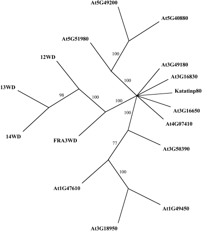
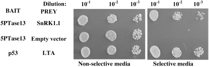
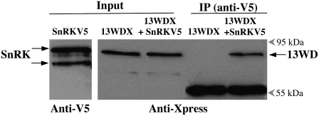
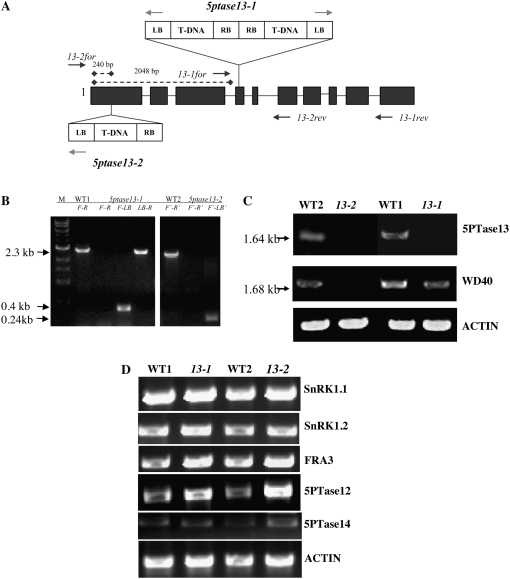
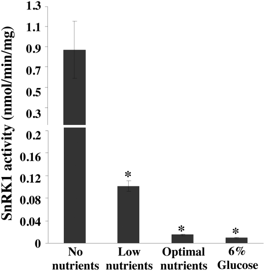
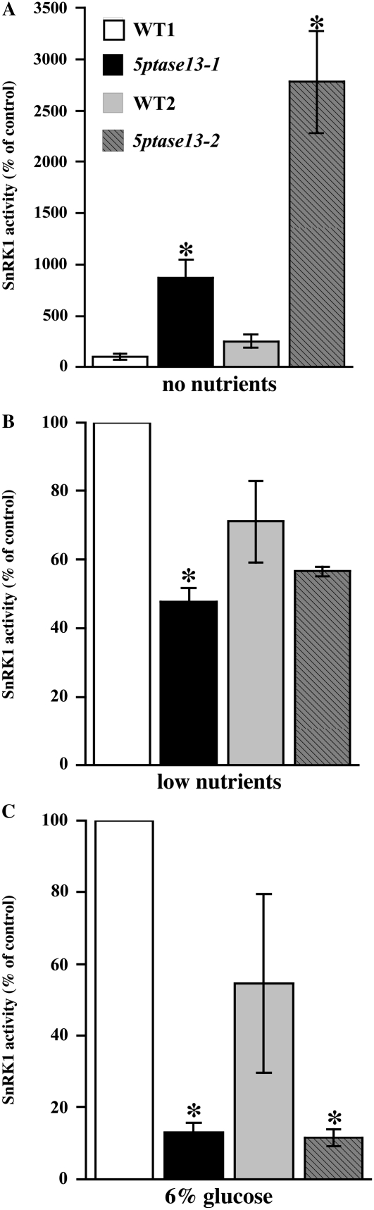
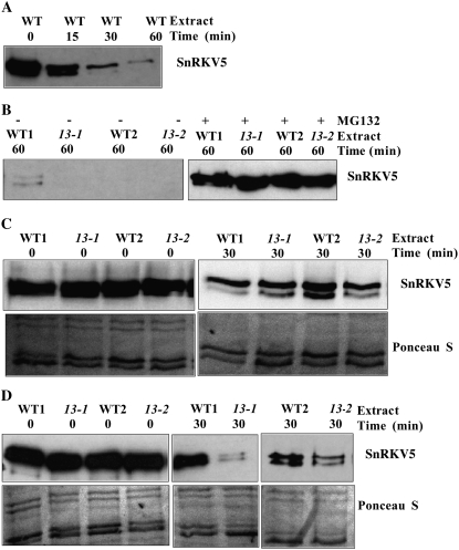
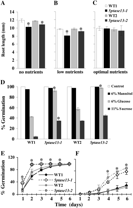
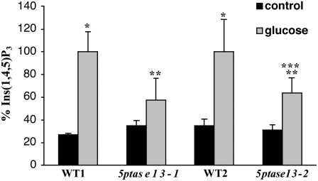
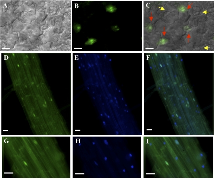
In plants, myoinositol signaling pathways have been associated with several stress, developmental, and physiological processes, but the regulation of these pathways is largely unknown. In our efforts to better understand myoinositol signaling pathways in plants, we have found that the WD40 repeat region of a myoinositol polyphosphate 5-phosphatase (5PTase13; At1g05630) interacts with the sucrose nonfermenting-1-related kinase (SnRK1.1) in the yeast two-hybrid system and in vitro. Plant SnRK1 proteins (also known as AKIN10/11) have been described as central integrators of sugar, metabolic, stress, and developmental signals. Using mutants defective in 5PTase13, we show that 5PTase13 can act as a regulator of SnRK1 activity and that regulation differs with different nutrient availability. Specifically, we show that under low-nutrient or -sugar conditions, 5PTase13 acts as a positive regulator of SnRK1 activity. In contrast, under severe starvation conditions, 5PTase13 acts as a negative regulator of SnRK1 activity. To delineate the regulatory interaction that occurs between 5PTase13 and SnRK1.1, we used a cell-free degradation assay and found that 5PTase13 is required to reduce the amount of SnRK1.1 targeted for proteasomal destruction under low-nutrient conditions. This regulation most likely involves a 5PTase13-SnRK1.1 interaction within the nucleus, as a 5PTase13:green fluorescent protein was localized to the nucleus. We also show that a loss of function in 5PTase13 leads to nutrient level-dependent reduction of root growth, along with abscisic acid (ABA) and sugar insensitivity. 5ptase13 mutants accumulate less inositol 1,4,5-trisphosphate in response to sugar stress and have alterations in ABA-regulated gene expression, both of which are consistent with the known role of inositol 1,4,5-trisphosphate in ABA-mediated signaling. We propose that by forming a protein complex with SnRK1.1 protein, 5PTase13 plays a regulatory role linking inositol, sugar, and stress signaling.


Figures

Figure 1.
Figure 1.
Phylogenetic tree of the WD40 regions from 5PTases and other WD40-containing Arabidopsis proteins. ClustalW and PAUP4.0 were used to generate a tree. Bootstrapping (1,000 times) yielded the confidence level indicated at each branch. The results show that the WD40 region of 5PTase14 (14WD) is the closest relative to 5PTase13 (13WD). Furthermore, the tree indicates that the 5PTase WD40 regions (12WD, 13WD, 14WD, and FRA3WD) are more closely related to each other than to other WD40-containing proteins.
Figure 2.
Figure 2.
Yeast two-hybrid screen. An Arabidopsis cDNA prey library was screened using the WD40 region of 5PTase13 as a bait in the yeast two-hybrid system. Over 1 million yeast transformants were screened for reporter gene expression, which resulted in the isolation of a prey plasmid expressing the C-terminal domain of SnRK1.1. Serially diluted yeast strains were spotted onto agar plates that are either nonselective or selective for expression of the two-hybrid reporter genes.
Figure 3.
Figure 3.
13WD40-Xpress protein interacts with SnRK1.1-V5 in vitro. Immunoprecipitation (IP) was carried out using an anti-V5 antibody bound to a protein A-Sepharose (PAS). Purified and dialyzed proteins (13WDX and SnRKV5) were incubated for 2 h at room temperature in immunoprecipitation buffer before transferring to the antiV5:PAS complex. The resulting immunoprecipitation fractions were analyzed using protein gel blotting. Lane 1, Input fraction of SnRKV5 probed with an anti-V5 antibody; lane 2, input fraction of 13WDX; lane 3, input fraction of 13WDX in combination with SnRKV5; lane 4, bound fraction of 13WDX alone; lane 5, bound fraction of 13WDX in combination with SnRKV5. Lanes 2 through 5 were probed with an anti-Xpress antibody. The 55-kD band is the immunoglobin heavy chain component of the anti-V5 antibody. The blot images are representative of four independent experiments.
Figure 4.
Figure 4.
T-DNA insertions and gene expression in 5ptase13 mutant lines. A, T-DNA insertions in the 5PTase13 gene. Exons are shown as dark gray boxes; light gray arrows indicate primers used to amplify the LB of the T-DNA; dark gray arrows indicate positions of gene-specific primers. RB, Right border. B, PCR performed with gene-specific and LB primers confirms that both lines are homozygous. Gene-specific primers that flank the T-DNA insertion (primers 13-1for [F] and 13-1rev [R]) in conjunction with the T-DNA LB primer amplify 0.4- and 2.3-kb fragments in 5ptase13-1 mutants, indicating the presence of two LB sequences in proximity to the 5PTase13 gene. C and D, Expression of genes in 5ptase13 and wild-type seedlings. Total RNA (1–2 μg) was isolated, and semiquantitative RT-PCR was performed with the indicated primers (Supplemental Table S4). C, Verification of the loss of 5PTase13 expression in 2-week-old leaves from soil-grown plants. D, SnRK1 and 5PTase expression from 7-d-old wild-type and 5ptase13 (13-1 and 13-2) seedlings grown on 0.5× MS salts and 0.8% agar in the dark. The experiment was independently repeated two times.
Figure 5.
Figure 5.
SnRK1 activity varies with different nutrient conditions. Seeds from wild-type (WT2-CS60000) plants were grown on 0.8% agar, no nutrients, low nutrients (0.5× MS salts), optimal nutrients (0.5× MS salts, 3% Suc), or 6% Glc (0.5× MS salts, 6% Glc) under low light for 7 d. The seeds grown in 6% Glc were allowed to germinate in 0.5× MS medium for 4 d and then transferred to 6% Glc medium for 3 d. SnRK1 activity with an SPS peptide was measured from crude plant extracts precipitated with ammonium sulfate according to the method described (Radchuk et al., 2006). Bars represent means ± se of three replicates. The experiment was independently repeated two times. *, P < 0.05 compared with no nutrients.
Figure 6.
Figure 6.
SnRK1 activity in 5ptase13 mutants. SnRK1 activity was measured in crude plant extracts from 7-d-old seedlings precipitated with ammonium sulfate according to the method described (Radchuk et al., 2006) using an SPS peptide. Values for SnRK1 activity (nmol inorganic phosphate min−1 mg−1 protein) are normalized to WT1. Extracts are from seedlings grown in no nutrients (A), low nutrients (0.5× MS salts; B), or 6% Glc (0.5× MS salts, 6% Glc; C). Bars represent means ± se of three replicates. The experiment was independently repeated two times. *, P < 0.05 compared with the wild type.
Figure 7.
Figure 7.
Degradation of V5-tagged SnRK1.1 in cell extracts from wild-type (WT) and 5ptase13 seedlings grown under different nutrient conditions. SnRK1.1-V5 protein (SnRKV5; 500 ng) was incubated in extracts (30 μg) prepared from 7-d-old light-grown wild-type and 5ptase13 seedlings for the indicated times at 30°C and was analyzed by western blotting with an anti-V5 antibody. A and B, Extracts from seedlings grown on low nutrients were incubated in the presence or absence of 10 μm MG132. C, SnRKV5 protein degradation in extracts prepared from seedlings grown with no nutrients. D, SnRKV5 protein degradation in extracts prepared from seedlings grown with low nutrients (0.5× MS salts). Ponceau S-stained filters are shown as controls for equivalent loading. The blot images are representative of two independent experiments.
Figure 8.
Figure 8.
Phenotypes of 5ptase13 mutants. A to C, Roles of 5PTase13 in root development under different nutrient conditions. Wild-type and 5ptase13 mutant (13-1 and 13-2) seeds were grown in low light for 6 d on 0.8% agar plates and no nutrients (A), low nutrients (0.5× MS salts; B), or optimal nutrients (0.5× MS salts, 3% Suc; C). The root length for each group of seedlings was measured on days 2, 4, and 6. The results represent the root length measured after 4 d. Values are means ± se (n ≥ 40). The experiment was independently repeated two times. D, Comparison of germination of wild-type and 5ptase13 seeds grown on 0.5× MS salts and 0.8% agar that were untreated (control) or treated with 6% Glc, 6% mannitol, or 11% Suc after 7 d in the dark. Values are means ± se (n = 50). The data are representative of three independent experiments. E and F, Wild-type and 5ptase13 mutant seeds were germinated in the light for 6 d on 0.5× MS salts, 0.8% agar, and either 1 μm ABA (E) or 3 μm ABA (F). The germination rate was scored for each group of seeds starting from day 1. Values are means ± se (n = 50). Significant differences from wild-type germination were noted at days 1 to 5 for 5ptase13-1 (1 μm ABA) and days 4 to 6 for both mutants (3 μm ABA). The experiment was independently repeated two times. *, P < 0.05 compared with the wild type.
Figure 9.
Figure 9.
A, Expression of Glc- and ABA-inducible genes from dark-grown 4-d-old wild-type and 5ptase13 seedlings that were either untreated (control; 0.5× MS salts, 0.8% agar) or treated with 6% Glc (0.5× MS salts, 0.8% agar) in the dark for 3 d. The experiment was independently repeated two times. B, Verification of overexpression of 5PTase13 in complemented lines and a 5PTase13:GFP line. Total RNA (1–2 μg) was isolated, and semiquantitative RT-PCR was performed with the indicated primers (Supplemental Table S4). C, The 5PTase13:GFP gene complements the ABA-insensitive phenotype of 5ptase13-1 mutants. Seeds from WT1, 5ptase13-1, and 5ptase13-1 containing a 5PTase13:GFP transgene were germinated and grown in the light for 6 d on 0.5× MS salts, 0.8% agar, and 3 μm ABA. The germination rate was scored for each group of seeds at day 3. Values are means ± se (n = 50). The experiment was independently repeated two times. *, P < 0.05 compared with the 5ptase13-1 mutant.
Figure 10.
Figure 10.
Comparison of mass InsP3 levels in wild-type and 5ptase13 seedlings. Dark-grown 4-d-old wild-type and 5ptase13 seedlings were untreated (control; 0.5× MS salts, 0.8% agar) or treated with 6% Glc and 0.5× MS salts for 3 d. The seedlings were frozen in liquid nitrogen, ground, and analyzed for mass InsP3 levels as described in “Materials and Methods.” Values for InsP3 (pmol g−1) were normalized to the matched wild-type Glc-treated sample; raw values are found in “Materials and Methods.” Bars represent means ± se of three to five replicates. The experiment was repeated three times. *, P < 0.05 compared with the control, untreated sample; **, P < 0.10 compared with the matched wild-type Glc sample; ***, P < 0.10 compared with the control, untreated sample.
Figure 11.
Figure 11.
Subcellular localization of GFP-tagged 5PTase13 in Arabidopsis seedlings. GFP-tagged 5PTase13 (13:GFP) was expressed in the 5ptase13-1 mutant background, and the subcellular location in cotyledons (A–C) and roots (D–I) from 7-d-old seedlings was examined with fluorescence deconvolution microscopy. A, Differential interference contrast. B, D, and G, GFP fluorescence. C, Overlay of A and B. E and H, Seedlings were stained with DAPI and examined with a standard UV fluorescence filter set. F and I, Overlay of GFP and DAPI fluorescence. Note that red arrows in C indicate nuclei from guard cells expressing 13:GFP, while yellow arrows correspond to nuclei lacking expression. Bars = 20 μm.

Comment in

References

    1. Alonso JM, Stepanova AN, Leisse TJ, Kim CJ, Chen H, Shinn P, Stevenson DK, Zimmerman J, Barajas P, Cheuk R, et al (2003) Genome-wide insertional mutagenesis of Arabidopsis thaliana. Science 301 653–657 - PubMed
    1. Andersson MX, Kourtchenko O, Dangl JL, Mackey D, Ellerstrom M (2006) Phospholipase-dependent signalling during the AvrRpm1- and AvrRpt2-induced disease resistance responses in Arabidopsis thaliana. Plant J 47 947–959 - PubMed
    1. Arenas-Huertero F, Arroyo A, Zhou L, Sheen J, Leon P (2000) Analysis of Arabidopsis glucose insensitive mutants, gin5 and gin6, reveals a central role of the plant hormone ABA in the regulation of plant vegetative development by sugar. Genes Dev 14 2085–2096 - PMC - PubMed
    1. Astle MV, Horan KA, Ooms LM, Mitchell CA (2007) The inositol polyphosphate 5-phosphatases: traffic controllers, waistline watchers and tumour suppressors? Biochem Soc Symp 74 161–181 - PubMed
    1. Baena-Gonzalez E, Rolland F, Thevelein JM, Sheen J (2007) A central integrator of transcription networks in plant stress and energy signalling. Nature 448 938–942 - PubMed

Publication types

MeSH terms