

Structure of nicotinic acid mononucleotide adenylyltransferase from Bacillus anthracis
- PMID: 18931430
- PMCID: PMC2564882
- DOI: 10.1107/S1744309108029102
Structure of nicotinic acid mononucleotide adenylyltransferase from Bacillus anthracis
Abstract
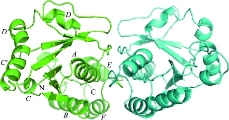
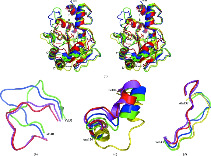
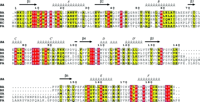
Nicotinic acid mononucleotide adenylyltransferase (NaMNAT; EC 2.7.7.18) is the penultimate enzyme in the biosynthesis of NAD(+) and catalyzes the adenylation of nicotinic acid mononucleotide (NaMN) by ATP to form nicotinic acid adenine dinucleotide (NaAD). This enzyme is regarded as a suitable candidate for antibacterial drug development; as such, Bacillus anthracis NaMNAT (BA NaMNAT) was heterologously expressed in Escherichia coli for the purpose of inhibitor discovery and crystallography. The crystal structure of BA NaMNAT was determined by molecular replacement, revealing two dimers per asymmetric unit, and was refined to an R factor and R(free) of 0.228 and 0.263, respectively, at 2.3 A resolution. The structure is very similar to that of B. subtilis NaMNAT (BS NaMNAT), which is also a dimer, and another independently solved structure of BA NaMNAT recently released from the PDB along with two ligated forms. Comparison of these and other less related bacterial NaMNAT structures support the presence of considerable conformational heterogeneity and flexibility in three loops surrounding the substrate-binding area.
Figures

References
-
- Berger, F., Ramirez-Hernandez, M. H. & Ziegler, M. (2004). Trends Biochem. Sci.29, 111–118. - PubMed
-
- Brünger, A. T., Adams, P. D., Clore, G. M., DeLano, W. L., Gros, P., Grosse-Kunstleve, R. W., Jiang, J.-S., Kuszewski, J., Nilges, M., Pannu, N. S., Read, R. J., Rice, L. M., Simonson, T. & Warren, G. L. (1998). Acta Cryst. D54, 905–921. - PubMed
-
- Collaborative Computational Project, Number 4 (1994). Acta Cryst. D50, 760–763. - PubMed
-
- Emsley, P. & Cowtan, K. (2004). Acta Cryst. D60, 2126–2132. - PubMed
Publication types
MeSH terms
Substances
Grants and funding
LinkOut - more resources
Full Text Sources
Other Literature Sources
