. 2008 Nov;15(11):1138-46.
doi: 10.1038/nsmb.1504. Epub 2008 Oct 19.

FANCI phosphorylation functions as a molecular switch to turn on the Fanconi anemia pathway

Affiliations

FANCI phosphorylation functions as a molecular switch to turn on the Fanconi anemia pathway

Masamichi Ishiai et al. Nat Struct Mol Biol. 2008 Nov.

Abstract

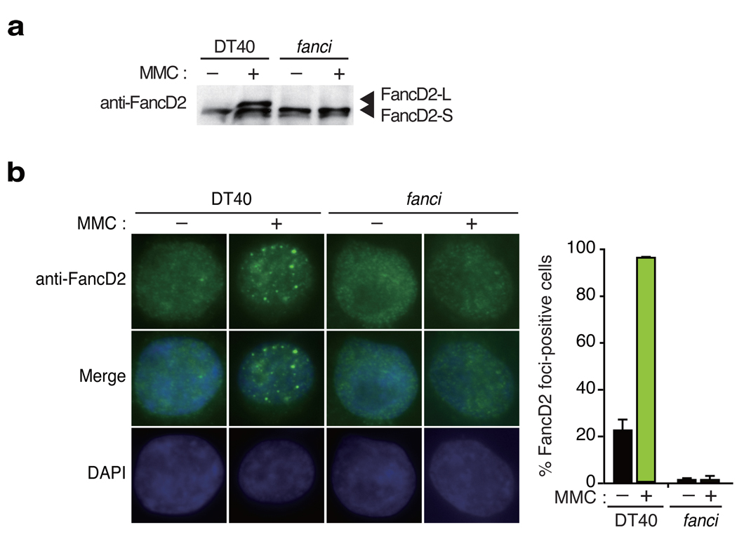
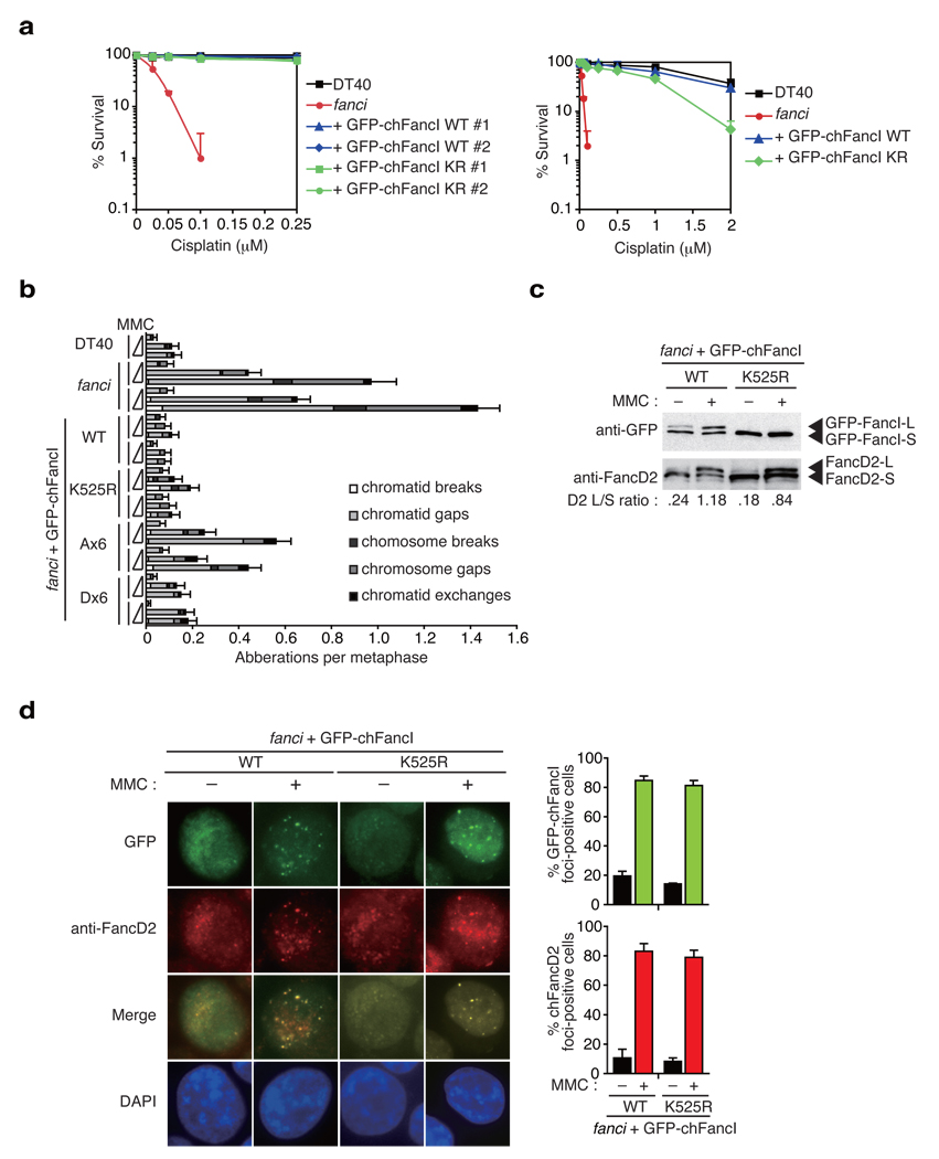
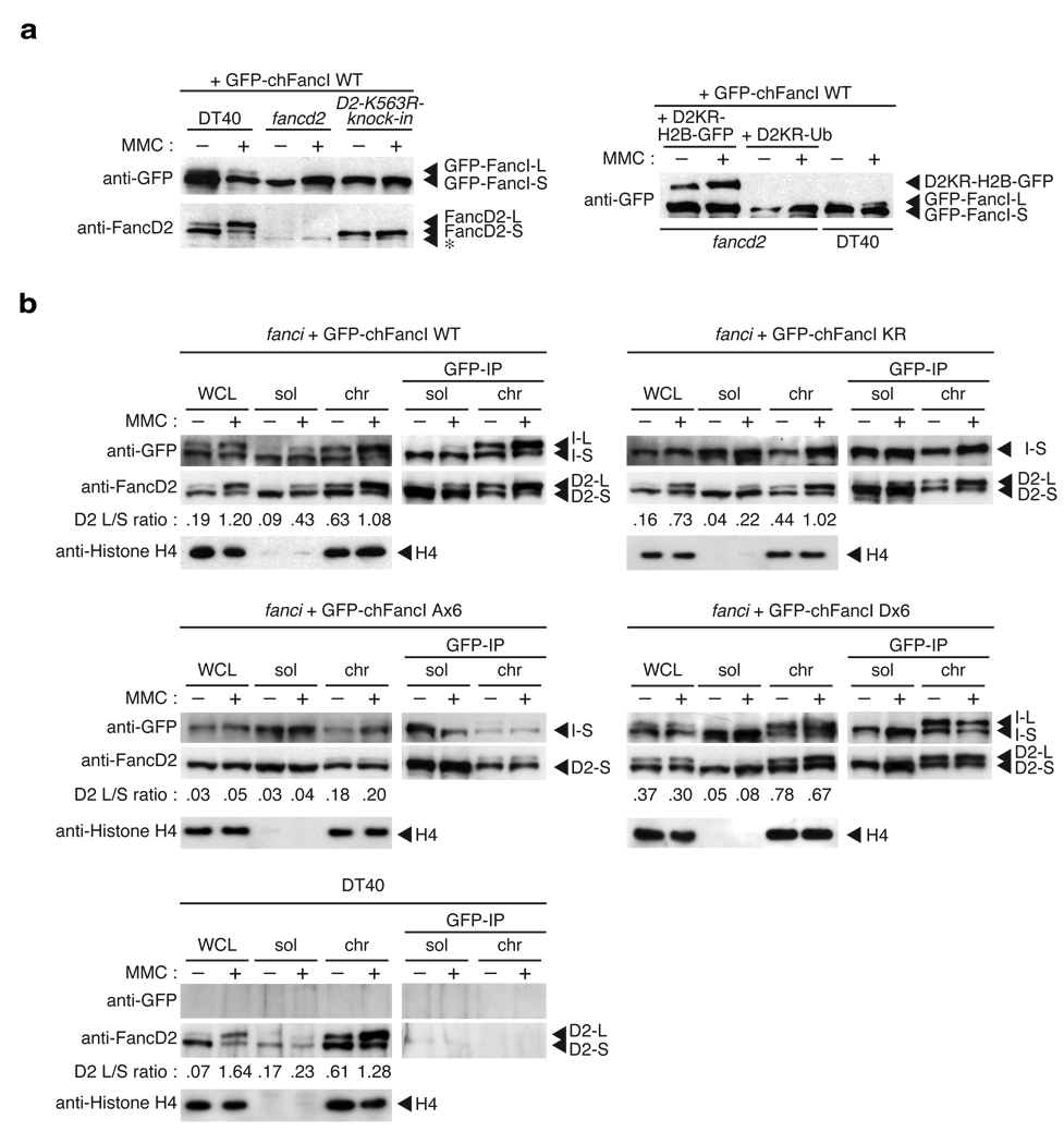
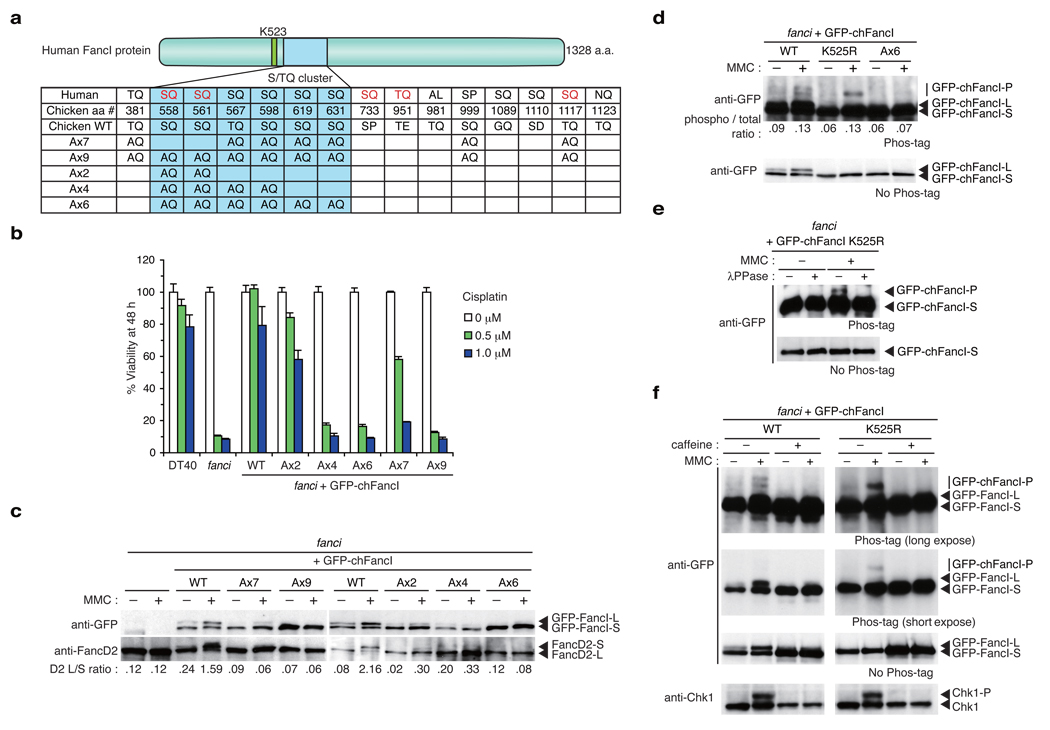
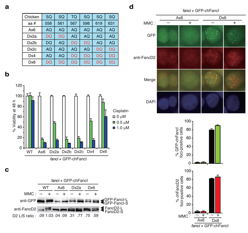
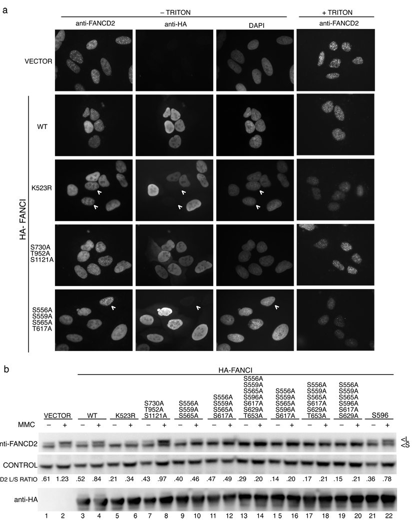
In response to DNA damage or replication fork stress, the Fanconi anemia pathway is activated, leading to monoubiquitination of FANCD2 and FANCI and their colocalization in foci. Here we show that, in the chicken DT40 cell system, multiple alanine-substitution mutations in six conserved and clustered Ser/Thr-Gln motifs of FANCI largely abrogate monoubiquitination and focus formation of both FANCI and FANCD2, resulting in loss of DNA repair function. Conversely, FANCI carrying phosphomimic mutations on the same six residues induces constitutive monoubiquitination and focus formation of FANCI and FANCD2, and protects against cell killing and chromosome breakage by DNA interstrand cross-linking agents. We propose that the multiple phosphorylation of FANCI serves as a molecular switch in activation of the Fanconi anemia pathway. Mutational analysis of putative phosphorylation sites in human FANCI indicates that this switch is evolutionarily conserved.


Figures

Figure 1
Figure 1. Gene targeting of FANCI resulted in loss of FancD2 monoubiquitination and focus formation
(a) Western blot analysis of FancD2 monoubiquitination induced by MMC treatment. (b) FancD2 localization to nuclear foci by indirect fluorescence using anti-FancD2 antibodies and DAPI counter staining. Representative images of wild type (WT) or fanci cells with or without MMC treatment stained for endogenous FancD2 are shown. The mean and standard error (s.e.m.) of %FancD2 foci-positive cells in three independent experiments are shown in the bar graph. More than 100 cells were scored in each experiment, and cells containing more than 4 bright foci were defined as foci-positive.
Figure 2
Figure 2. FancI monoubiquitination plays a minor role in inducing FA pathway activation
(a) Cisplatin sensitivity of cells with indicated genotypes were evaluated by colony survival. fanci cells were transfected with GFP-chFancI WT or K525R expression vector. The experiment was repeated at least three times, and a representative data set with mean and standard deviation of the triplicate cultures are shown. (b) Chromosome analysis of cells with the indicated genotypes. Along with wild type (DT40) and two clones of fanci cells, two independent clones of fanci cells expressing GFP-chFancI WT or mutant proteins were examined. Cells were treated with the increasing concentrations of MMC (0, 20, and 40 ng/ml) for 24 h. At least 50 metaphases were scored blindly for each preparation. Error bars indicate standard error (s.e.m.). (c) Monoubiquitination of FancD2 and FancI. Whole cell extracts were prepared from cells with the indicated genotypes and analyzed using antibodies against GFP or FancD2. (d) GFP-chFancI or FancD2 localization to DNA damage-induced nuclear foci. Representative images of fanci cells expressing GFP-chFancI wild type (WT) or the K525R mutant with or without MMC treatment are shown. The graph shows the mean and s.e.m. of %FancD2 or %GFP-chFancI foci-positive cells in fanci cells with GFP-FancI WT or K525R in three independent experiments. Cells with more than four bright foci were defined as foci-positive. More than 100 cells were scored for each data.
Figure 3
Figure 3. FancI monoubiquitination depends on FandD2 monoubiquitination but their physical interaction is constitutive
(a) Genetic requirements for FancI monoubiquitination. Wild type DT40 cells, fancd2 cells, cells with the FANCD2 monoubiuqitination site K563R knockin mutation (D2-K563R-knock-in), and fancd2 cells carrying D2KR-H2B-GFP or D2KR-Ub fusions were stably transfected with GFP-chFancI wild type (WT), and treated with or without MMC. Whole cells lysates were blotted with anti-GFP or anti-FancD2 antibodies. Asterisk (*) indicates a non-specific band. (b) Co-immunoprecipitation between FancD2 and FancI. Indicated cells were stimulated with MMC, and fractionated into soluble and chromatin fractions. Immunoprecipitation was carried out using anti-GFP antibody beads. Whole cell lysates (WCL), fractions (5% of the input), and immunoprecipitates were separated by SDS-PAGE and blotted with antibodies as indicated. As a negative control for anti-GFP immunoprecipitation, wild type DT40 not expressing GFP-chFancI was similarly fractionated and analyzed. L or S indicates L-form or S-form, respectively.
Figure 4
Figure 4. Phosphorylation sites in FancI are required for FA pathway activation and DNA repair
(a) S/TQ sites in human and chicken FancI proteins were listed with corresponding sites in the other protein. Red lettering indicate sites whose IR-induced phosphorylation was detected by mass spectrometry. Locations of multiple alanine substitutions in mutant chFancI proteins are indicated. (b) Cell survival in cisplatin containing medium was assayed using PI staining and FACSCalibur (Becton-Dickinson). (c) Monoubiquitination of GFP-chFancI and chFancD2 in response to MMC. Whole cell lysates were separated by SDS-PAGE and blotted using antibodies against GFP and chFancD2. (d) Phosphorylation of GFP-chFancI in response to MMC. Whole cell lysates were separated by SDS-PAGE using a gel containing Phos-tag-acrylamide, and blotted using anti-GFP antibodies. Shifted bands are indicated as GFP-chFancI-P. (e) GFP-chFancI was immunoprecipitated from the indicated cells, and treated with λ-phosphatase (λPPase), and detected as in d. (f) Cells were treated with caffeine (20 mM) and/or MMC (500 ng ml−1) or 6 h or left untreated, and analyzed by Phos-tag-containing gel and Western blotting as in d using antibodies against GFP or Chk1. For Phos-tag western, long and short exposures are shown.
Figure 5
Figure 5. Phospho-mimic mutants induce constitutive activation of the FA pathway
(a) Phospho-mimic mutations in the S/TQ cluster in chFancI. (b) Phospho-mimic mutations protected fanci cells against DNA damage induced by cisplatin. Cell survival was assessed as in Fig. 4b. (c) Constitutive monoubiquitination of GFP-chFancI and chFancD2. Whole cell lysates were separated and blotted using antibodies against GFP or chFancD2. (d) GFP-chFancI Ax6 or Dx6 localization in cells with or without DNA damage. Fixed cells were stained with anti-FancD2 antibodies and detected using fluorescence microscopy. The mean and standard error (s.e.m.) of %FancD2 or %GFP-chFancI foci-positive cells in three independent experiments are shown in the bar graph. Cells with more than four bright foci were defined as foci-positive. More than 100 cells were scored for each sample.
Figure 6
Figure 6. Analysis of phosphorylation mutants in human FANCI
(a) Localization of FancD2 in U2OS cells expressing the indicated HA-tagged FANCI alleles. Cells were treated with 1 µM MMC and 24 h later were costained with an antibody against FANCD2 and the HA-tag. The white arrowheads indicate cells that show no expression of HA-tagged FancI serving as an internal control for FANCD2 staining. Triton treatment removes the majority of the nucleoplasmic FANCD2 allowing for a better visualization of FANCD2 foci. (b) Western analysis of FANCD2 in U2OS cells expressing HA-tagged FANCI alleles. Cells expressing the indicated alleles of FANCI were treated with 1 µM MMC and collected 24 h later. L indicates the long (monoubiquitinated) and S is the short form of FANCD2.

Comment in

References

    1. Hoeijmakers JH. Genome maintenance mechanisms for preventing cancer. Nature. 2001;411:366–374. - PubMed
    1. Nijnik A, et al. DNA repair is limiting for haematopoietic stem cells during ageing. Nature. 2007;447:686–690. - PubMed
    1. Ito K, et al. Regulation of oxidative stress by ATM is required for self-renewal of haematopoietic stem cells. Nature. 2004;431:997–1002. - PubMed
    1. Rossi DJ, et al. Deficiencies in DNA damage repair limit the function of haematopoietic stem cells with age. Nature. 2007;447:725–729. - PubMed
    1. D'Andrea AD, Grompe M. The Fanconi anaemia/BRCA pathway. Nat. Rev. Cancer. 2003;3:23–34. - PubMed

Publication types

MeSH terms

Substances