. 2008 Dec;9(12):1407-14.
doi: 10.1038/ni.1669. Epub 2008 Oct 19.

Proteolytic cleavage in an endolysosomal compartment is required for activation of Toll-like receptor 9

Affiliations

Proteolytic cleavage in an endolysosomal compartment is required for activation of Toll-like receptor 9

Boyoun Park et al. Nat Immunol. 2008 Dec.

Abstract

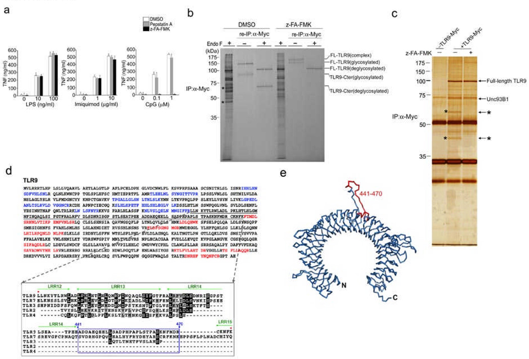
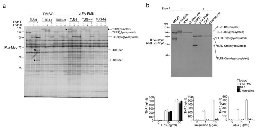
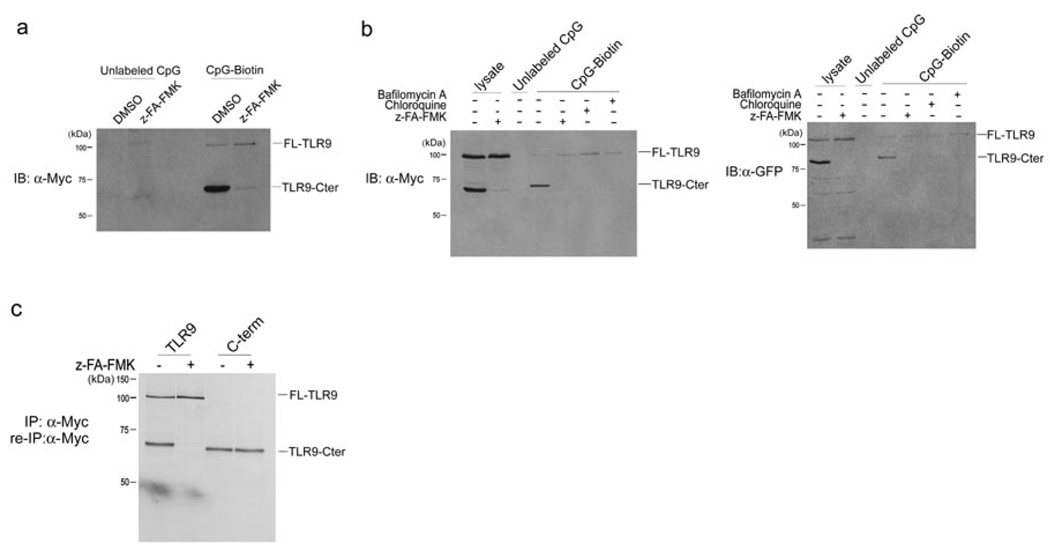
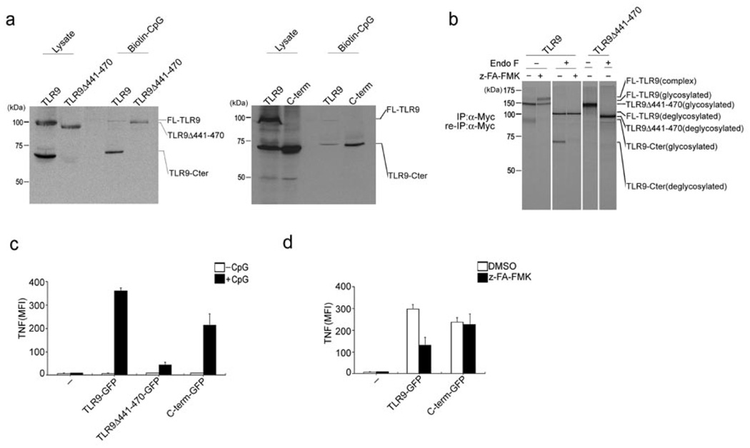
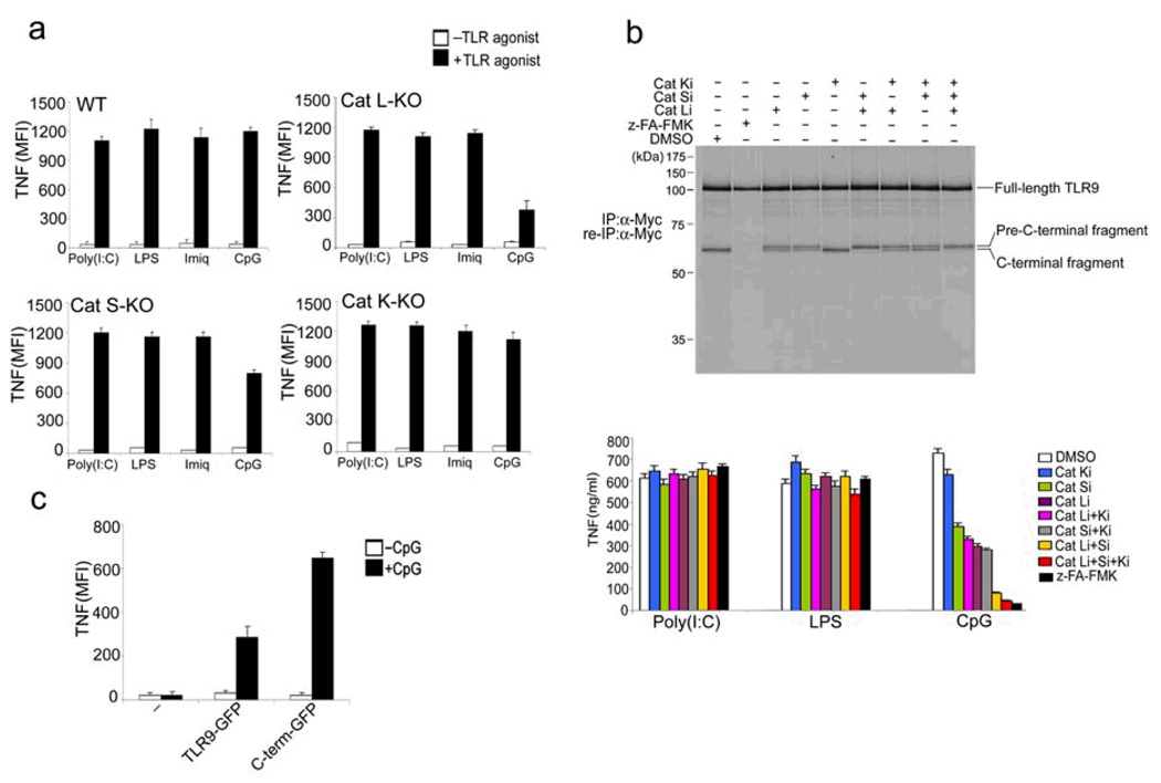
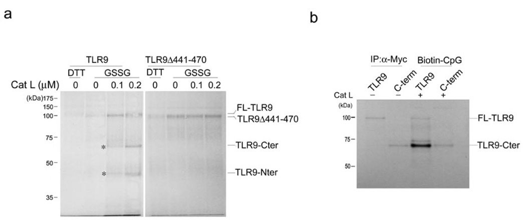
Toll-like receptors (TLRs) activate the innate immune system in response to pathogens. Here we show that TLR9 proteolytic cleavage is a prerequisite for TLR9 signaling. Inhibition of lysosomal proteolysis rendered TLR9 inactive. The carboxy-terminal fragment of TLR9 thus generated included a portion of the TLR9 ectodomain, as well as the transmembrane and cytoplasmic domains. This cleavage fragment bound to the TLR9 ligand CpG DNA and, when expressed in Tlr9(-/-) dendritic cells, restored CpG DNA-induced cytokine production. Although cathepsin L generated the requisite TLR9 cleavage products in a cell-free in vitro system, several proteases influenced TLR9 cleavage in intact cells. Lysosomal proteolysis thus contributes to innate immunity by facilitating specific cleavage of TLR9.


Figures

Figure 1
Figure 1
TLR9 is cleaved into two distinct polypeptides by cathepsins. (a) RAW macrophages were treated with either pepstatin A, z-FA-FMK or DMSO, followed by incubation with LPS, Imiquimod or CpG DNA. (b) RAW macrophages expressing C-terminally Myc-tagged TLR9 were pre-treated with DMSO or z-FA-FMK. Radioactively labeled proteins were subjected to immunoprecipitation with anti-Myc. One tenth of the immunoprecipitates were resolved by SDS-PAGE. After denaturation, the remainder was subjected to re-immunoprecipitation with anti-Myc and treated with EndoF where indicated. Asterisks depict 45 kDa and 65 kDa TLR9 cleavage fragments. We analyzed duplicate samples for each condition. FL: full length; Cter: C-terminal fragment (c) RAW macrophages expressing TLR9-Myc were treated with DMSO (−) or z-FA-FMK (+), anti-Myc immunoprecipitated proteins were visualized by silver staining. Polypeptides of 65 and 45 kDa (*) were analyzed by LC/MS/MS. (d) Top, peptides identified by LC/MS/MS from (c) are highlighted in blue (N-terminal) and red (C-terminal) in the murine Tlr9 sequence. No peptides were identified in the region encompassing residues 378–475 (underlined). Bottom, alignment of the region encompassing the cleavage site(s) of TLR9 (378–475) with sequences of other indicated TLRs. Residues 441 to 470 (boxed in blue) are part of a flexible loop. Leucine rich repeats (LRR) are highlighted in green. (e) Ribbon representation of a model of the TLR9 ectodomain based on the crystal structure of the TLR3 ectodomain. The predicted cathepsin cleavage site (441–470) is highlighted in red. Data are representative of four (a) or two (b–c) independent experiments (a;average, s.d.).
Figure 2
Figure 2
Trafficking of TLR9 to the endolysosomal compartment is required for its fragmentation. (a) RAW macrophages stably expressing Myc-tagged TLR chimeras or Myc-tagged wild-type TLR9 were treated with DMSO or 10 µM z-FA-FMK for 12 h, metabolically labeled for 1.5 h and chased for 6 h. TLR proteins were recovered by immunoprecipitation with an anti-Myc, treated with glycosidases EndoH or EndoF where indicated and resolved by SDS-PAGE. The arrowhead indicates mature EndoH resistant TLR9, and the asterisks denote the C-terminal TLR9 cleavage fragments. (b) Top, RAW macrophages expressing TLR9-Myc were incubated for 10 h with DMSO (lanes 1 and 5), 10 µM z-FA-FMK (lanes 2 and 6) and for 4 h with 5 µg/ml bafilomycin (lanes 3 and 7) or 5 µM chloroquine (lanes 4 and 8) and metabolically labeled for 1 h, followed by a chase period of 6 h. Immunoprecipitation and reimmunoprecipitation were performed with an anti-Myc. Immunoprecipitates were subjected to treatment with EndoF where indicated. Bottom, RAW macrophages were incubated with DMSO or 10 µM z-FA-FMK for 12 h, 5 µg/ml bafilomycin or 5 µM chloroquine for 4 h, stimulated with increasing concentrations of LPS, Imiquimod or CpG DNA for 2 h, and TNF secretion was analyzed by ELISA. Data are representative of three independent experiments (b;average, s.d.).
Figure 3
Figure 3
The C-terminal TLR9 fragment directly interacts with CpG DNA. (a) RAW macrophages expressing TLR9-Myc were pretreated with DMSO or 10 µM z-FA-FMK for 12 h and then incubated with 3 µM unlabeled or biotinylated CpG DNA for 3 h at 37°C. CpG DNA and materials bound to it were recovered on streptavidin agarose, resolved by SDS-PAGE and probed with anti-Myc. (b) RAW macrophages expressing TLR9-Myc or TLR9-GFP were treated with DMSO or 10 µM z-FA-FMK for 12 h, followed by incubation with 5 µg/ml bafilomycin or 5 µM chloroquine for 4 h and incubated with 3 µM biotinylated or unlabeled CpG DNA for 3 h at 37°C. CpG DNA and materials bound to it were retrieved on streptavidin agarose, subjected to digestion with EndoF, resolved by SDS-PAGE next to total input lysate and probed with anti-Myc or anti-GFP. (c) RAW macrophages expressing either wild-type TLR9-Myc or Myc-tagged C-terminal fragment of TLR9 (471–1032) were treated with z-FA-FMK (+) or DMSO (−) and metabolically labeled. Lysates were subjected to immunoprecipitation with anti-Myc, digested with EndoF and resolved by SDS-PAGE. Data are representative of three independent experiments.
Figure 4
Figure 4
The C-terminal TLR9 fragment is the active form responsible for binding CpG DNA and subsequent TLR9 signal transduction. (a) RAW macrophages stably expressing TLR9Δ441–470 or the C-terminal TLR9 fragment (471–1032) were incubated with 3 µM biotinylated CpG DNA for 3 h at 37°C. CpG DNA and materials bound to it were precipitated with streptavidin agarose, subjected to digestion with EndoF and precipitates and input lysates were immunoblotted with anti-Myc. (b) Myc-tagged wild-type TLR9 and Myc-tagged TLR9Δ441–470 were immunoprecipitated and reimmunoprecipitated with anti-Myc from DMSO- or z-FA-FMK-treated and metabolically labeled RAW macrophages, digested with EndoF where indicated, and visualized by SDS-PAGE. (c,d) BMDCs from Tlr9−/− mice were retrovirally transduced with vectors encoding GFP-tagged wild-type TLR9, TLR9Δ441–470 or the C-terminal TLR9 fragment (471–1032). (c) Cells were stimulated with CpG DNA (1 µM) for 4 h in the presence of brefeldin A at day 5 of BMDC culture. Cells were fixed and stained with anti-TNF, and TNF-expressing GFP+ cells were quantified by flow cytometry. Data were generated from three independent experiments and are expressed as mean fluorescence intensity (MFI). (d) Cells were stimulated with CpG (1 µM) for 4 h in the presence of DMSO or 1 µM z-FA-FMK for 8 h at day 5 of BMDC culture. TNF expression was measured as in (c). Data are representative of three independent experiments (c,d; average, s.d.).
Figure 5
Figure 5
Multiple lysosomal proteases are required for TLR9 cleavage. (a) BMDCs from wild-type mice or mice lacking cathepsin L (Cat L-KO), cathepsin S (Cat S-KO) or cathepsin K (Cat K-KO) were stimulated with poly I:C (100 µg/ml), LPS (1 µg/ml), Imiquimod (10 µg/ml) or CpG DNA (1 µM) for 4 h in the presence of brefeldin A at day 6 of BMDC culture. Cells were fixed and stained with anti-TNF, and intracellular TNF was measured by flow cytometry. (b) Top, RAW macrophages expressing TLR9-Myc were pretreated for 12 h with DMSO, z-FA-FMK (10 µM) or the selective cathepsin L inhibitors Clik195 and Clik148 (Cat Li, 10 µM), the cathepsin S inhibitor LHVS (Cat Si, 10 nM), the cathepsin K inhibitor II (Cat Ki, 1 µM), or combinations thereof. Cells were metabolically labeled for 1.5 h followed by a 5 h chase period. TLR9 was immunoprecipitated and re-immunoprecipitated from lysates with anti-Myc, and was digested with EndoF. Bottom, RAW macrophages were treated for 12 h with the inhibitors listed and stimulated for 2 h with the indicated TLR agonists. Secreted TNF was analysed by ELISA. (c) BMDCs from Cat L-KO mice were retrovirally transduced with vectors encoding GFP-tagged wild-type TLR9 or GFP-tagged C-terminal TLR9 fragment (471–1032) or were left untransduced (−) at day 1 of BMDC culture. At day 6, cells were stimulated with CpG (1µM) for 4 h in the presence of brefeldin A, fixed and stained with anti-TNF. TNF was measured by flow cytometry in GFP+ (transduced) cells. Data are representative of two (a,c) or three (b) independent experiments (a-c; average, s.d.).
Figure 6
Figure 6
Cathepsin L cleaves TLR9 in vitro but fails to cleave the TLR9 deletion mutant lacking the region encompassing the putative cathepsin cleavage site(s). (a) Wild-type TLR9 or the TLR9 deletion mutant TLR9Δ441–470 were transcribed and translated in vitro in the presence of microsomes and 35S-methionine and either 2 mM DTT or 2 mM oxidized glutathione (GSSG). Microsomes were pelleted, lysed and recombinant cathepsin L was added for 2 h at 37°C. 10% of the total volume of the in vitro transcription and translation reaction was loaded per lane and separated by SDS-PAGE. Asterisks indicate the C- and N-terminal fragment of TLR9. (b) Myc-tagged wild-type TLR9 or C-terminal TLR9 fragment (471–1032) were transcribed and translated in vitro in the presence of microsomes and 35S-methionine. Microsomes were pelleted, lysed and 10% was subjected to immunoprecipitation with anti-Myc antibody, digested with EndoF and resolved by SDS-PAGE. 90% was incubated with 0.2 µM recombinant cathepsin L for 2 h at 37°C followed by a 2 h incubation with 5 µM biotinylated CpG DNA. Samples were then incubated with streptavidin agarose, digested with EndoF and resolved by SDS-PAGE. Data are representative of two independent experiments.

References

    1. Takeda K, Kaisho T, Akira S. Toll-like receptors. Annu Rev Immunol. 2003;21:335–376. - PubMed
    1. Janeway CA, Jr, Medzhitov R. Innate immune recognition. Annu Rev Immunol. 2002;20:197–216. - PubMed
    1. Kawai T, Akira S. TLR signaling. Cell Death Differ. 2006;13:816–825. - PubMed
    1. Takeda K, Akira S. Toll-like receptors in innate immunity. Int Immunol. 2005;17:1–14. - PubMed
    1. Poltorak A, et al. Defective LPS signaling in C3H/HeJ and C57BL/10ScCr mice: mutations in Tlr4 gene. Science. 1998;282:2085–2028. - PubMed

Publication types

MeSH terms