

Permeability of the rhesus monkey oocyte membrane to water and common cryoprotectants
- PMID: 18932214
- PMCID: PMC4141568
- DOI: 10.1002/mrd.20956
Permeability of the rhesus monkey oocyte membrane to water and common cryoprotectants
Abstract
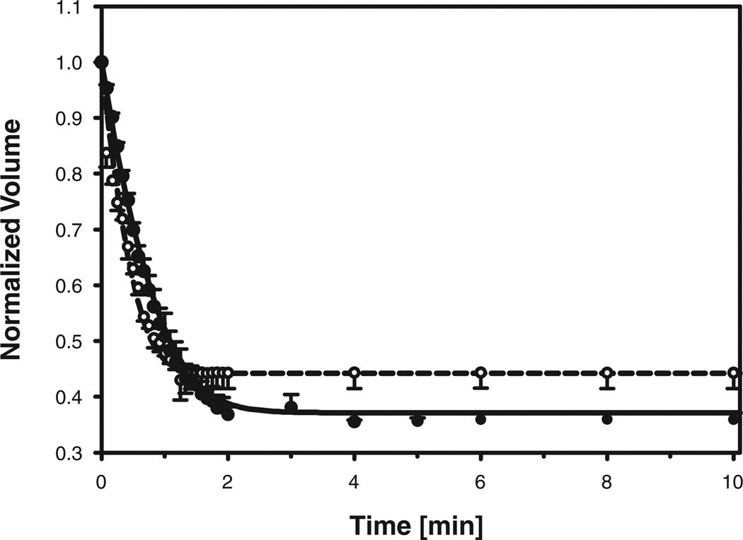
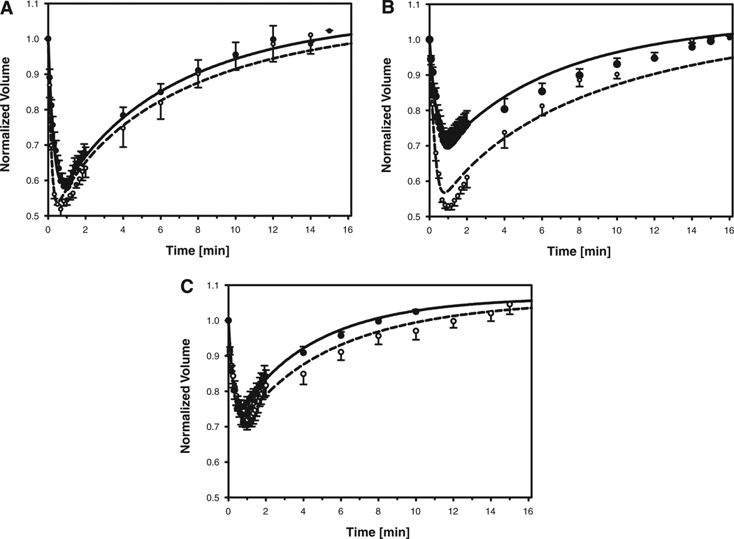
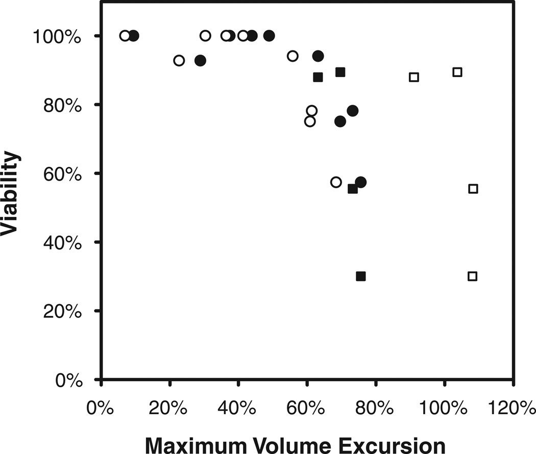
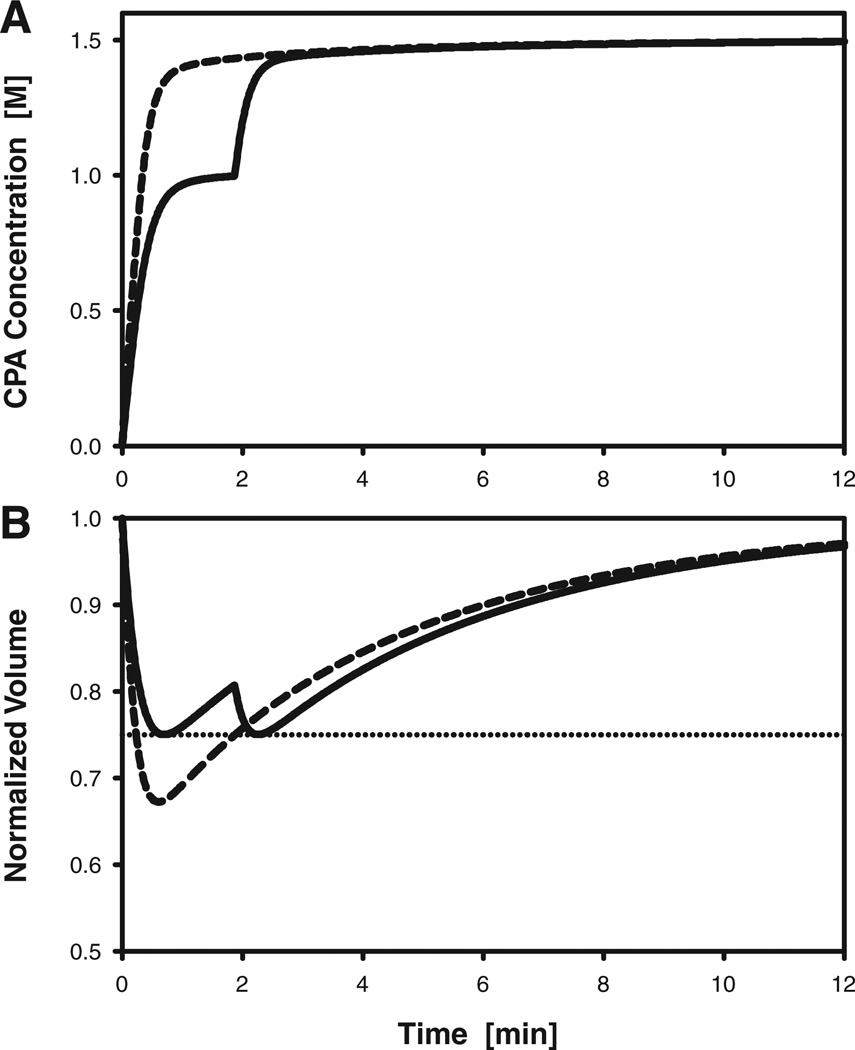
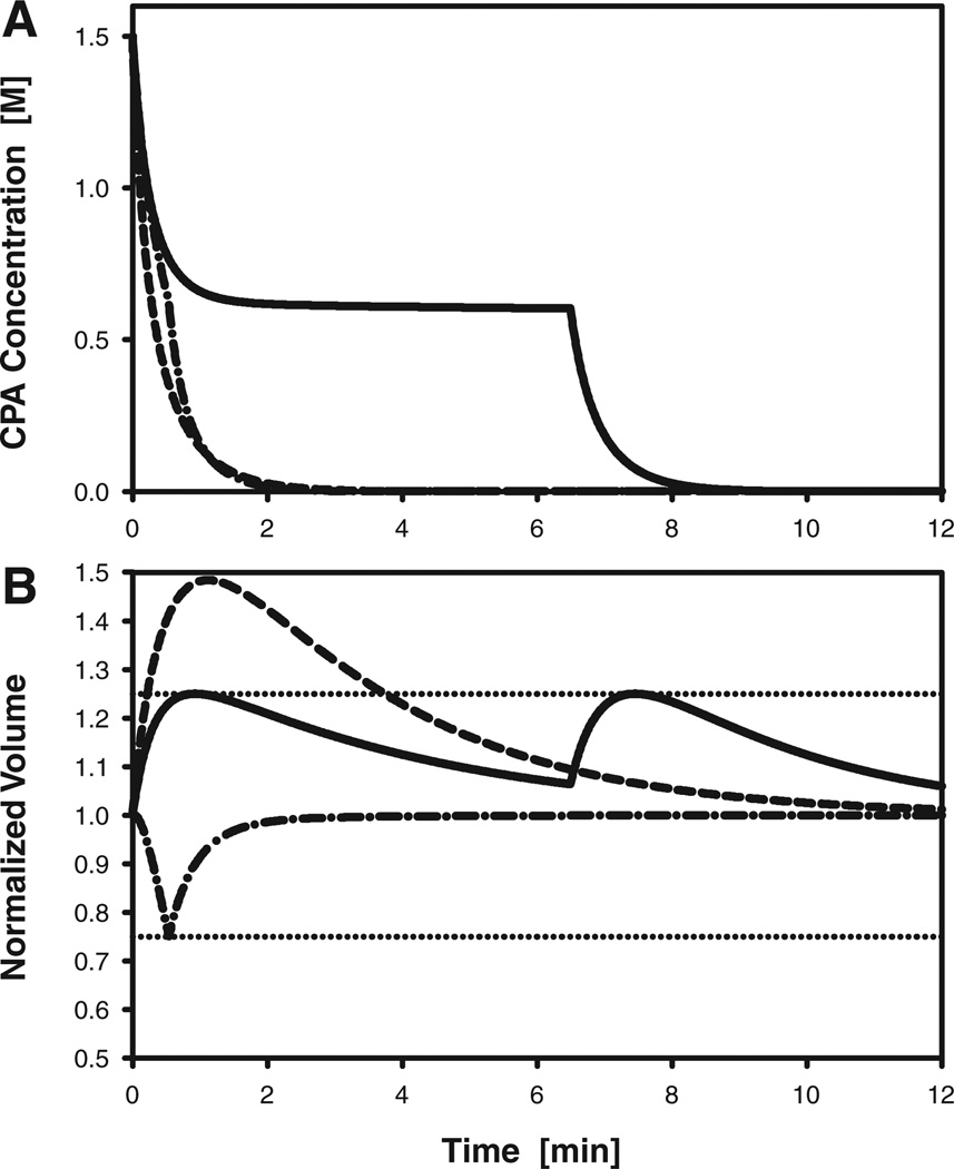
Successful cryopreservation of oocytes of the rhesus monkey (Macaca mulatta) would facilitate the use of this valuable animal model in research on reproduction and development, while providing a stepping stone towards human oocyte cryopreservation and the conservation of endangered primate species. To enable rational design of cryopreservation techniques for rhesus monkey oocytes, we have determined their osmotic and permeability characteristics in the presence of dimethylsulfoxide (DMSO), ethylene glycol (EG), and propylene glycol (PROH), three widely used cryoprotectants. Using nonlinear regression to fit a membrane transport model to measurements of dynamic cell volume changes, we estimated the hydraulic conductivity (L(p)) and cryoprotectant permeability (P(s)) of mature and immature oocytes at 23.5 degrees C. Mature oocyte membranes were most permeable to PROH (P(s) = 0.56 +/- 0.05 microm/sec) and least permeable to DMSO (P(s) = 0.24 +/- 0.02 microm/sec); the permeability to EG was 0.34 +/- 0.07 microm/sec. In the absence of penetrating cryoprotectants, mature oocytes had L(p) = 0.55 +/- 0.05 microm/min/atm, whereas the hydraulic conductivity increased to 1.01 +/- 0.10, 0.61 +/- 0.07, or 0.86 +/- 0.06 microm/min/atm when mature oocytes were exposed to DMSO, EG, or PROH, respectively. The osmotically inactive volume (V(b)) in mature oocytes was 19.7 +/- 2.4% of the isotonic cell volume. The only statistically significant difference between mature and immature oocytes was a larger hydraulic conductivity in immature oocytes that were exposed to DMSO. The biophysical parameters measured in this study were used to demonstrate the design of cryoprotectant loading and dilution protocols by computer-aided optimization.
(c) 2008 Wiley-Liss, Inc.
Figures
Similar articles
-
Determination of oocyte membrane permeability coefficients and their application to cryopreservation in a rabbit model.Cryobiology. 2009 Oct;59(2):127-34. doi: 10.1016/j.cryobiol.2009.06.002. Epub 2009 Jun 13. Cryobiology. 2009. PMID: 19527701
-
Studies on membrane permeability of zebrafish (Danio rerio) oocytes in the presence of different cryoprotectants.Cryobiology. 2005 Jun;50(3):285-93. doi: 10.1016/j.cryobiol.2005.02.007. Epub 2005 Apr 1. Cryobiology. 2005. PMID: 15925580
-
Effect of developmental stage on bovine oocyte plasma membrane water and cryoprotectant permeability characteristics.Mol Reprod Dev. 1998 Apr;49(4):408-15. doi: 10.1002/(SICI)1098-2795(199804)49:4<408::AID-MRD8>3.0.CO;2-R. Mol Reprod Dev. 1998. PMID: 9508092 Review.
-
Osmotic tolerance and membrane permeability characteristics of rhesus monkey (Macaca mulatta) spermatozoa.Cryobiology. 2005 Aug;51(1):1-14. doi: 10.1016/j.cryobiol.2005.04.004. Cryobiology. 2005. PMID: 15922321
-
The need for novel cryoprotectants and cryopreservation protocols: Insights into the importance of biophysical investigation and cell permeability.Biochim Biophys Acta Gen Subj. 2021 Jan;1865(1):129749. doi: 10.1016/j.bbagen.2020.129749. Epub 2020 Sep 25. Biochim Biophys Acta Gen Subj. 2021. PMID: 32980500 Review.
Cited by
-
Mathematical Modeling and Optimization of Cryopreservation in Single Cells.Methods Mol Biol. 2021;2180:129-172. doi: 10.1007/978-1-0716-0783-1_4. Methods Mol Biol. 2021. PMID: 32797410
-
Detection of volume changes in calcein-stained cells using confocal microscopy.J Fluoresc. 2013 May;23(3):393-8. doi: 10.1007/s10895-013-1202-1. Epub 2013 Mar 7. J Fluoresc. 2013. PMID: 23468245
-
Controlled loading of cryoprotectants (CPAs) to oocyte with linear and complex CPA profiles on a microfluidic platform.Lab Chip. 2011 Oct 21;11(20):3530-7. doi: 10.1039/c1lc20377k. Epub 2011 Sep 1. Lab Chip. 2011. PMID: 21887438 Free PMC article.
-
Membrane permeability of the human granulocyte to water, dimethyl sulfoxide, glycerol, propylene glycol and ethylene glycol.Cryobiology. 2014 Feb;68(1):35-42. doi: 10.1016/j.cryobiol.2013.11.004. Epub 2013 Nov 20. Cryobiology. 2014. PMID: 24269528 Free PMC article.
-
Toxicity Minimized Cryoprotectant Addition and Removal Procedures for Adherent Endothelial Cells.PLoS One. 2015 Nov 25;10(11):e0142828. doi: 10.1371/journal.pone.0142828. eCollection 2015. PLoS One. 2015. PMID: 26605546 Free PMC article.
References
-
- Agca Y, Liu J, Peter AT, Critser ES, Critser JK. Effect of developmental stage on bovine oocyte plasma membrane water and cryoprotectant permeability characteristics. Mol Reprod Dev. 1998;49:408–415. - PubMed
-
- Agca Y, Liu J, Critser ES, Critser JK. Fundamental cryobiology of rat immature and mature oocytes: Hydraulic conductivity in the presence of Me2SO, Me2SO permeability, and their activation energies. J Exp Zool. 2000;286:523–533. - PubMed
-
- Al-Hasani S, Diedrich K, van der Ven H, Reinecke A, Hartje M, Krebs D. Cryopreservation of human oocytes. Hum Reprod. 1987;2:695–700. - PubMed
-
- Arnaud FG, Pegg DE. Permeation of glycerol and propane-1,2-diol into human platelets. Cryobiology. 1990;27:107–118. - PubMed
-
- Ashwood-Smith MJ, Morris GW, Fowler R, Appleton TC, Ashorn R. Physical factors are involved in the destruction of embryos and oocytes during freezing and thawing procedures. Hum Reprod. 1988;3:795–802. - PubMed
Publication types
MeSH terms
Substances
Grants and funding
LinkOut - more resources
Full Text Sources
Research Materials
Miscellaneous
