

Review
doi: 10.1016/j.semcdb.2008.09.006.
Epub 2008 Sep 30.
ADAM function in embryogenesis
Affiliations
- PMID: 18935966
- PMCID: PMC2693894
- DOI: 10.1016/j.semcdb.2008.09.006
Item in Clipboard
Review
ADAM function in embryogenesis
Semin Cell Dev Biol.
2009 Apr.
Abstract
Cleavage of proteins inserted into the plasma membrane (shedding) is an essential process controlling many biological functions including cell signaling, cell adhesion and migration as well as proliferation and differentiation. ADAM surface metalloproteases have been shown to play an essential role in these processes. Gene inactivation during embryonic development have provided evidence of the central role of ADAM proteins in nematodes, flies, frogs, birds and mammals. The relative contribution of four subfamilies of ADAM proteins to developmental processes is the focus of this review.
Figures

(I) ADAMs are single-pass transmembrane proteins composed of a pro-domain (P) a metalloprotease domain (M), a disintegrin domain (D) a cysteine-rich domain (C) an EGF repeat (E) and a cytoplasmic domain (Cy). (II) The pro-domain of ADAMs is removed by furin cleavage either during the transit through the trans golgie network or at the cell surface producing the metalloprotease active form. (III) In some cases, a second cleavage occurs removing the metalloprotease domain and potentially “unmasking” the disintegrin domain to promote integrin binding [63, 73]. (IV) Additional shedding has been reported for both ADAM13 and ADAM33 whereby the metalloprotease domain is shed with the disintegrin and cysteine-rich domains [75, 80]. (V) A similar soluble active metalloprotease can be obtained by alternative splicing as shown for ADAM9s and ADAM12s [67, 100]. (VI) Alternative splicing can also generate a proteolytically inactive ADAM, anchored in the membrane as shown for ADAM19 [101].
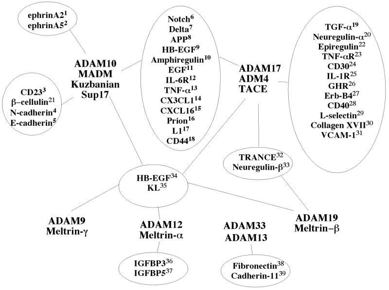
Diagram of selected ADAMs and substrates. The potential overlap between different ADAMs is presented. The number of potential substrates and the overlap makes the analysis of phenotype very difficult. 1 [55], 2 [23], 3 [102], 4 [49], 5 [50], 6 [26], 7 [35], 8 [103], 9 [104], 10 [105], 11 [71], 12 [106], 13 [107], 14 [108, 109], 15 [110, 111], 16 [112], 17 [78], 18 [113, 114], 19 [115], 20 [116], 21 [51], 22 [71], 23 [115], 24 [117], 25 [118], 26 [119], 27 [120], 28 [121], 29 [115, 122], 30 [123], 31 [124], 32 [125-127], 33 [83, 128, 129], 34 [83, 130-132], 35 [127, 133], 36 [134, 135], 37 [134], 38 [27], 39 McCusker et al., submitted.

Example of the strategy employed in Xenopus to test the function and potential substrate of ADAM proteases during development. The ADAM protein translation is inhibited by injection of a Morpolino oligonucleotide into the fertilized egg (A). At the blastula stage, mRNA encoding the wild type protein or various mutant lacking selected domains can be injected in a cell that will give rise to a specific tissue, for example the cranial neural crest cells (B). A lineage tracer is co-injected to compare the behavior of these cells to the rest of the embryo missing the ADAM protein. If an appropriate substrate is identified (C), the putative cleavage product (EC) can be produced and injected to test its ability to rescue the loss of ADAM phenotype. Phenotypical analysis can be performed at the tailbud stage (D) using the lineage tracer and tissue specific marker to determine the position and differentiation of the injected cells progeny. Similar techniques can be used in genetic models using tissue specific promoter rather than targeted injection.
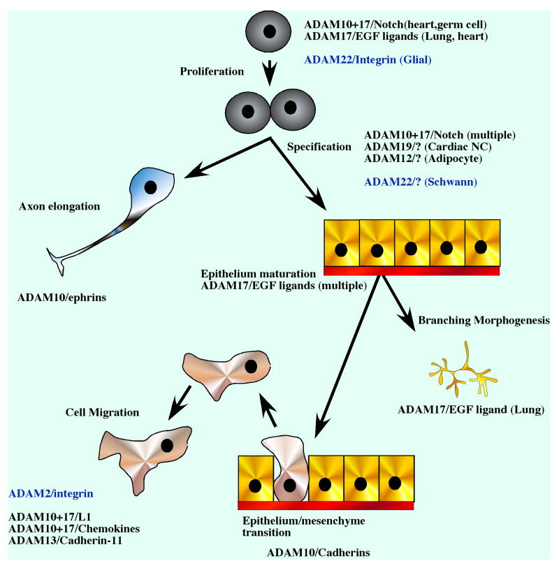
ADAM proteins are involved at every step of embryogenesis. They can control cell proliferation, cell specification and differentiation as well as various aspects of morphogenesis. ADAMs that have been implicated with each step are indicated together with the substrate if it is known. In parenthesis the cell type or tissue in which the function was shown is indicated. Non-proteolytic ADAMs are in blue.
References
-
- Blobel CP, Wolfsberg TG, Turck CW, Myles DG, Primakoff P, White JM. A potential fusion peptide and an integrin ligand domain in a protein active in sperm-egg fusion. Nature. 1992;356:248–252. - PubMed
-
- Muga A, Neugebauer W, Hirama T, Surewicz WK. Membrane interaction and conformational properties of the putative fusion peptide of PH-30, a protein active in sperm-egg fusion. Biochemistry. 1994;33:4444–4448. - PubMed
Publication types
MeSH terms
Substances
Grants and funding
LinkOut - more resources
Full Text Sources
Other Literature Sources
