. 2008 Dec 15;586(24):5911-29.
doi: 10.1113/jphysiol.2008.163154. Epub 2008 Oct 20.

Role of the hyperpolarization-activated current Ih in somatosensory neurons

Affiliations

Role of the hyperpolarization-activated current Ih in somatosensory neurons

Aliakmal Momin et al. J Physiol. .

Abstract

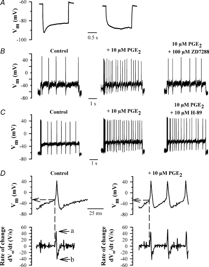
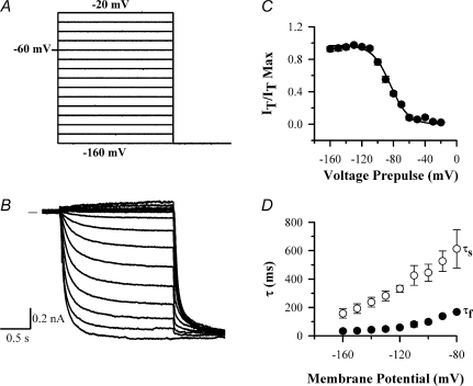
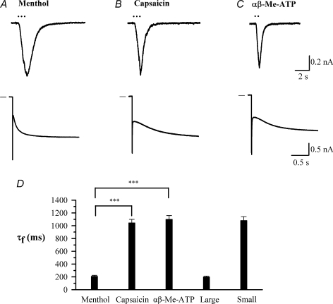
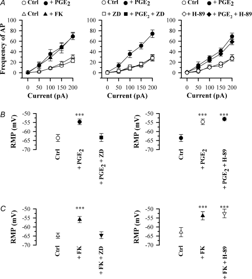
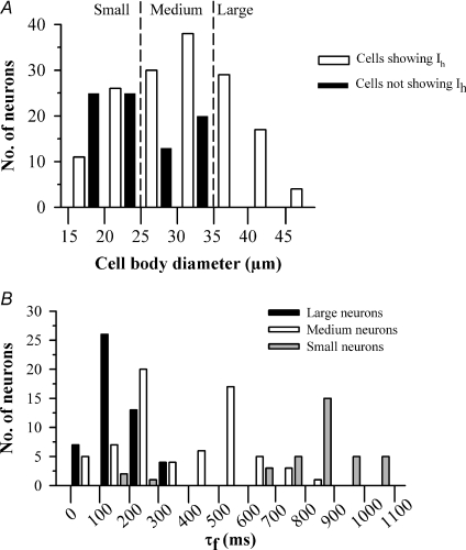
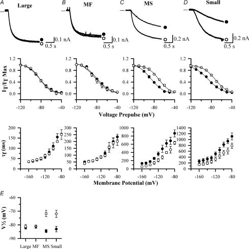
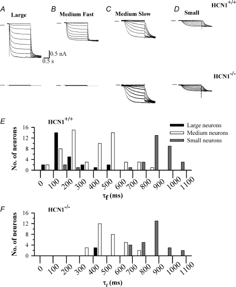
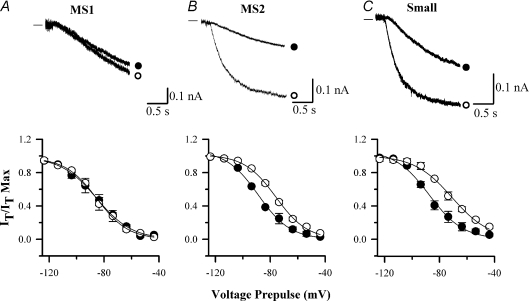
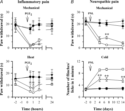
The hyperpolarization-activated current (I(h)) is an inward current activated by hyperpolarization from the resting potential and is an important modulator of action potential firing frequency in many excitable cells. Four hyperpolarization-activated, cyclic nucleotide-modulated subunits, HCN1-4, can form I(h) ion channels. In the present study we investigated the function of I(h) in primary somatosensory neurons. Neuronal firing in response to current injection was promoted by elevating intracellular cAMP levels and inhibited by blockers of I(h), suggesting that I(h) plays a critical role in modulating firing frequency. The properties of I(h) in three size classes of sensory neurons were next investigated. In large neurons I(h) was fast activating and insensitive to elevations in cAMP, consistent with expression of HCN1. I(h) was ablated in most large neurons in HCN1(-/-) mice. In small neurons a slower activating, cAMP-sensitive I(h) was observed, as expected for expression of HCN2 and/or HCN4. Consistent with this, I(h) in small neurons was unchanged in HCN1(-/-) mice. In a neuropathic pain model HCN1(-/-) mice exhibited substantially less cold allodynia than wild-type littermates, suggesting an important role for HCN1 in neuropathic pain. This work shows that I(h) is an important modulator of action potential generation in somatosensory neurons.


Figures

Figure 1
Figure 1. Enhancement by PGE2 of action potential firing frequency in small DRG neurons
A, time-dependent rectification or voltage ‘sag’ of membrane potential in small (< 25 μm diameter) rat DRG neurons. Representative traces of membrane potential following injection of a 500 pA hyperpolarizing current pulse for 2 s, showing a neuron expressing Ih (left) or not expressing Ih (right). A neuron was considered to express Ih if the amplitude of the voltage sag exceeded 10% of the initial peak voltage excursion. B, representative traces showing action potential firing produced by a 100 pA depolarizing current in control conditions (left), in the presence of 10 μm PGE2 (centre), and in the presence of 10 μm PGE2 plus 100 μm ZD7288 (right). C, same as B but in control (left), in the presence of 10 μm PGE2 (centre) and in the presence of 10 μm PGE2 plus 10 μm H-89 (right). Data in B and C were obtained from two different neurons. D, upper panels: action potentials, taken from traces in B, at an expanded time scale, in control conditions (left) and in the presence of 10 μm PGE2 (right). Lower panels show the 1st derivative (dVm/dt) of the voltage trace shown above. Arrows show maximum rate of rise (‘a’), maximum rate of fall (‘b’).
Figure 3
Figure 3. Voltage-clamp traces of Ih recorded from a large neuron using the caesium-subtraction technique
A, voltage protocol; Ih was activated from a holding potential of −60 mV in 2 s pulses from −20 mV to −160 mV in 10 mV increments, followed by a final step to −160 mV. B, Cs-subtracted traces, following subtraction of current traces in which Ih had been completely inhibited by application of 5 mm CsCl. Zero current level shown by horizontal line in this and other traces. C, activation of Ih as a function of membrane voltage, obtained from the magnitude of the Ih tail current at the start of the −160 mV test potential. Trace fitted to points with parameters V½=−81.4 ± 1.3 mV, slope factor s = 11.6 ± 1.0 mV. D, time constants as a function of membrane voltage. Ih current traces were fitted with sum of two exponentials as described in Methods to obtain a fast (τf, •) and slow time constant (τs, ○).
Figure 5
Figure 5. A subpopulation of small rat DRG neurons expresses TRPM8 and has fast-activating Ih
A–C, sample inward currents activated by a 1 s exposure to menthol (50 μm), capsaicin (1 μm) and α,β-methyl ATP (αβ-Me-ATP; 10 μm) in three different small neurons (< 25 μm); and Ih activated in the same neurons by a voltage step from −60 mV to −90 mV. The cell responding to menthol did not respond to either capsaicin or αβ-Me-ATP. D, time constants of the fast component of Ih in each class of neuron compared with the overall time constants for large (> 35 μm) and small (< 25 μm) neurons. ***P < 0.001 for the comparisons indicated. Note that data in this figure were obtained without the use of the Cs-subtraction technique, and values of time constants are therefore not directly comparable with those given in other figures which were all obtained using Cs-subtraction.
Figure 2
Figure 2. Effect of PGE2, forskolin (FK), ZD7288 (ZD) and H-89 on action potential firing in small (< 25 μm) rat DRG neurons
A, frequency of AP firing in response to increasing injected current strength in the following conditions: left panel, control conditions (○) and in 10 μm PGE2 (•, n = 19), or (in a separate series of experiments) in control (Δ) and in 50 μm forskolin (FK) (▲, n = 11); centre panel, control (○), 10 μm PGE2 (•), 100 μm ZD7288 (□), 10 μm PGE2 and 100 μm ZD7288 (▪, n = 13); right panel, control (○), 10 μm PGE2 (•),10 μm H-89 (◊), 10 μm PGE2 and 10 μm H-89 (♦, n = 6). B, effect on mean resting membrane potential (RMP) of 10 μm PGE2 alone and with 100 μm ZD7288 (left panels, n = 6) and alone and with 10 μm H-89 (right panels, n = 5). C, similar series of experiments in which 50 μm forskolin (FK) was used in place of PGE2 (left panels, n = 6; right panels, n = 5). Significance levels compared to control: ***P < 0.001.
Figure 4
Figure 4. Properties of Ih in 3 size classes of rat DRG neurons
A, histogram showing numbers of neurons which show Ih (amplitude of Ih at −60 mV > 50 pA, open bars) or do not show Ih (amplitude of Ih at −160 mV < 50 pA, black bars) in relation to cell body diameter. B, histogram showing τf values, obtained at a membrane potential of −90 mV, in the three classes of rat neurons: large (black bars, > 35 μm); medium (open bars, 25−35 μm); and small (grey bars, < 25 μm).
Figure 6
Figure 6. Effect of elevation of cAMP on properties of Ih in rat DRG neurons of different sizes
A, B, C and D show properties of activation of Ih in rat large (A), medium fast (MF, B), medium slow (MS, C) and small DRG neurons (D). Upper panels show representative current traces in response to a voltage pulse from −60 mV to −90 mV in the absence (•) and presence (○) of 50 μm FK. Middle panels show steady-state activation curves of Ih as a function of membrane voltage over the range −120 mV to −40 mV in the absence (•) and presence (○) of 50 μm FK. Lower panels show τf in absence (•) and presence (○) of 50 μm FK. E, effect of 50 μm FK on values of V½ in the four populations of rat DRG neurons shown in A–D. Absence and presence of 50 μm FK is shown by filled or open symbols, respectively. Experiments repeated n = 10–15 times in each neuronal population.
Figure 7
Figure 7. Recordings of Ih from mouse HCN1+/+ and HCN1−/− DRG neurons
A, B, C and D show representative recordings of Ih in large (A), medium fast (B), medium slow (C) and small DRG neurons (D), from HCN1+/+ (upper panel) and HCN1−/− mice (lower panel). Ih was activated from a holding potential of −60 mV in 10 mV steps followed by a final step to −140 mV. E, histogram showing τf values derived at a membrane potential of −90 mV in the three classes of mouse HCN1+/+ neurons. F, similar histogram for HCN1−/− neurons.
Figure 8
Figure 8. cAMP sensitivity of Ih in mouse HCN1−/− neurons
A, B and C are representative recordings of Ih activation in medium slow 1 (A, MS1), medium slow 2 (B, MS2) and small DRG neurons (C) from HCN1−/− mice. Upper panel: Ih was activated by a voltage pulse from −60 mV to −90 mV in the absence (•) and presence (○) of 50 μm FK. Lower panel: activation curves of Ih as a function of membrane voltage in the absence (•) and presence (○) of 50 μm FK. Time constants in the MS1 and MS2 classes are similar but MS1 neurons are cAMP insensitive. Experiments repeated n = 10–15 times in each neuronal population.
Figure 9
Figure 9. Hyperalgesia and allodynia in response to mechanical and thermal stimuli in inflammatory and neuropathic pain models in HCN1+/+ and HCN1−/− mice
A, response to mechanical (2.5 g, upper panel) and radiant heat stimuli (lower panel) in HCN1+/+ and HCN1−/− mice following PGE2-induced inflammation. Symbols show the following: •, HCN1+/+, saline; ○, HCN1+/+, PGE2; ▼, HCN1−/−, saline; ○, HCN1−/−, PGE2. Different groups of n = 5 mice used for each time series. B, response to mechanical stimuli (upper panel ordinate shows paw withdrawal time to constant 2.5 g stimulus) and cold stimuli (lower panel, ordinate shows mean number of flinches/licks in 1 min following application of a drop of acetone) in HCN1+/+ and HCN1−/− mice following partial sciatic nerve ligation (PNL). Symbols are: •, HCN1+/+, sham; ○, HCN1+/+, operated; ▼, HCN1−/−, sham; Δ, HCN1−/−, operated. Same mice used for both panels; n = 6 for sham and n = 10 for operated. Tests of significance conducted between data obtained in HCN1+/+ and HCN1−/− mice; *P < 0.05, **P < 0.01.

Comment in

References

    1. Akopian AN, Souslova V, England S, Okuse K, Ogata N, Ure J, Smith A, Kerr BJ, McMahon SB, Boyce S, Hill R, Stanfa LC, Dickenson AH, Wood JN. The tetrodotoxinresistant sodium channel SNS has a specialized function in pain pathways. Nat Neurosci. 1999;2:541–548. - PubMed
    1. Altomare C, Terragni B, Brioschi C, Milanesi R, Pagliuca C, Viscomi C, Moroni A, Baruscotti M, DiFrancesco D. Heteromeric HCN1-HCN4 channels: a comparison with native pacemaker channels from the rabbit sinoatrial node. J Physiol. 2003;549:347–359. - PMC - PubMed
    1. Carlton SM, Lekan HA, Kim SH, Chung JM. Behavioral manifestations of an experimental model for peripheral neuropathy produced by spinal nerve ligation in the primate. Pain. 1994;56:155–166. - PubMed
    1. Cesare P, McNaughton PA. A novel heat-activated current in nociceptive neurons, and its sensitization by bradykinin. Proc Natl Acad Sci U S A. 1996;93:15435–15439. - PMC - PubMed
    1. Chan CS, Shigemoto R, Mercer JN, Surmeier DJ. HCN2 and HCN1 channels govern the regularity of autonomous pacemaking and synaptic resetting in globus pallidus neurons. J Neurosci. 2004;24:9921–9932. - PMC - PubMed

Publication types

MeSH terms

LinkOut - more resources