. 2009 Feb;50(2):707-16.
doi: 10.1167/iovs.08-2499. Epub 2008 Oct 20.

Memantine blocks mitochondrial OPA1 and cytochrome c release and subsequent apoptotic cell death in glaucomatous retina

Affiliations

Memantine blocks mitochondrial OPA1 and cytochrome c release and subsequent apoptotic cell death in glaucomatous retina

Won-Kyu Ju et al. Invest Ophthalmol Vis Sci. 2009 Feb.

Abstract

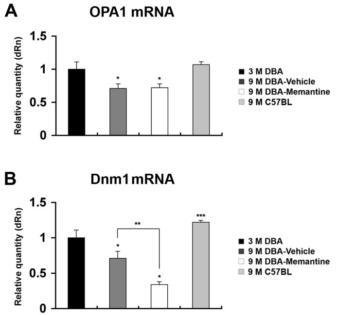
Purpose: To determine whether intraocular pressure (IOP) elevation alters OPA1 expression and triggers OPA1 release, as well as whether the uncompetitive N-methyl-d-aspartate (NMDA) glutamate receptor antagonist memantine blocks OPA1 release and subsequent apoptotic cell death in glaucomatous DBA/2J mouse retina.

Methods: Preglaucomatous DBA/2J mice received memantine (5 mg/kg, intraperitoneal injection, twice daily for 3 months) and IOP in the eyes was measured monthly. RGC loss was counted after FluoroGold labeling. OPA1, Dnm1, Bcl-2, and Bax mRNA were measured by qPCR. OPA1 protein was assessed by immunohistochemistry and Western blot. Apoptotic cell death was assessed by TUNEL staining.

Results: Memantine treatment significantly increased RGC survival in glaucomatous DBA/2J mice and increased the 75-kDa OPA1 isoform, but did not alter the 80- and 90-kDa isoforms. The isoforms of OPA1 were significantly increased in the cytosol of the vehicle-treated glaucomatous retinas but were significantly decreased in memantine-treated glaucomatous retinas. OPA1 immunoreactivity was decreased in the photoreceptors of both vehicle- and memantine-treated glaucomatous retinas, but was increased in the outer plexiform layer of only the memantine-treated glaucomatous retinas. Memantine blocked apoptotic cell death in the GCL, increased Bcl-2 gene expression, and decreased Bax gene expression.

Conclusions: OPA1 release from mitochondria in glaucomatous mouse retina is inhibited by blockade of glutamate receptor activation. Because this OPA1 effect was accompanied by increased Bcl-2 expression, decreased Bax expression, and apoptosis blockade, glutamate receptor activation in the glaucomatous retina may involve a distinct mitochondria-mediated cell death pathway.


Figures

Figure 1
Figure 1. IOP elevation in glaucomatous DBA/2J mice following memantine treatment
The average IOP (A), the representative actual IOP values (B), and the body weight (C) in vehicle and memantine-treated eyes of mice aged 7, 8, and 9 months.
Figure 2
Figure 2. RGC survival in glaucomatous DBA/2J mice following memantine treatment
The retinal flatmounts of 3 month-old DBA/2J mice (A and B), vehicle-treated 9 month-old glaucomatous DBA/2J mice (C and D), and mematine-treated 9 month-old glaucomatous DBA/2J mice (E and F). Note that high magnification showed a significant increase of RGC survival following memantine treatment compared to vehicle treatment. The quantitative analysis of RGC survival (G). Error bar represents the standard deviation (*Significant at P<0.05 compared with 3 month-old DBA/2J mice or vehicle-treated 9 month-old glaucomatous DBA/2J mice, n=4 retinal flatmounts/mice/group). Scale bars = 50 μm (A, C and E); 100 μm (B, D and F).
Figure 3
Figure 3. Alterations of OPA1 and Dnm1 gene expression in the retinas of glaucomatous DBA/2J mice following memantine treatment
(A) OPA1 mRNA was significantly decreased in the retinas of both vehicle- and memantine-treated 9 month-old glaucomatous DBA/2J mice compared to 3 month-old DBA/2J mice. However, memantine treatment did not show a difference of OPA1 mRNA expression in the retinas compared to vehicle-treated 9 month-old glaucomatous DBA/2J mice. In addition, there was no significant difference of OPA1 mRNA expression in retinas between 3 month-old DBA/2J mice and 9 month-old C57BL mice. (B) Dnm1 mRNA was significantly decreased in the retinas of both vehicle- and memantine-treated 9 month-old glaucomatous DBA/2J mice compared to 3 month-old DBA/2J mice. However, memantine treatment significantly decreased Dnm1 mRNA expression in the retinas compared to vehicle-treated 9 month-old glaucomatous DBA/2J mice. In addition, Dnm1 mRNA expression was significantly increased in 9 month-old non-glaucomatous C57BL/6 mice compared to 3 month-old DBA/2J mice. Error bars represent the standard deviation (*Significant at P<0.05 compared with 3 month-old DBA/2J mice; **significant at P<0.05 compared with vehicle-treated 9 month-old glaucomatous DBA/2J mice; ***significant at P<0.05 compared with 3 month-old DBA/2J mice, n=4 retinas per pool).
Figure 4
Figure 4. Alteration of total OPA1 expression and distribution in the retinas of glaucomatous DBA/2J mice following memantine treatment
(A) The OPA1 protein bands show the positions of 5 major isoforms of OPA1 (>100 kDa; L2 and L3; 90 kDa:L1; 80 kDa:S1) in the retina of 3 month-old DBA/2J mouse. However, the isoforme of OPA1 protein bands (75 kDa:S2) was increased in both vehicle- and memantine-treated 9 month-old glaucomatous DBA/2J mouse retina compared to 3 month-old DBA/2J mouse retina. (B) Relative intensity of chemiluminescence for each protein band was normalized using actin as cytosolic fraction calibrator. Error bars represent the standard deviation (*Significant at P<0.05 compared with 3 month-old DBA/2J mice, n=4 retinas per pool). (C–F) When the primary antibody for OPA1 was omitted, there was no binding of secondary antibody (C). OPA1 immunoreactivity in 3 month-old DBA/2J mouse retina (D), in vehicle-treated 9 month-old glaucomatous DBA/2J mouse retina (E), and in memantine-treated 9 month-old glaucomatous DBA/2J mouse retina (F). (G–I) OPA1 (G, green) and Fluoro-Gold (H, red) double labeling. Neurons containing OPA1 immunreactivity were co-labeled by Fluoro-Gold (arrow heads), indicating that RGCs in the GCL contained OPA1 protein. V, vehicle; M, memantine; PR, photoreceptor; ONL, outer nuclear layer; OPL, outer plexiform layer; INL, inner nuclear layer; IPL, inner plexiform layer; GCL, ganglion cell layer. Scale bar = 20μm (C–I).
Figure 5
Figure 5. Memantine prevents OPA1 and cytochrome c release in the retinas of glaucomatous DBA/2J mice
(A) OPA1 Western blot analysis of cytosolic and mitochondrial fractions from whole retinas. The OPA1 protein bands show the positions of 4 major isoforms of OPA1 (>100 kDa; L2 and L3; 90 kDa:L1; 80 kDa:S1). There was a fifth isoform of OPA1 protein band (75 kDa:S2) in the cytosolic fraction of the retinas in 9 month-old glaucomatous DBA/2J mice with vehicle and memantine treatment. The cytochrome c protein bands show the positions of 17 kDa form of cytochrome c in cytosolic and mitochondrial fraction. (B) Relative intensity of chemiluminescence for each protein band was normalized using actin (42 kDa) as cytosolic fraction calibrator and voltage dependent anion channel (VDAC, 31 kDa) as mitochondrial fraction calibrator. In vehicle-treated 9 month-old glaucomatous DBA/2J mice compared to 3 month-old DBA/2J mice, the L1–L3 and S2 isoforms of OPA1 were significantly increased in the cytosolic fraction but the L and S1 isoforms of OPA1 were significantly decreased in the mitochondrial fraction. In addition, the cytochrome c protein bands were significantly increased in the cytosolic fraction but decreased in the mitochondrial fraction. In memantie-treated 9 month-old glaucomatous DBA/2J mice compared to vehicle-treated 9 month-old glaucomatous DBA/2J mice, the L1–L3 and S2 isoforms of OPA1 were significantly decreased in the cytosolic fraction but the L and S1 isoforms of OPA1 were significantly increased in the mitochondrial fraction. In addition, the cytochrome c protein bands were significantly decreased in the cytosolic fraction but increased in the mitochondrial fraction. Error bar represents the standard deviation (*Significant at P<0.05 compared with 3 month-old DBA/2J mice or vehicle-treated 9 month-old glaucomatous DBA/2J mice, n=4 retinas per pool). V, vehicle; M, memantine.
Figure 6
Figure 6. Memantine blocks apoptotic cell death, and induces increase of Bcl-2 gene expression and decrease of Bax gene expression in the retinas of glaucomatous DBA/2J mice
(A) There were no TUNEL-positive cells in the retina of 3 month-old DBA/2J mouse. (B) Apoptotic cell death was prominent present in the GCL of the retina in vehicle-treated 9 month-old glaucomatous DBA/2J mouse. (C) Memantine treatment prevents apoptotic cell death in the GCL of the retina in vehicle-treated 9 month-old glaucomatous DBA/2J mouse. (D) Vehicle treatment did not alter Bcl-2 mRNA expression in the retinas of 9 month-old glaucomatous DBA/2J mice compared to 3 month-old DBA/2J mice. However, memantine treatment significantly increased Bcl-2 mRNA expression in the retinas of 9 month-old glaucomatous DBA/2J mice compared to vehicle treatment. (E) Vehicle treatment significantly increased Bax mRNA expression in the retinas of 9 month-old glaucomatous DBA/2J mice compared to 3 month-old DBA/2J mice. However, memantine treatment significantly decreased Bax mRNA expression in the retinas of 9 month-old glaucomatous DBA/2J mice compared to vehicle treatment. Error bars represent the standard deviation (*Significant at P<0.05 compared with 3 month-old DBA/2J mice or vehicle-treated 9 month-old glaucomatous DBA/2J mice, n=4 retinas per pool). PR, photoreceptor; ONL, outer nuclear layer; OPL, outer plexiform layer; INL, inner nuclear layer; IPL, inner plexiform layer; GCL, ganglion cell layer. Scale bar = 20μm (A–C).

References

    1. Weinreb RN, Khaw PT. Primary open-angle glaucoma. Lancet. 2004;363(9422):1711–1720. - PubMed
    1. Lipton SA. Possible role for memantine in protecting retinal ganglion cells from glaucomatous damage. Surv Ophthalmol. 2003;48(Suppl 1):S38–46. - PubMed
    1. Beal MF. Aging, energy, and oxidative stress in neurodegenerative diseases. Ann Neurol. 1995;38(3):357–366. - PubMed
    1. Mattson MP, Pedersen WA, Duan W, Culmsee C, Camandola S. Cellular and molecular mechanisms underlying perturbed energy metabolism and neuronal degeneration in Alzheimer’s and Parkinson’s diseases. Ann N Y Acad Sci. 1999;893:154–175. - PubMed
    1. Nicholls DG. Mitochondrial dysfunction and glutamate excitotoxicity studied in primary neuronal cultures. Curr Mol Med. 2000;4(2):149–177. - PubMed

Publication types

MeSH terms