. 2009 Jan;29(1):214-28.
doi: 10.1128/MCB.01377-08. Epub 2008 Oct 20.

Fragile X mental retardation protein FMRP binds mRNAs in the nucleus

Affiliations

Fragile X mental retardation protein FMRP binds mRNAs in the nucleus

Miri Kim et al. Mol Cell Biol. 2009 Jan.

Abstract

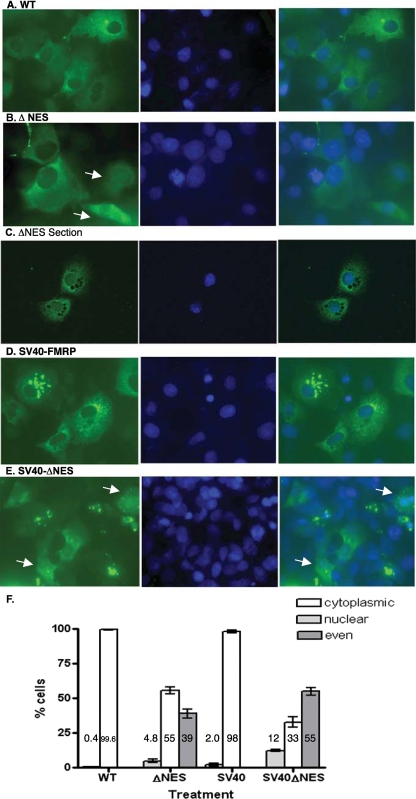
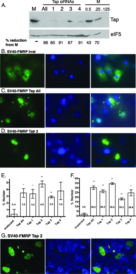
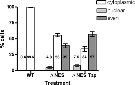
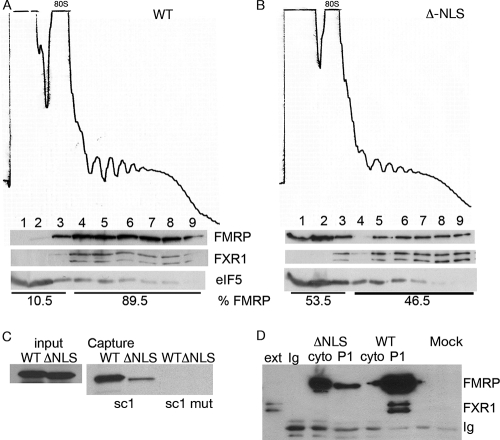
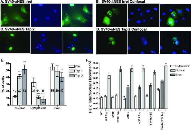
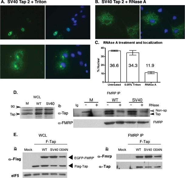
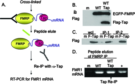
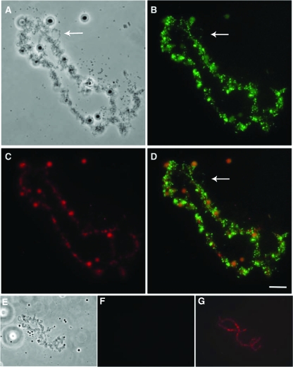
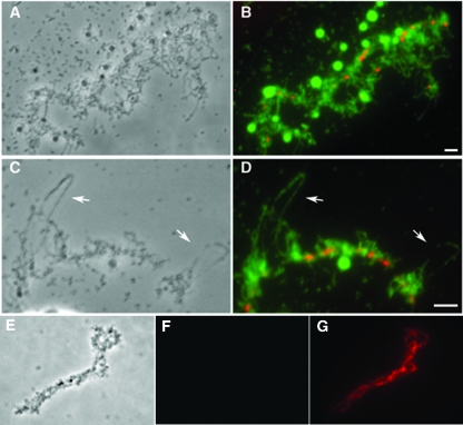
The fragile X mental retardation protein FMRP is an RNA binding protein that associates with a large collection of mRNAs. Since FMRP was previously shown to be a nucleocytoplasmic shuttling protein, we examined the hypothesis that FMRP binds its cargo mRNAs in the nucleus. The enhanced green fluorescent protein-tagged FMRP construct (EGFP-FMRP) expressed in Cos-7 cells was efficiently exported from the nucleus in the absence of its nuclear export sequence and in the presence of a strong nuclear localization sequence (the simian virus 40 [SV40] NLS), suggesting an efficient mechanism for nuclear export. We hypothesized that nuclear FMRP exits the nucleus through its bound mRNAs. Using silencing RNAs to the bulk mRNA exporter Tap/NXF1, we observed a significantly increased number of cells containing EGFP-FMRP in the nucleus, which was further augmented by removal of FMRP's nuclear export sequence. Nuclear-retained SV40-FMRP could be released upon treatment with RNase. Further, Tap/NXF1 coimmunoprecipitated with EGFP-FMRP in an RNA-dependent manner and contained the FMR1 mRNA. To determine whether FMRP binds pre-mRNAs cotranscriptionally, we expressed hemagglutinin-SV40 FMRP in amphibian oocytes and found it, as well as endogenous Xenopus FMRP, on the active transcription units of lampbrush chromosomes. Collectively, our data provide the first lines of evidence that FMRP binds mRNA in the nucleus.


Figures

FIG. 1.
FIG. 1.
FMRP is efficiently exported from the nucleus in the absence of an NES and in the presence of the SV40 NLS. Cos-7 cells were plated and transfected with constructs expressing EGFP-FMRP (WT) (A); EGFP-FMRP with the NES deleted (ΔNES) (B, C); EGFP-SV40-FMRP (SV40-FMRP) (D); and EGFP-SV40-ΔNES (SV40-ΔNES), fixed in DAPI-containing mounting medium and imaged for the expression of EGFP-FMRP (green, left panels) and nuclei (blue DAPI stain, middle panels) and for the merged EGFP and DAPI images (right panels) (E). The arrows in panel B indicate cells expressing ΔNES in the nucleus. Panel C shows ΔNES-expressing cells examined by optical sectioning as described in Materials and Methods. The arrows in panel E indicate cells expressing comparable amounts of SV40-ΔNES in the nucleus and cytoplasm. (F) Cos-7 cells expressing EGFP-FMRP (WT), ΔNES, SV40-FMRP (SV40), and SV40-ΔNES were scored for the percentage of cells with a nuclear accumulation of FMRP. The results of individual experiments, where over 100 cells were scored each time, were averaged to find the percentage of cells with nuclear accumulation of FMRP for each construct. The WT and SV40 constructs were scored for primarily nuclear FMRP, while ΔNES and EGFP-SV40-ΔNES were scored for cells that demonstrated primarily nuclear, primarily cytoplasmic, or evenly distributed FMRP between the nucleus and cytoplasm (even).
FIG. 2.
FIG. 2.
The reduction of Tap/NXF1 expression results in the nuclear accumulation of EGFP-FMRP and SV40-FMRP. (A) Cos-7 cells were mock treated with an irrelevant siRNA (M) or treated with a mixture of the four siRNAs against Tap/NXF1 (All) at a final concentration of 100 nM or individually with each of the four Tap/NXF1 siRNAs (1 to 4). Forty-eight hours later, the lysates were prepared and 75 μg of each were resolved on 7.5% SDS-polyacrylamide gel electrophoresis gels. The last three lanes contain dilutions of the mock: 50% (37.5 μg), 25% (18.8 μg), and 12.5% (9.4 μg). The blot was probed with affinity-purified anti-Tap antisera and reprobed with anti-eIF-5 as a loading control. The amount of Tap per lane was calculated using NIH Image and shown as the percent reduction from the mock. (B to D) Cos-7 cells were transfected with SV40 FMRP and the siRNAs indicated as follows: an irrelevant siRNA (B), a mixture of the four Tap/NXF1 siRNAs (Tap All) (C), and Tap/NXF1-2 siRNA (D). The cells were imaged for the expression of EGFP (green, left panels) and nuclei (blue DAPI stain, middle panels), and the EGFP and DAPI images were merged (right panels). (E, F) Cos-7 cells transfected with either EGFP-FMRP (E) or EGFP-SV40-FMRP (F) and treated with either the irrelevant siRNA, a mixture of the four Tap/NXF1 siRNAs (Tap All), or the individual Tap/NXF1 siRNAs (Tap 1 to 4) for 48 h and scored for the percentage of cells with a primarily nuclear accumulation of FMRP. The percentage of cells expressing FMRP in the nucleus is indicated by each bar. The results were plotted using GraphPad Prism 4. Significance was calculated using a one-tailed Student's t test. A single star indicates a P of <0.05, and two stars indicate a P of <0.01. (G) Cos-7 cells were transfected with SV40 FMRP and the Tap/NXF1-2 siRNA. Twenty-four hours later, the cytoplasmic counterstain CellTrace BODIPY TR methyl ester (Invitrogen) was added, and the cells were live imaged with a confocal microscope at ×63 magnification with oil for EGFP (green, left panels) and cytoplasm stain (blue, middle panels), and the EGFP and BODIPY images were merged (right panels).
FIG. 3.
FIG. 3.
The endogenous NLS of FMRP directs a significant amount of EGFP-ΔNES into the nucleus. Cos-7 cells were transfected with either EGFP-FMRP (WT) or ΔNES with irrelevant siRNA (ΔNES) or with Tap/NXF1-2 siRNA (ΔNES Tap). The cells were imaged and scored for nuclear, cytoplasmic, or even distribution between the nucleus and cytoplasm. Five independent experiments were scored and plotted using GraphPad Prism 4.
FIG. 4.
FIG. 4.
Removal of the NLS impairs the function of FMRP. Immortalized Fmr1 knockout fibroblast cells (STEK) (57a) were transfected with EGFP-FMRP (WT) (A) or EGFP-FMRP in which the endogenous NLS has been removed (ΔNLS) (B), treated with cycloheximide, and fractionated on a linear 15 to 45% sucrose gradient. Profiles are shown as the absorbance at 254 nm, and the position of the 80S monosome is indicated at the top of each gradient. Fractions were analyzed on 7.5% gels, transferred to polyvinylidene difluoride and probed with the anti-FMRP antibody 1a to visualize transgene expression (top row), FXR1 (middle row), or eIF5 (bottom row). The amount of FMRP in each fraction was quantified using NIH Image. Removal of the NLS led to a loss of ∼50% of the polyribosome-associated FMRP. (C) EGFP-FMRP or ΔNLS expressed in pSport were transcribed and translated in vitro and used in a biotinylated RNA capture assay (75). sc1 is an RNA encoding a G quartet, and the sc1 mutant (sc1 mut) is an RNA encoding a G quartet mutant unable to bind the RGG box of FMRP (18). (D) STEK cells transfected with either EGFP-FMRP (WT) or ΔNLS transgenes were fractionated into either a postnuclear supernatant (cyto) or pelleted again (P1). FMRP was immunoprecipitated from each fraction and blotted for FXR1 or FMRP as indicated on the right. STEK extract was loaded as a control (ext), and the immunoprecipitating immunoglobulin chains (Ig) are indicated.
FIG. 5.
FIG. 5.
Tap/NXF1 knockdown increases the nuclear accumulation of SV40-ΔNES. (A to D) Cos-7 cells were plated and transfected with SV40-ΔNES and the siRNAs indicated and imaged for the expression of EGFP-SV40-ΔNES (green, left panels) and nuclei (blue DAPI stain, middle panels); the EGFP and DAPI images were merged (right panels) by inverted fluorescence microscopy (A, C) and by confocal microscopy (B, D). (E) Three independent experiments were scored for cells that expressed transgene primarily in the nucleus (nuclear), primarily in the cytoplasm (cytoplasmic), or evenly distributed between the nucleus and cytoplasm (even) after treatment with an irrelevant siRNA (irrel) or Tap 1 or 2 siRNA. The average percentage of cells is given at the bottom of the bar. The results were plotted using GraphPad Prism 4. Significance was calculated using a one-tailed Student's t test. A single star indicates a P of <0.05, two stars indicate a P of <0.01, and three stars indicate a P of <0.005. (F) Consistency of cell scoring was evaluated by calculating the ratio of total nuclear fluorescence to total cellular fluorescence and performing a one-way analysis of variance using an α value of 0.05. Cells scored as cytoplasmic had an average nuclear fluorescence of 12.93% ± 2.12%, while cells scored as even had an average nuclear fluorescence of 23.37% ± 4.57% and nuclear cells had average nuclear fluorescence of 38.29% ± 3.65%. All P values were less than 0.05, and many were less than 0.001, with the exception of the SV40-ΔNES cells treated with Tap/NXF1.
FIG. 6.
FIG. 6.
The retention of EGFP-FMRP in the nucleus in the absence of Tap/NXF1 is RNA dependent. Cos-7 cells were plated and transfected with SV40-FMRP and Tap/NXF1-2 siRNA, permeabilized with 0.05% Triton (A) (top, cells expressing SV40-FMRP in the nucleus; bottom, cells expressing SV40-FMRP in the cytoplasm), and treated with RNase (B). (C) The percentage of cells expressing FMRP in the nucleus is indicated by each bar. The results were plotted using GraphPad Prism 4. (D) Cos-7 cells were either untransfected (M) or transfected with EGFP-FMRP (WT) or SV40-FMRP (SV40) (WCL, whole cell lysate [50 μg/lane]), immunoprecipitated (IP) with the anti-FMRP antibody 7G1-1, washed with buffer containing RNase (+) or not (−) and immunoblotted for endogenous Tap/NXF1 (Tap) coimmunoprecipitation and FMRP transgene (bottom). Non-sp, reactivity to an irrelevant protein; Ig, immunoprecipitating antibody alone; ib, immunoblot. (E) Cos-7 cells were either mock transfected (M) or transfected with Flag-Tap/NXF1 (F-Tap) alone or with Flag-Tap/NXF1 and one of the following constructs: FMRP (WT), SV40-FMRP (SV40), or I304N. Cells were treated with 0.5% formaldehyde and sonicated as described in Materials and Methods. Left panel, 35 μg of the lysates were loaded, resolved, and probed for transfection efficiency with the anti-Flag antibody (M2; Sigma) to visualize EGFP-FMRP expression (these constructs contain the Flag epitope) and Flag-Tap/NXF1; the eIF5 immunoblot (ib) shows equal loading. Right panel, immunoprecipitation with the anti-murine FMRP antibody 7G1-1. Immunoprecipitated FMRP was visualized using the anti-FMRP antibody K1, and Flag-Tap coimmunoprecipitation was visualized by a rabbit anti-Tap antibody.
FIG. 7.
FIG. 7.
Tap/NXF1 associates with FMRP in a complex with FMR1 mRNA. (A) Schematic showing the reimmunoprecipitation experiment. Transfected cells were treated with formaldehyde (cross-linked) and then immunoprecipitated with the anti-FMRP antibody 7G1-1. Immunoprecipitated complexes were eluted with the 7G1-1 peptide and reimmunoprecipitated (Re-IP) with an anti-Tap antibody. RNA was extracted and analyzed from the complex. (B) Cos-7 cells were mock-transfected (m) or transfected with EGFP-FMRP (WT) or EGFP-FMRP and Flag-Tap (WT Tap) and immunoblotted (ib) with an anti-Flag antibody (Flag). (C) The anti-Tap/NXF1 antibodies immunoprecipitate with ∼15% efficiency. Cos-7 cells were mock transfected (m) or transfected with Flag-Tap (F-Tap); 50 μg was loaded per lane (wcl, whole cell lysate). Two different anti-Tap/NXF1 antibodies (IP-1 and IP-2) were used to immunoprecipitate extracts from mock or Flag-Tap-expressing cells. (D) FMRP immunoprecipitations (IP) from mock-treated Cos-7 cells (m), EGFP-FMRP-expressing Cos-7 cells (WT), or EGFP-FMRP- and Flag-Tap/NXF1-expressing Cos-7 cells (WT Tap) were peptide eluted. RNA was extracted from mock and WT peptide elutions. The peptide elution from WT Tap was reimmunoprecipitated with the anti-Tap/NXF1 antibody from which the RNA was extracted. First-strand synthesis was performed, followed by PCR for FMR1 mRNA. +, RT-PCR from Cos-7 cell total RNA.
FIG. 8.
FIG. 8.
Nuclear EGFP-SV40-Flag-FMRP is not recognized by the anti-FMRP antibody 1a. Cos-7 cells were transfected with SV40-FMRP and Tap siRNAs (Tap All), fixed, and permeabilized as described previously (57). The cells were stained with either the anti-FMRP antibody 1a (22) (A) or the anti-Flag antibody (M2) (B). Nuclear EGFP SV40-FMRP was observed using the green channel (left images) and by Flag staining (middle image in panel B); however, it was not detected by antibody 1a (middle image in panel A). Right images show merged DAPI staining with EGFP and anti-mouse rhodamine (red).
FIG. 9.
FIG. 9.
FMRP associates with LBCs in the nucleus. Stage V Xenopus laevis oocytes were injected with HA-SV40-WT FMRP cRNA. Nuclear spreads were prepared 24 to 48 h after injection, and the localization of FMRP was visualized using fluorescence microscopy. (A) The phase contrast image shows an LBC and associated proteins. (B) Chromosomal spreads were incubated with anti-HA antibody and visualized by fluorescence microscopy. FMRP is localized along the DNA axis and is also seen along chromosomal loops (arrows) in green. (C) Chromosomes were counterstained using DAPI and false colored in red. (D) Merged images of chromosomes and FMRP localization demonstrate that FMRP is associated with the LBCs and along a subset of chromosomal loops. The scale bar represents 5 mm. (E) Uninjected control oocytes were prepared in parallel to injected oocytes. The phase contrast image shows a single chromosome. (F) Anti-HA antibody staining. (G) Counterstaining using DAPI and false coloring in red.
FIG. 10.
FIG. 10.
Endogenous FMRP associates with nascent transcripts in the nucleus. Xenopus oocytes were stained with the anti-Xenopus FMRP antibody K1. (A) A ×40 magnification of a single chromosome visualized in phase contrast. (B) FMRP (green) is localized with the LBCs. The DNA axis is shown in red. (C) A ×100 magnification shows single-stranded loops (indicated by arrows) of DNA extending off the axis in phase contrast. Scale bar represents 2 mm. (D) FMRP (green) is localized with the LBCs. The DNA axis is shown in red. Phase contrast of an LBC (E), stained with rabbit preimmune antisera (F), and counterstained using DAPI and false colored in red (G).

Similar articles

Cited by

References

    1. Adinolfi, S., C. Bagni, G. Musco, T. Gibson, L. Mazzarella, and A. Pastore. 1999. Dissecting FMR1, the protein responsible for fragile X syndrome, in its structural and functional domains. RNA 51248-1258. - PMC - PubMed
    1. Adinolfi, S., A. Ramos, S. R. Martin, F. Dal Piaz, P. Pucci, B. Bardoni, J. L. Mandel, and A. Pastore. 2003. The N-terminus of the fragile X mental retardation protein contains a novel domain involved in dimerization and RNA binding. Biochemistry 4210437-10444. - PubMed
    1. Antar, L. N., R. Afroz, J. B. Dictenberg, R. C. Carroll, and G. J. Bassell. 2004. Metabotropic glutamate receptor activation regulates fragile X mental retardation protein and Fmr1 mRNA localization differentially in dendrites and at synapses. J. Neurosci. 242648-2655. - PMC - PubMed
    1. Arttamangkul, S., V. Alvarez-Maubecin, G. Thomas, J. T. Williams, and D. K. Grandy. 2000. Binding and internalization of fluorescent opioid peptide conjugates in living cells. Mol. Pharmacol. 581570-1580. - PubMed
    1. Ashley, C. T., J. S. Sutcliffe, C. B. Kunst, H. A. Leiner, E. E. Eichler, D. L. Nelson, and S. T. Warren. 1993. Human and murine FMR-1: alternative splicing and translational initiation downstream of the CGG-repeat. Nat. Genet. 4244-251. - PubMed

Publication types

MeSH terms

Substances