. 2009 Jan;29(1):266-80.
doi: 10.1128/MCB.00315-08. Epub 2008 Oct 20.

The STAGA subunit ADA2b is an important regulator of human GCN5 catalysis

Affiliations

The STAGA subunit ADA2b is an important regulator of human GCN5 catalysis

Armin M Gamper et al. Mol Cell Biol. 2009 Jan.

Abstract

Human STAGA is a multisubunit transcriptional coactivator containing the histone acetyltransferase GCN5L. Previous studies of the related yeast SAGA complex have shown that the yeast Gcn5, Ada2, and Ada3 components form a heterotrimer that is important for the enzymatic function of SAGA. Here, we report that ADA2a and ADA2b, two human homologues of yeast Ada2, each have the ability to form a heterotrimer with ADA3 and GCN5L but that only the ADA2b homologue is found in STAGA. By comparing the patterns of acetylation of several substrates, we found context-dependent requirements for ADA2b and ADA3 for the efficient acetylation of histone tails by GCN5. With human proteins, unlike yeast proteins, the acetylation of free core histones by GCN5 is unaffected by ADA2b or ADA3. In contrast, the acetylation of mononucleosomal substrates by GCN5 is enhanced by ADA2b, with no significant additional effect of ADA3, and the efficient acetylation of nucleosomal arrays (chromatin) by GCN5 requires both ADA2b and ADA3. Thus, ADA2b and ADA3 appear to act at two different levels of histone organization within chromatin to facilitate GCN5 function. Interestingly, although ADA2a forms a complex(es) with GCN5 and ADA3 both in vitro and in vivo, ADA2a-containing complexes are unable to acetylate nucleosomal H3. We have also shown the preferential recruitment of ADA2b, relative to ADA2a, to p53-dependent genes. This finding indicates that the previously demonstrated presence and function of GCN5 on these promoters reflect the action of STAGA and that the ADA2a and ADA2b paralogues have nonredundant functional roles.


Figures

FIG. 1.
FIG. 1.
STAGA subunits increase the ability of GCN5 to acetylate nucleosomes. (A) Analysis of purified STAGA. The complex was purified from a HeLa cell line that stably expresses FLAG-HA-tagged SPT3 (fha:SPT3) and analyzed by SDS-PAGE with silver staining. M, molecular size markers. The numbers in parentheses refer to the original TAF nomenclature designations. (B) Analysis of purified GCN5L and GCN5S. FLAG-tagged GCN5L (L) and GCN5S (S) were purified from Sf9 cells and analyzed by SDS-PAGE with Coomassie blue staining. Arrowheads indicate the positions of the protein bands. (C) Comparison of the HAT activities of STAGA and non-complex-associated GCN5 isoforms with histones as the substrate. Molar amounts were based on comparative immunoblots with an anti-GCN5 antibody. −, negative control. (D) Comparison of the HAT activities of STAGA and non-complex-associated GCN5L with mononucleosomes as the substrate. (E) Schematic of ADA2a and ADA2b, with overall sequence relationships of 26% identity and 50% homology. Levels of identity and homology between the zinc finger, SANT, and SWIRM domains are indicated at the bottom. Numbers above the diagrams are amino acid positions.
FIG. 2.
FIG. 2.
ADA2a and ADA2b both interact with GCN5 and ADA3, but only ADA2b is a subunit of STAGA. (A) Coprecipitation of endogenous GCN5L with FLAG-HA-tagged ADA2a (fha:ADA2a) or ADA2b overexpressed in H1299 cells. Inputs (IN) and anti-FLAG (M2 agarose) immunoprecipitates (IP) were analyzed by immunoblotting with the antibodies indicated on the right. −, negative control. (B) Interaction of in vitro-translated radiolabeled GCN5L or ADA3 (detected by SDS-PAGE and autoradiography) with GST-fused full-length ADA2 proteins. (C) Analysis of purified GCN5-ADA2a and GCN5-ADA2b complexes. FLAG-tagged GCN5L (f:GCN5L) was expressed in Sf9 cells either alone (lane 1) or with ADA2a (lane 2) or ADA2b (lane 3), purified (along with associated proteins) on M2 agarose, and analyzed by SDS-PAGE with Coomassie blue staining. Asterisks denote contaminants copurified with GCN5L under the conditions used. (D) Analysis of purified ADA3. His-tagged ADA3 (his6ADA3) was purified from bacteria and analyzed by SDS-PAGE with Coomassie blue staining. (E) Presence of ADA2b, but not ADA2a, in STAGA. GCN5-ADA2a and GCN5-ADA2b heterodimers, purified STAGA (from a stable FLAG-HA-tagged SPT3 cell line), and the purified PCAF complex were analyzed by immunoblotting with antibodies against GCN5, ADA2a, ADA2b, and TRRAP. The STAGA and PCAF complexes were normalized on the basis of the TRRAP contents. HeLa NE, HeLa cell nuclear extract.
FIG. 3.
FIG. 3.
ADA2a and ADA2b are exclusive components of distinct complexes. (A) Presence of ADA3 in both STAGA and an ADA2a-containing complex(es). An anti-FLAG (M2 agarose) immunoprecipitate from a HeLa cell line that stably expresses FLAG-HA-tagged ADA3 (fha:ADA3) was probed for ADA2a and the indicated STAGA subunits by immunoblotting. IP, immunoprecipitates. (B) Differential patterns of the presence of ADA2b and ADA2a in complexes. Anti-FLAG (M2 agarose) immunoprecipitates from HeLa cell lines that stably express FLAG-HA-tagged SPT3, ADA2b, ADA3, or STAF65γ were probed for ADA2a and the indicated STAGA subunits by immunoblotting. ADA2a coprecipitates with ADA3 only. The asterisk denotes a nonspecific signal. (C) Exclusive presence of ADA2b and that of ADA2a in distinct complexes. Anti-FLAG (M2 agarose) immunoprecipitates from U2OS cell lines that stably express FLAG-HA-tagged ADA2a or ADA2b were probed for ADA2a and the indicated STAGA subunits by immunoblotting. No ADA2b was detected in the anti-ADA2a precipitate. (D and E) Size fractionation of anti-ADA3 and anti-GCN5S immunoprecipitates. Anti-FLAG (M2 agarose) immunoprecipitates from HeLa cell lines that stably express FLAG-HA-tagged ADA3 (D) or GCN5S (E) were fractionated on a Superose 6 column. Fractions were tested for the presence of ADA2a and various STAGA subunits by immunoblotting.
FIG. 4.
FIG. 4.
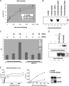
ADA2b, but not ADA2a, increases the acetylation of nucleosomes by GCN5L. (A) Lack of influence of ADA2a and ADA2b on the acetylation of free core histones by GCN5L. HAT assays with free core histones from HeLa cells were conducted with increasing amounts of FLAG-tagged GCN5L (f:GCN5) and FLAG-tagged GCN5L-ADA2a or GCN5L-ADA2b heterodimers. Filter binding assays were used to quantify tritium incorporation from [3H]AcCoA. (B) Requirement of ADA2b for GCN5L to efficiently acetylate nucleosomes. HAT assays with mononucleosome substrates were conducted with FLAG-tagged GCN5L, GCN5L-ADA2a, and GCN5L-ADA2b in the absence or presence of ADA3. Radiolabeled (acetylated histone) products were monitored by SDS-PAGE and autoradiography. −, negative control. (C) Effect of H1 on ADA2b-enhanced acetylation of mononucleosomes by GCN5. Mononucleosomes with or without histone H1 (shown with Coomassie blue staining in the insert) were employed as the substrate for FLAG-tagged GCN5L, GCN5L-ADA2a, or GCN5L-ADA2b, and acetylation was measured by filter binding assays. The level of acetylation relative to that of mononucleosomes by GCN5L alone is represented. +, present; −, absent. (D) Differential patterns of acetylation of mononucleosomes by endogenous ADA2a and ADA2b complexes. Anti-FLAG (M2 agarose) immunoprecipitates (IP) from extracts of U2OS cells that stably express FLAG-HA-tagged ADA2a (fha:ADA2a) or ADA2b were normalized on the basis of the GCN5L contents, and the immunoprecipitates were tested for their ability to acetylate free core histones and mononucleosomes. −, negative controls. (E) Kinetic analyses of GCN5L and the GCN5L-ADA2b heterodimer. Equal amounts of GCN5L in the free form (•) and in the form of a heterodimer with ADA2b (▪) were assayed with various concentrations of free histones or mononucleosomes in a time-dependent manner. Initial rates of histone acetylation (v0; expressed as acetyl transfers per GCN5 molecule per second) were determined by filter binding assays and plotted versus the substrate concentration. A Michaelis-Menten curve was fitted to the data for histone acetylation. The quantitation of substrates was based on the data for histone H3, and the quantitation of GCN5 and the GCN5L-ADA2b heterodimer was by immunoblotting (right panel).
FIG. 5.
FIG. 5.
ADA3 and GCN5L interaction sites on ADA2b. (A) Interaction of the carboxy terminus of ADA3 with ADA2b. Beads with GST alone or GST-fused ADA2b were incubated with various in vitro-translated amino-terminal deletion mutant ADA3 proteins, and bound proteins were analyzed by SDS-PAGE and autoradiography. Numbers to the right of the panel indicate the amino acids present in the ADA3 proteins. (B) Binding of ADA3 to two ADA2b regions. Beads with GST or GST-fused deletion mutant ADA2b proteins (indicated by amino acid numbers) were incubated with in vitro-translated ADA3. SW, SWIRM domain; ΔSW, mutant protein ADA2b(1-345), lacking the SWIRM domain. (C) Interaction of GCN5L with a SANT domain-containing region of ADA2b. Beads with GST or GST-fused deletion mutant ADA2b proteins were incubated with in vitro-translated GCN5L. (D) Summary of ADA2b domain interactions with GCN5L and ADA3. Numbers indicate amino acid positions.
FIG. 6.
FIG. 6.
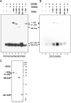
Effect of ADA3 deletions on ADA3 and nucleosomal histone acetylation by the GCN5L-ADA2b heterodimer. (A) Results of HAT assays with mononucleosome substrates. Mononucleosomes were incubated with GCN5L or a GCN5L-ADA2b heterodimer in the absence (−) or presence (+) of equal amounts of different ADA3 truncation mutant proteins. Reaction products were separated by SDS-PAGE and visualized by autoradiography. The gel was exposed for a prolonged period to emphasize the acetylation of ADA3 by GCN5L. Numbers above the gel indicate the amino acids present in the ADA3 proteins. (B) Results of HAT assays with chromatin. Reactions were carried out as described in the legend to panel A but with the reconstituted chromatin substrate. (C) Analysis of purified ADA3 proteins. The ADA3 wild type (1-432) and the deletion mutant proteins indicated on the left were expressed and purified from E. coli and analyzed by SDS-PAGE with Coomassie blue staining.
FIG. 7.
FIG. 7.
The SWIRM domain is dispensable for mononucleosome and chromatin acetylation, but point mutation of the SWIRM domain can lead to decreased acetylation of mononucleosomes. (A) Acetylation of mononuclesomes and free core histones. Substrates were incubated with the indicated combinations of GCN5L or GCN5L heterodimers with full-length (FL) or SWIRM deletion (Δ) forms of ADA2b and ADA3. Acetylated proteins were resolved by SDS-PAGE and visualized by fluorography. +, present; −, absent. (B) Acetylation of chromatin. The same protein combinations described in the legend to panel A were used for the acetylation of chromatin (lanes 3 to 9). Lanes 1 and 2 show the acetylation of chromatin by GCN5L-ADA2a with and without ADA3. (C) Analysis of GCN5L-ADA2b heterodimers. (Left panel) Heterodimers of untagged GCN5L and FLAG-tagged wild-type ADA2b (left lane) or ADA2b with the SWIRM domain deleted (ADA2bΔ; right lane) were copurified from Sf9 cells and analyzed by SDS-PAGE with Coomassie blue staining. (Right panel) Immunoblot of FLAG-tagged GCN5L (lane 1) and heterodimers of untagged GCN5L with intact (lane 2) or SWIRM deletion (lane 3) forms of FLAG-tagged ADA2b employed in the analyses presented in panels A and B. (D) Effect of ADA2b mutation R404A on the acetylation of nucleosomal histones by GCN5. Equal amounts of heterodimers of untagged GCN5L and FLAG-tagged wild-type ADA2b (WT) or SWIRM domain K403A (K) or R404A (R) mutant forms were copurified from Sf9 cells and tested for their abilities to acetylate free core histones and mononucleosomes. 2X indicates that the concentrations of GCNSL-ADA2b heterodimer used in lanes 5 to 7 were twice those used in the corresponding lanes 1 to 3. (E) Comparison of the SWIRM domain sequences of six human proteins. The evolutionary distance among human SWIRM domains, determined by the degree of nucleotide substitution, is shown as a dendrogram. Only SWIRM domain sequences encompassing the alpha helix (shown as a bold bar labeled α5) that is thought to interact with DNA are shown. Two amino acids with positive charges discussed in the text are highlighted. Numbers indicate amino acid positions.
FIG. 8.
FIG. 8.
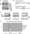
ADA2a and ADA2b are differentially recruited to activated p53 target gene promoters. (A) Immunoblot of U2OS-derived cells that stably express FLAG-HA-tagged (fha) ADA2a and ADA2b. Equal amounts of whole-cell extracts were resolved by SDS-PAGE, blotted, and probed for GCN5L, the large subunit (RBP1) of RNA polymerase II, and FLAG-HA-tagged ADA2 proteins. +, UV irradiated; −, untreated; IIo and IIa, forms of RNA polymerase II. (B) Diagram of p21 and GADD45 genes and locations of primers used for ChIP assays. p53RE1 and p53RE2, p53 response elements 1 and 2. (C) Results of ChIP assays showing that ADA2b, but not ADA2a, is recruited to p53 response elements after UV induction. Chromatin from control or UV-irradiated U2OS cells expressing tagged ADA2a or ADA2b was subjected to ChIP assays using anti-HA antibodies and primers designed to score binding to the p53 response elements of the p21 and GADD45 gene promoters. A region downstream of the p21 gene served as a control. Representative ethidium bromide-stained gels are shown. (D) Quantitative PCR analysis of anti-HA-based chromatin immunoprecipitates from untreated and UV-irradiated U2OS cells that stably express FLAG-HA-tagged ADA2a or ADA2b. The binding to p53 response elements of several p53-dependent promoters was tested. Average values from multiple independent ChIP experiments are shown. All ChIP values were normalized to the amount of input and are represented in comparison to the signal for a control site downstream of the p21 gene promoter (p21 CON) after UV exposure. −, no UV treatment.

References

    1. An, W., J. Kim, and R. G. Roeder. 2004. Ordered cooperative functions of PRMT1, p300, and CARM1 in transcriptional activation by p53. Cell 117735-748. - PubMed
    1. Aravind, L., and L. M. Iyer. 2002. The SWIRM domain: a conserved module found in chromosomal proteins points to novel chromatin-modifying activities. Genome Biol. 3RESEARCH0039. - PMC - PubMed
    1. Balasubramanian, R., M. G. Pray-Grant, W. Selleck, P. A. Grant, and S. Tan. 2002. Role of the Ada2 and Ada3 transcriptional coactivators in histone acetylation. J. Biol. Chem. 2777989-7995. - PubMed
    1. Barlev, N. A., A. V. Emelyanov, P. Castagnino, P. Zegerman, A. J. Bannister, M. A. Sepulveda, F. Robert, L. Tora, T. Kouzarides, B. K. Birshtein, and S. L. Berger. 2003. A novel human Ada2 homologue functions with Gcn5 or Brg1 to coactivate transcription. Mol. Cell. Biol. 236944-6957. - PMC - PubMed
    1. Barlev, N. A., L. Liu, N. H. Chehab, K. Mansfield, K. G. Harris, T. D. Halazonetis, and S. L. Berger. 2001. Acetylation of p53 activates transcription through recruitment of coactivators/histone acetyltransferases. Mol. Cell 81243-1254. - PubMed

Publication types

MeSH terms