. 2009 Jan;77(1):374-86.
doi: 10.1128/IAI.01081-08. Epub 2008 Oct 20.

The PmrA/PmrB two-component system of Legionella pneumophila is a global regulator required for intracellular replication within macrophages and protozoa

Affiliations

The PmrA/PmrB two-component system of Legionella pneumophila is a global regulator required for intracellular replication within macrophages and protozoa

Souhaila Al-Khodor et al. Infect Immun. 2009 Jan.

Abstract

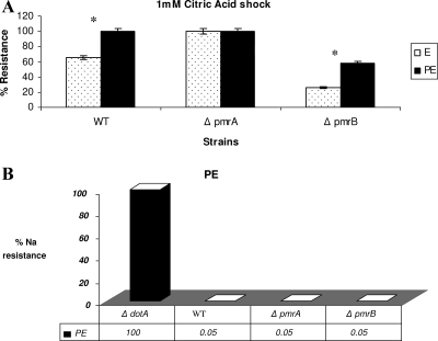
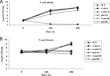
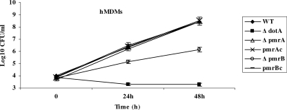
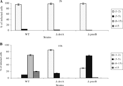
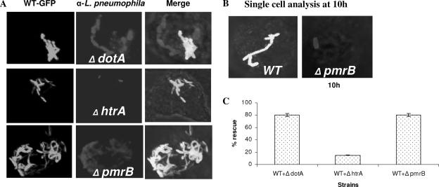
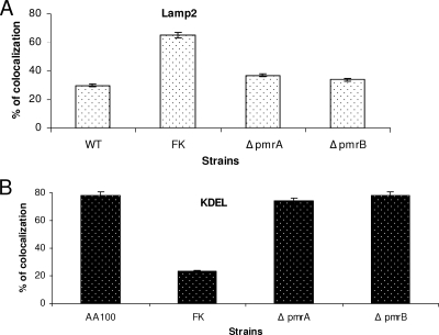
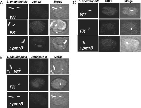
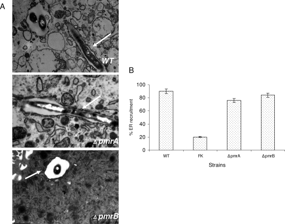
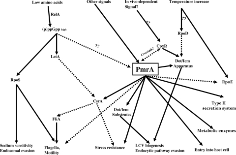
To examine the role of the PmrA/PmrB two-component system (TCS) of Legionella pneumophila in global gene regulation and in intracellular infection, we constructed pmrA and pmrB isogenic mutants by allelic exchange. Genome-wide microarray gene expression analyses of the pmrA and pmrB mutants at both the exponential and the postexponential phases have shown that the PmrA/PmrB TCS has a global effect on the expression of 279 genes classified into nine groups of genes encoding eukaryotic-like proteins, Dot/Icm apparatus and secreted effectors, type II-secreted proteins, regulators of the postexponential phase, stress response genes, flagellar biosynthesis genes, metabolic genes, and genes of unknown function. Forty-one genes were differentially regulated in the pmrA or pmrB mutant, suggesting a possible cross talk with other TCSs. The pmrB mutant is more sensitive to low pH than the pmrA mutant and the wild-type strain, suggesting that acidity may trigger this TCS. The pmrB mutant exhibits a significant defect in intracellular proliferation within human macrophages, Acanthamoeba polyphaga, and the ciliate Tetrahymena pyriformis. In contrast, the pmrA mutant is defective only in the ciliate. Despite the intracellular growth defect within human macrophages, phagosomes harboring the pmrB mutant exclude late endosomal and lysosomal markers and are remodeled by the rough endoplasmic reticulum. Similar to the dot/icm mutants, the intracellular growth defect of the pmrB mutant is totally rescued in cis within communal phagosomes harboring the wild-type strain. We conclude that the PmrA/PmrB TCS has a global effect on gene expression and is required for the intracellular proliferation of L. pneumophila within human macrophages and protozoa. Differences in gene regulation and intracellular growth phenotypes between the pmrA and pmrB mutant suggests a cross talk with other TCSs.


Figures

FIG. 1.
FIG. 1.
Phenotypic characteristics of the pmrA and pmrB mutants under different stress conditions. For the different stress conditions, the cell pellet was resuspended in an equal volume solution of 0.1 M citric acid at pH 3 for acid stress (A) or 5 M sodium chloride (B) and compared to bacteria resuspended in M63 salts. Cells were incubated in a 37°C water bath for 30 min and then washed, serially diluted, and plated on BCYE agar plates to determine the number of resistant bacteria. The experiment was done three times, and the data are representative of one independent experiment. Asterisks represent a significant difference between the WT strain and the pmrB mutant.
FIG. 2.
FIG. 2.
Intracellular growth kinetics of the pmrA and pmrB mutants of L. pneumophila within protozoa. The intracellular growth kinetics of the pmrA and pmrB mutant in A. polyphaga (A) and T. pyriformis (B) were determined. The infection was carried out in triplicates for 1 h, followed by 1 h of gentamicin treatment to kill extracellular bacteria in case of A. polyphaga. The infected monolayers were lysed at different time intervals and plated onto agar plates for colony enumeration. The experiment was done three times, and the data are representative of one independent experiment. Error bars represent standard deviations, but some were too small to appear in the figure.
FIG. 3.
FIG. 3.
Intracellular growth kinetics of the pmrA and pmrB mutants of L. pneumophila within macrophages. Intracellular growth kinetics of the WT AA100 strain and the dotA, pmrA, and pmrB mutants in hMDMs. pmrAc and pmrBc represent the pmrA and pmrB mutant strains complemented with the WT copy of the gene on the pBC plasmid. The infection was carried out in triplicates for 1 h at an MOI of 10, followed by 1 h of gentamicin treatment to kill the extracellular bacteria. The infected monolayers were hypotonically lysed at the indicated time points after infection and plated onto agar plates for colony enumeration. The data are representative of three independent experiments, and error bars represent the standard deviations.
FIG. 4.
FIG. 4.
Single cell analyses of replicative phagosomes. At 2 and 10 h postinfection of hMDMs, 100 infected cells were analyzed by CLSM for the formation of replicative phagosomes. Representative quantitation of the number of bacteria/cell at 2 h (A) and 10 h (B) is shown. The dotA mutant was used as a negative control. Infected cells from multiple coverslips were examined in each experiment. The results are representative of three independent experiments performed in triplicates. Error bars represent the standard deviations.
FIG. 5.
FIG. 5.
in cis rescue of intracellular growth of the pmrB mutant within communal phagosomes harboring the WT strain in hMDMs. The hMDMs were simultaneously infected with the GFP-positive L. pneumophila strain AA100 (WT) and one of the mutants—dotA, htrA, or pmrB—followed by fixation at 10 h after infection (see Materials and Methods). Macrophages harboring phagosomes containing both strains (GFP-WT and the mutants) were scored. Representative confocal images and quantitation are shown in panels A and C, respectively. Panel B shows the replication status of single infection by the WT strain and the pmrB mutant at 10 h postinfection. The results are representative of three independent experiments performed in triplicates. Error bars represent the standard deviation.
FIG. 6.
FIG. 6.
Quantitative analysis of intracellular trafficking of the pmrA and pmrB mutants within hMDMs. Quantitation of infected hMDMs for colocalization of the bacterial phagosome for the WT strain AA100 and the pmrA and pmrB mutants with the late endosomal marker LAMP-2 (A) at 2 h postinfection and the ER marker KDEL at 4 h (B) was performed. Formalin-killed (FK) bacteria were used as a negative control. At least 100 infected cells from multiple coverslips were examined in each experiment by CLSM. The results shown are representative of three independent experiments performed in triplicates. The data represent means ± the standard deviation. There was no significant difference in trafficking of the WT strain and the mutants.
FIG. 7.
FIG. 7.
Intracellular trafficking of the pmrA and pmrB mutants of L. pneumophila within hMDMs. Representative confocal microscopy images of infected hMDMs show colocalization of the bacterial phagosome with the late endosomal marker LAMP-2 (A), the lysosomal enzyme cathepsin D (B), and the ER marker KDEL (C). The bacteria and the LAMP-2, cathepsin D, and KDEL markers were detected by specific antibodies. Formalin-killed (FK) bacteria were used as a negative control. The results shown are representative of three independent experiments performed in triplicates.
FIG. 8.
FIG. 8.
The L. pneumophila PmrA/PmrB mutants are contained within ER-derived phagosomes. (A) Cells were examined by TEM for the presence of the RER studded phagosome at 6 h. The ER is indicated by arrows in the representative electron micrographs shown. (B) Quantitative results for the pmrA and pmrB mutants compared to the WT strain AA100 and the formalin-killed (FK) WT strain. The results are expressed as percentage of 100 phagosomes surrounded by the RER. The experiment was done three times in triplicate, and error bars represent the standard deviations. There was no significant difference in trafficking of the WT strain and the mutants.
FIG. 9.
FIG. 9.
Working model of the regulatory cascade governing the life cycle phenotypic switch of L. pneumophila. Solid lines indicate positive regulation. Dashed lines indicate negative regulation (see Discussion for details).

References

    1. Abu Kwaik, Y. 1998. Induced expression of the Legionella pneumophila gene encoding a 20-kilodalton protein during intracellular infection. Infect. Immun. 66203-212. - PMC - PubMed
    1. Abu Kwaik, Y., B. I. Eisenstein, and N. C. Engleberg. 1993. Phenotypic modulation by Legionella pneumophila upon infection of macrophages. Infect. Immun. 611320-1329. - PMC - PubMed
    1. Abu Kwaik, Y., L. Y. Gao, B. J. Stone, C. Venkataraman, and O. S. Harb. 1998. Invasion of protozoa by Legionella pneumophila and its role in bacterial ecology and pathogenesis. Appl. Environ. Microbiol. 643127-3133. - PMC - PubMed
    1. Abu-Zant, A., R. Asare, J. E. Graham, and Y. Abu Kwaik. 2006. Role for RpoS but not RelA of Legionella pneumophila in modulation of phagosome biogenesis and adaptation to the phagosomal microenvironment. Infect. Immun. 743021-3026. - PMC - PubMed
    1. Abu-Zant, A., M. Santic, M. Molmeret, S. Jones, J. Helbig, and Y. Abu Kwaik. 2005. Incomplete activation of macrophage apoptosis during intracellular replication of Legionella pneumophila. Infect. Immun. 735339-5349. - PMC - PubMed

Publication types

MeSH terms