. 2008 Nov 4;118(19):1979-88.
doi: 10.1161/CIRCULATIONAHA.108.788331. Epub 2008 Oct 20.

Cardiac-specific overexpression of caveolin-3 induces endogenous cardiac protection by mimicking ischemic preconditioning

Affiliations

Cardiac-specific overexpression of caveolin-3 induces endogenous cardiac protection by mimicking ischemic preconditioning

Yasuo M Tsutsumi et al. Circulation. .

Abstract

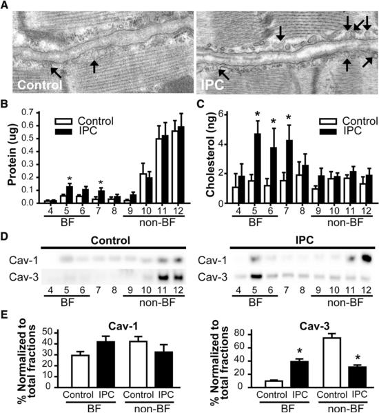
Background: Caveolae, lipid-rich microdomains of the sarcolemma, localize and enrich cardiac-protective signaling molecules. Caveolin-3 (Cav-3), the dominant isoform in cardiac myocytes, is a determinant of caveolar formation. We hypothesized that cardiac myocyte-specific overexpression of Cav-3 would enhance the formation of caveolae and augment cardiac protection in vivo.

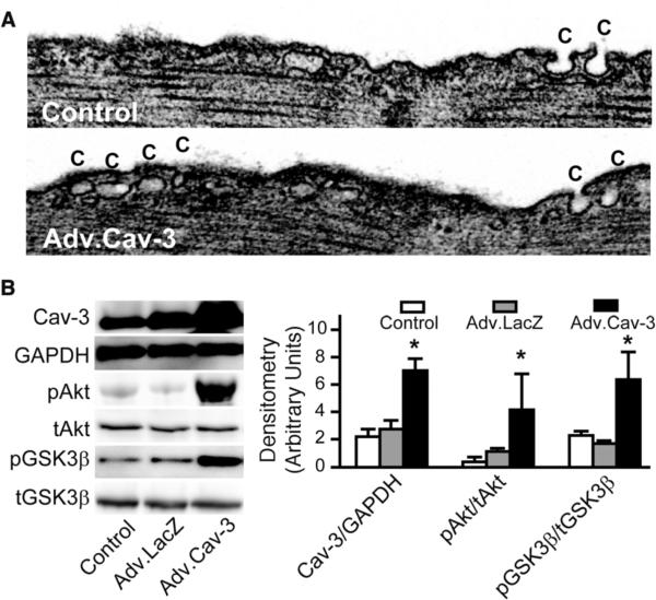
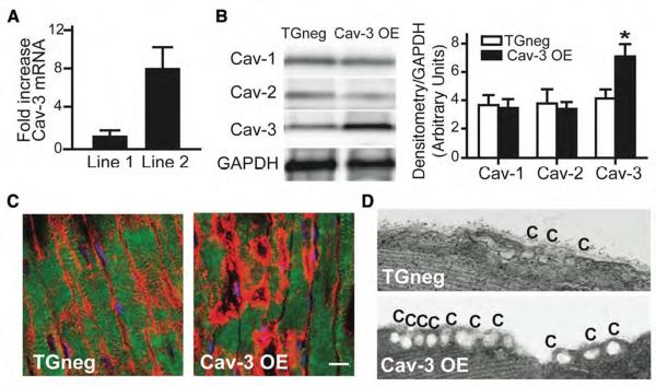
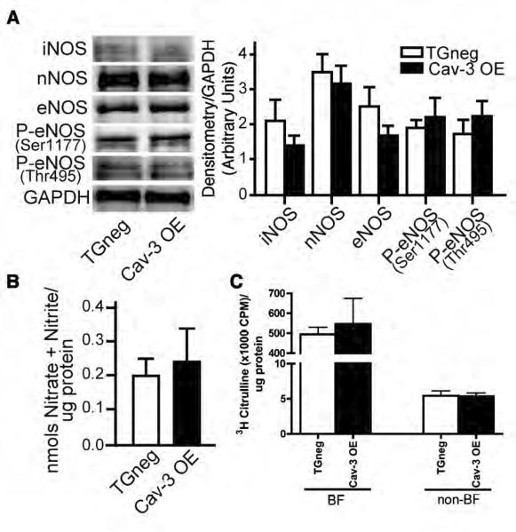
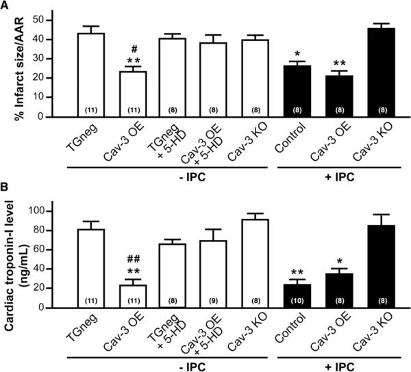
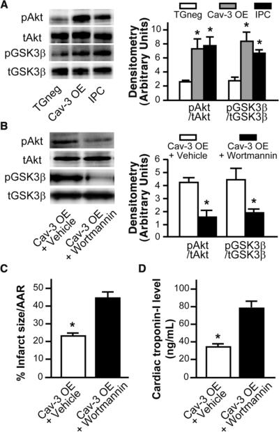
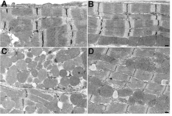
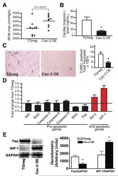
Methods and results: Ischemic preconditioning in vivo increased the formation of caveolae. Adenovirus for Cav-3 increased caveolar formation and phosphorylation of survival kinases in cardiac myocytes. A transgenic mouse with cardiac myocyte-specific overexpression of Cav-3 (Cav-3 OE) showed enhanced formation of caveolae on the sarcolemma. Cav-3 OE mice subjected to ischemia/reperfusion injury had a significantly reduced infarct size relative to transgene-negative mice. Endogenous cardiac protection in Cav-3 OE mice was similar to wild-type mice undergoing ischemic preconditioning; no increased protection was observed in preconditioned Cav-3 OE mice. Cav-3 knockout mice did not show endogenous protection and showed no protection in response to ischemic preconditioning. Cav-3 OE mouse hearts had increased basal Akt and glycogen synthase kinase-3beta phosphorylation comparable to wild-type mice exposed to ischemic preconditioning. Wortmannin, a phosphoinositide 3-kinase inhibitor, attenuated basal phosphorylation of Akt and glycogen synthase kinase-3beta and blocked cardiac protection in Cav-3 OE mice. Cav-3 OE mice had improved functional recovery and reduced apoptosis at 24 hours of reperfusion.

Conclusions: Expression of caveolin-3 is both necessary and sufficient for cardiac protection, a conclusion that unites long-standing ultrastructural and molecular observations in the ischemic heart. The present results indicate that increased expression of caveolins, apparently via actions that depend on phosphoinositide 3-kinase, has the potential to protect hearts exposed to ischemia/reperfusion injury.


Figures

Figure 1
Figure 1. IPC increases the expression of caveolae and the enrichment of protein and cholesterol in buoyant fractions
Hearts were subjected to a 5 min IPC stimulus. Control wild-type animals underwent no treatment. (A) Electron microscopy showed an increase in number of caveolae compared to control hearts (arrow). (B) and (C) Excised control and hearts subjected to IPC underwent sucrose density fractionation. Protein and cholesterol assays showed increased protein and cholesterol in buoyant fractions (BF) after IPC (*P < 0.05). (D) and (E) Fractions were probed for Cav-1 and Cav-3. Cav-3, but not Cav-1, was increased in buoyant fractions after IPC (representative immunoblots are shown) and confirmed by densitometry normalized to total fraction amounts (E). *P <0.05 relative to respective control. Data are from 6 mice per group.
Figure 2
Figure 2. Cav-3 adenovirus increases caveolae expression in adult cardiac myocytes (ACM)
(A) ACM incubated with an adenovirus (Adv) encoding full-length mouse Cav-3 (Adv.Cav-3) for 72 h increased caveolae (C) number. (B) ACM exposed to no virus, Adv.LacZ or Adv.Cav-3 for 72 h. were lysed and immunoblotted (left panel). Adv.Cav-3-treated ACM have increased expression of Cav-3 protein as well as increased phosphorylated phospho Akt and GSK3β compared to control or LacZ-treated ACM (n = 4 for Akt and GSK3β and n=6 for Cav-3) (right panel). *P < 0.05 vs. Adv.LacZ.
Figure 3
Figure 3. Cardiac myocyte-specific Cav-3 OE mice: caveolin and caveolae
(A) Real-time PCR of Cav-3 mRNA expression in two founder lines. Line 2 had 8-fold increased Cav-3 mRNA expression compared to Line 1. Data are represented relative to TGneg and normalized to GAPDH expression (n = 4). (B) Immunoblot of Cav-1, -2, and -3 in whole heart homogenates from Cav-3 OE and TGneg mice (left panel). Densitometry was normalized to expression of GAPDH and showed a significant increase in Cav-3 protein in whole heart homogenates (right panel; *P = 0.011, n=7 for TGneg and n=9 for Cav-3 OE). (C) Immunohistochemistry showed increased Cav-3 (red pixels) in sarcolemma of cardiac myocytes from Cav-3 OE vs. TGneg mice. The bar denotes 10 μm. (D) Electron microscopy shows increased caveolae (C) in cardiac myocytes from Cav-3 OE vs. TGneg mice.
Figure 4
Figure 4. NOS expression and activity
(A) Immunoblot analysis of basal expression of NOS and phosphorylated eNOS in TGneg and Cav-3 OE mice (left panel). Densitometry was normalized to GAPDH (right panel). No differences were observed in expression of any NOS isoforms. Data are from 5 mice per group. (B) Basal NOS activity was measured in TGneg and Cav-3 OE murine whole heart homogenates. No differences in basal NOS activity was observed between groups. (C) Hearts from TGneg and Cav-3 OE mice were homogenized in triton-X and fractionated on a discontinuous sucrose density gradient to separate buoyant (caveolae) and non-buoyant (non-caveolar membrane) fractions (BF and non-BF, respectively). NOS activity was measured in BF and non-BF using a [3H]-arginine assay. No difference in NOS activity was observed in the two separate fractions in TGneg vs. Cav-3 OE mice. Data are from 6 mice per group.
Figure 5
Figure 5. Cardiac protection in Cav-3 OE mice
Mice were subjected to ischemia-reperfusion injury. (A) Infarct size (percent of AAR) was reduced by IPC in control animals; however, Cav-3 OE mice were protected to similar levels with and without IPC. Cav-3 KO mice could not be protected with IPC. Treatment of Cav-3 OE mice with 5-hydroxydecanoate (5-HD; 10 mg/kg i.v.), a mitochondrial KATP channel inhibitor, abolished protection. TGneg treated with 5-HD had similar infarct size to controls. *P < 0.05, **P < 0.001 vs. TGneg mice, and #P < 0.05 vs. Cav-3 OE + 5-HD. Group sizes are indicated on the individual bars in parentheses. (B) Serum cardiac troponin-I, a marker of cardiac myocyte damage, was measured following 2 h of reperfusion. *P < 0.05, **P < 0.001 vs. TGneg mice, and ##P < 0.01 vs. Cav-3 OE + 5-HD. Group sizes are indicated on the individual bars parentheses.
Figure 6
Figure 6. Role of survival kinases in protection of Cav-3 OE mice
(A) Whole heart homogenates showed elevated phosphorylation of Akt and GSK3β in IPC-treated mice and Cav-3 OE mice when compared to TGneg mice (n = 6 for TGneg, n=5 Cav-3 OE, and n=6 for IPC). Total Akt or GSK3β was used to assess protein loading. * P < 0.05 vs. TGneg mice. (B-D) To determine the role of PI3K/Akt/GSK3β in the endogenous protection, Cav-3 OE mice were treated with a PI3K inhibitor, wortmannin (15μg/kg), 15 minutes prior to index ischemia-reperfusion. DMSO vehicle treated Cav-3 OE served as controls (n=7). Wortmannin treatment resulted in decreased basal phosphorylation of Akt and GSK3β (B). Additionally, wortmannin treatment attenuated the endogenous protection seen in vehicle treated Cav-3 OE mice with respect to infarct size (C) and cardiac troponin-I (D), n=7 for both groups in C and D.
Figure 7
Figure 7. Electron micrograph of area at risk from the hearts of TGneg and Cav-3 OE mice following ischemia-reperfusion
(A) and (B) No tissue swelling or structural change were seen in sham groups from TGneg and Cav-3 OE mice. (C) After ischemia-reperfusion in TGneg mice, myofibrils were distended, Z-lines were irregular and unclear, and mitochondria were swollen and contained amorphous matrix densities. (D) Cav-3 OE mice had fewer damaged myocytes after ischemia-reperfusion: Myofibrils had well-arranged Z-lines and organized mitochondria.
Figure 8
Figure 8. Cardiac function and cell death in mice 24 h after ischemic injury
Following 30 min of ischemia, mice were allowed to recover for 24 h. (A) Cav-3OE mice displayed better cardiac function, as measured by dP/dt max (4619 ± 226 vs. 3717 ± 275 mmHg/s, n = 9, P = 0.011). (B) Cardiac troponin-I levels were significantly decreased in Cav-3 OE mice (n = 7 of TGneg and n=8 for Cav-3 OE, *P = 0.002 vs. TGneg). (C) Apoptosis in the AAR, determined using TUNEL staining, in 24 h reperfused hearts (arrow, representative images). Nuclei staining positive for TUNEL, quantified as a percent of total nuclei, were significantly decreased in hearts from Cav-3 OE vs. TGneg mice (right panel, n=4 for TGneg and n=5 for Cav-3 OE). *P = 0.003 vs. TGneg mice. (D) Real-time PCR analysis of pro- and anti-apoptotic gene expression in AAR 24 h after reperfusion of Cav-3 OE mice and TGneg mice (n=5). (E) Immunoblot analysis shows decreased Fas (pro-apoptotic) and increased IAP-1 (anti-apoptotic) protein expression in agreement with mRNA data presented in (D) (n=4).

References

    1. Noble R. The development of resistance by rats and guinea pigs to amounts of trauma usually fatal. Am J Physiol. 1943;38:346–351.
    1. Murry CE, Jennings RB, Reimer KA. Preconditioning with ischemia: a delay of lethal cell injury in ischemic myocardium. Circulation. 1986;74:1124–1136. - PubMed
    1. Hausenloy DJ, Tsang A, Yellon DM. The reperfusion injury salvage kinase pathway: a common target for both ischemic preconditioning and postconditioning. Trends Cardiovasc Med. 2005;15:69–75. - PubMed
    1. Steinberg SF, Brunton LL. Compartmentation of g protein-coupled signaling pathways in cardiac myocytes. Annu Rev Pharmacol Toxicol. 2001;41:751–773. - PubMed
    1. Williams TM, Lisanti MP. The caveolin proteins. Genome Biol. 2004;5:214. - PMC - PubMed

Publication types

MeSH terms