. 2009 Jan;87(1):81-90.
doi: 10.1038/icb.2008.70. Epub 2008 Oct 21.

Inflammatory caspases are critical for enhanced cell death in the target tissue of Sjögren's syndrome before disease onset

Affiliations

Inflammatory caspases are critical for enhanced cell death in the target tissue of Sjögren's syndrome before disease onset

Marievic Bulosan et al. Immunol Cell Biol. 2009 Jan.

Abstract

To date, little is known about why exocrine glands are subject to immune cell infiltrations in Sjögren's syndrome (SjS). Studies with SjS-prone C57BL/6.NOD-Aec1Aec2 mice showed altered glandular homeostasis in the submandibular glands (SMX) at 8 weeks before disease onset and suggested the potential involvement of inflammatory caspases (caspase-11 and -1). To determine whether inflammatory caspases are critical for the increased epithelial cell death before SjS-like disease, we investigated molecular events involving caspase-11/caspase-1 axis. Our results revealed concurrent upregulation of caspase-11 in macrophages, STAT-1 activity, caspase-1 activity and apoptotic epithelial cells in the SMX of C57BL/6.NOD-Aec1Aec2 at 8 weeks. Caspase-1, a critical factor for interleukin (IL)-1beta and IL-18 secretion, resulted in an elevated level of IL-18 in saliva. Interestingly, TUNEL-positive cells in the SMX of C57BL/6.NOD-Aec1Aec2 were not colocalized with caspase-11, indicating that caspase-11 functions in a noncell autonomous manner. Increased apoptosis of a human salivary gland (HSG) cell line occurred only in the presence of lipopolysaccharide (LPS-) and interferon (IFN)-gamma-stimulated human monocytic THP-1 cells, which was reversed when caspase-1 in THP-1 cells was targeted by siRNA. Taken together, our study discovered that inflammatory caspases are essential in promoting a pro-inflammatory microenvironment and influencing increased epithelial cell death in the target tissues of SjS before disease onset.


Conflict of interest statement

Conflict of interest

Authors have no financial conflict of interest.

Figures

Figure 1
Figure 1. Increased caspase-11 expression in the SMX of the SjS-prone C57BL/6.NOD-Aec1Aec2 mouse prior to lymphocytic infiltration
A) Two NOD-derived genetic intervals, namely autoimmune exocrine loci (Aec) 1 and 2 on chromosomes 3 and 1, respectively, in the disease-prone-C57BL/6.NOD-Aec1Aec2 mouse are depicted. B) Elevated caspase-11, its major transcription factors Stat-1, and Nfkb1 in the salivary glands of C57BL/6.NOD-Aec1Aec2 were confirmed by semi-quantitative RT-PCR. C) Caspase-11 in the SMX was stained with FITC-labeled anti-mouse caspase-11 antibody in the C57BL/6.NOD-Aec1Aec2 at 8 weeks. Arrows indicate positive staining for caspase-11. Magnification: 20x. D) Double staining of caspase-11 with anti-Cd11c antibody (dendritic cells) and anti-F4/80 antibody (macrophages) revealed that cells positive for caspase-11 were also positive for both cell types. Arrows indicate double-stained cells, which are shown in yellow. Magnifications: 10x and 40x.
Figure 2
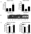
Figure 2. Concomitant increase in STAT-1 activity in the SMX of C57BL/6.NOD-Aec1Aec2 at 8 weeks
A) Increased STAT-1 activity was detected by EMSA in the glands isolated at 8 weeks. Inhibition of binding is shown with a cold probe (unlabelled probe) incubation. The right panel indicates bar graphs generated by densitometer analyses on EMSA results for NF-kappaB. B) Absence of STAT-1 and NF-kappaB activity in the LAC at 8 weeks was detected while elevated STAT-1 was shown in the SMX of C57BL/6.NOD-Aec1Aec2 and NOD/LtJ mouse. Increased amount of nuclear extract (ten micrograms) isolated from 0.5 gram of pooled glands (n=5–7 mice) was used per lane to enhance binding activity for NF-kappa B. The right panel indicates bar graphs generated by densitometer analyses on EMSA results for STAT-1. The experiments were carried out 3 times per molecule of interest. *p<0.05 in comparison with C57BL/6.
Figure 3
Figure 3. Activation of caspase-1-mediated pathway in C57BL/6.NOD-Aec1Aec2 prior to disease onset
A) Caspase-1 activity was up-regulated significantly in the SMX of C57BL/6.NOD-Aec1Aec2 at 8 weeks. Experiments were performed in triplicate. **p<0.01 in comparison with C57BL/6. B) Genes downstream of caspase-1 such as IL-1β and IL-18 in the C57BL/6.NOD-Aec1Aec2 mouse at 8 weeks were analyzed by RT-PCR. The C57BL/6.NOD-Aec1Aec2 mice were positive for these genes while the SMX from other mouse strains showed either negative or weak expression (n=5–7 female mice). Beta-actin was used as a control for normalization. C) IL-18 protein expression was elevated in the saliva from C57BL/6.NOD-Aec1Aec2 at 8 weeks by ELISA. Pooled saliva and sera were used for ELISA (n=5 female mice). *p<0.05 and **p<0.01 in comparison with the age-matched C57BL/6 mouse.
Figure 4
Figure 4. Increased epithelial cell death in the glands of disease-prone mice at 8 weeks and lack of direct co-localization of caspase-11 with TUNEL positive cells
A) TUNEL staining was performed on the pre-diseased salivary glands. NC, negative control; PC, positive control treated with nuclease; upper panel at 10x and lower panel at 40x magnifications. B) Percentages of TUNEL positive cells are shown as a bar graph. For each mouse, 3 slides were evaluated for TUNEL positive cells, which were counted using a cell counter. C) Caspase-3 positive cells (yellow arrows in b) were co-localized with TUNEL positive cells (red arrows). White arrows indicate caspase-11 positive cell. Magnification: 40x.
Figure 5
Figure 5. Inhibition of apoptotic cell death of HSG cells by caspase-1 knockdown in THP-1 cells
A) HSG cells were cultured in the absence or presence of LPS-and/or IFN-γ-stimulated THP-1 cells. After removing culture media or stimulated THP-1 cells from the culture, apoptotic HSG cells were analyzed by TUNEL assays. For the caspase-1 inhibition study, siRNA to caspase-1 was transfected into THP-1 cells prior to stimulation with LPS and IFN-γ and co-cultured with HSG cells. B) Percent TUNEL positive cells were presented as a bar graph. The experiment was repeated 3 times for reproducibility (**p<0.01). C) Knockdown efficiency of caspase-1 was compared with a housekeeping protein golgin-97 by western blotting and depicted as a bar graph. The experiment was repeated twice and caspase-1 protein level was normalized to golgin-97.
Figure 6
Figure 6. Schematic representation of the current working hypothesis
Inductive viral profile results in alterations in the target tissue via the activation of IFN-STAT and a subsequent induction of caspase-11. Up-regulated caspase-1 activity produces mature IL-1β and IL-18 from macrophages and dendritic cells, which may play a role in the activation of caspase-3 or induction of caspase-3-independent apoptotic factors in neighboring acinar and/or ductal cells. Elevated pro-inflammatory cytokines in the SMX enhances IFN- γ production by epithelial cells, resulting in further activation of macrophages. Areas where further investigation is needed for confirmation are depicted with dotted lines (i.e., questions as to if caspase-11 is a constituent of the NALP inflammasome or why IFN- γ and consequently STAT-1 are up-regulated in the SMX prior to disease onset). The assembled proteins (the inflammasome) in response to signal recognition by LRR of the NALP leads to the activation of caspase-1, depicted in the bracket. Our current findings are depicted in bold arrows. NALP, NACHT, LRR and PYD containing protein; LRR, leucine-rich repeats; ASC, apoptosis–associated speck-like protein containing a caspase-activating recruitment domain; STAT-1, Signal Transducers and Activators of Transcription; DC, Dendritic Cells; SMX, submandibular glands.

References

    1. Robinson CP, Yamamoto H, Peck AB, Humphreys-Beher MG. Genetically programmed development of salivary gland abnormalities in the NOD (nonobese diabetic)-scid mouse in the absence of detectable lymphocytic infiltration: a potential trigger for sialoadenitis of NOD mice. Clin Immunol Immunopathol. 1996;79(1):50–9. - PubMed
    1. Cha S, Brayer J, Gao J, Brown V, Killedar S, Yasunari U, et al. A dual role for interferon-gamma in the pathogenesis of Sjogren’s syndrome-like autoimmune exocrinopathy in the nonobese diabetic mouse. Scand J Immunol. 2004 Dec;60(6):552–65. - PubMed
    1. Cha S, van Blockland SC, Versnel MA, Homo-Delarche F, Nagashima H, Brayer J, et al. Abnormal organogenesis in salivary gland development may initiate adult onset of autoimmune exocrinopathy. Exp Clin Immunogenet. 2001;18(3):143–60. - PubMed
    1. Killedar SJ, Eckenrode SE, McIndoe RA, She JX, Nguyen CQ, Peck AB, et al. Early pathogenic events associated with Sjogren’s syndrome (SjS)-like disease of the nod mouse using microarray analysis. Lab Invest. 2007 Apr;87(4):398. - PubMed
    1. Hisahara S, Okano H, Miura M. Caspase-mediated oligodendrocyte cell death in the pathogenesis of autoimmune demyelination. Neurosci Res. 2003 Aug;46(4):387–97. - PubMed

Publication types

MeSH terms