Review
. 2008 Nov;9(11):1215-23.
doi: 10.1038/ni.f.216.

New developments in mast cell biology

Affiliations
Review

New developments in mast cell biology

Janet Kalesnikoff et al. Nat Immunol. 2008 Nov.

Abstract

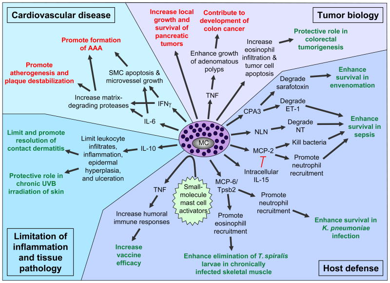
Mast cells can function as effector and immunoregulatory cells in immunoglobulin E-associated allergic disorders, as well as in certain innate and adaptive immune responses. This review focuses on exciting new developments in the field of mast cell biology published in the past year. We highlight advances in the understanding of FcvarepsilonRI-mediated signaling and mast cell-activation events, as well as in the use of genetic models to study mast cell function in vivo. Finally, we discuss newly identified functions for mast cells or individual mast cell products, such as proteases and interleukin 10, in host defense, cardiovascular disease and tumor biology and in settings in which mast cells have anti-inflammatory or immunosuppressive functions.


Figures

Figure 1
Figure 1
Simplified scheme of early FcεRI-mediated signaling events. Ag-induced crosslinking of FcεRI induces activation of Lyn, which phosphorylates FcεRI ITAMs (green) and activates Syk following ITAM binding, and Fyn, which phosphorylates the adaptor Gab2 to activate the PI3K pathway. Lyn and Syk phosphorylate many adaptor molecules, e.g., LAT and NTAL, and enzymes to regulate activation of the Ras, PLCγ, PI3K and other pathways. Grb2 and SOS activate the Ras/Erk pathway, which regulates transcription factor activation and arachidonic acid metabolism (through PLA2 activation). PLCγ can either be activated through the coordinated function of LAT/Gads/SLP-76/Vav and Btk or independently of LAT through a PI3K/Btk-dependent pathway. PLCγ activation regulates classical PKC activation (through DAG generation) and calcium responses (through the generation of IP3). IP3 binding to the IP3R triggers Ca2+ release from the ER; STIM1 couples ER Ca2+ store depletion with the activation of CRAC channels, leading to the influx of extracellular Ca2+ and ICRAC. The PI3K product, PI(3,4,5)P3, is an important lipid mediator that regulates the activity of various enzymes, e.g., Btk, Akt, PDK1, PLD and SK, and the formation of other lipid mediators, e.g., DAG and S1P. S1P can act intracellulary, to regulate Ca2+ influx and degranulation (independently of PLC and IP3), and extracellularly (following secretion from the cell) by binding to surface S1P1 or S1P2 receptors and thereby inducing cytoskeletal rearrangement or enhancing degranulation, respectively. The IKK complex consists of two catalytic subunits, IKKα/IKK1 and IKKβ/IKK2, and a regulatory subunit, NEMO/IKKγ; this complex phosphorylates IκB to activate the transcription factor NFκB. IKKβ/IKK2 also phosphorylates SNAP23 to facilitate SNARE complex formation. Arrows indicate the contributions of these signaling pathways toward mast cell degranulation, arachidonic acid metabolism, and cytokine/chemokine/growth factor production. Note: some arrows do not indicate direct interactions or targets. Bcl10, B cell lymphoma 10; Btk, Bruton’s tyrosine kinase; Ca2+, calcium; CaM, calmodulin; CRAC, Ca2+ release activated calcium channel; DAG, diacylglycerol; Gab2, Grb2 associated binding protein 2; GADS, Grb2 related adaptor downstream of Shc; ER, endoplasmic reticulum; Erk, extracellular signal-regulated kinase; ICRAC, Ca2+ release activated current; IκB, inhibitor of κB; IKK, IκB kinase; IP3, inositol 1,4,5-trisphosphate; IP3R, IP3 receptor; ITAM, immunoreceptor tyrosine based activation motif; LAT, linker for activation of T cells; MALT1, mucosa associated lymphoid tissue lymphoma translocation protein 1; NEMO, NFκB essential modulator; NFAT, nuclear factor of activated T cells; NFκB, nuclear factor κB; NTAL, non-T cell activation linker; PI3K, phosphoinositide 3-kinase; PI(3,4,5)P3, phosphatidylinositol 3,4,5-trisphosphate; PKC, protein kinase C; PL, phospholipase; RasGRP, Ras guanyl nucleotide-releasing protein; S1P, sphingosine 1 phosphate; SK, sphingosine kinase; SLP-76, SH2-domain containing leukocyte protein of 76 kDa; SOS, son of sevenless homolog; Sph, sphingosine; STIM1, stromal interaction molecule 1.
Figure 2
Figure 2
Negative regulation of FcεRI-mediated signaling events. FcεRI aggregation activates a number of proteins that negatively regulate the positive signaling pathways activated downstream of this receptor. For example, Lyn, which initiates both activating and inhibitory signals, negatively regulates Fyn activity and, thus, Gab2 phosphorylation. Other negative regulators include c-Cbl (which facilitates the ubiquitination of FcεRI, Lyn and Syk), the tyrosinse phosphatase SHP-1 (which dephosphorylates Syk), the lipid phosphatases SHIP (which catalyzes the hydrolysis of PI(3,4,5)P3 to PI(3,4)P2) and PTEN (which catalyzes the hydrolysis of PI(3,4,5)P3 to PI(4,5)P2), RasGAP (which enhances the intrinsic GTPase activity of Ras), RabGEF1 (which enhances FcεRI internalization and can bind to GTP-bound Ras), and RGS13 (which binds to the p85α subunit of PI3K and disrupts its association with Gab2 and Grb2). Ag-induced coaggregation of FcεRI with FcγRIIB inhibits FcεRI-induced signaling events and mast cell activation via Lyn mediated phosphorylation of the FcγRIIB ITIM (red) and the subsequent recruitment of SHIP and DOK1. Finally, ES-62, a glycoprotein secreted by filarial nematodes, forms a complex with TLR4 (which causes the sequesteration and subsequent proteosome-independent degradation of PKCα) to block FcεRI-induced PLD-coupled, SK-mediated Ca2+ flux and NFκB activation. DOK1, docking protein 1; Gab2, Grb2 associated binding protein 2; ITAM, immunoreceptor tyrosine based activation motif; ITIM, immunoreceptor tyrosine based inhibititory motif; NFκB, nuclear factor κB; PI3K, phosphoinositide 3-kinase; PKC, protein kinase C; PLD, phospholipase D; PTEN, phosphatase and tensin homolog; RabGEF, Rab5 guanine nucleotide exchange factor; RasGAP, GTPase activating protein; RGS, regulator of G protein signaling; SHIP, Src homology 2 (SH2) domain-containing inositol 5′-phosphatase; SHP-1, SH2 domain-containing tyrosine phosphatase-1; SK, sphingosine kinase; TLR4, toll like receptor 4.
Figure 3
Figure 3
Mast cell limit the pathology associated with CHS to urushiol. Cross-sections of ears (stained with Masson’s Trichrome) from WBB6F1-Kit+/+ (wild-type) mice (a,b), WBB6F1-KitW/W-v mice (c,d) or WBB6F1-KitW/W-v mice engrafted 8 weeks before the experiment with WT BMCMCs (WT BMCMC → KitW/W-v mice) (e,f) or Il10−/− BMCMCs (Il10−/− BMCMC → KitW/W-v mice) (g,h) were obtained 5 d after challenge with vehicle (100% acetone) only (a,c,e,g) or 5 mg/ml of urushiol (b,d,f,h). Focal full thickness necrosis of the epidermis and/or ulceration occurred in association with CHS responses to urushiol at 5 d after challenge in 8/10 of the mast cell-deficient WBB6F1-KitW/W-v mice and in 8/8 Il10−/− BMCMC → KitW/W-v mice but in none of the 10 wild-type or 7 WT BMCMC → KitW/W-v mice; *P < 0.05 by Chi-square test for all comparisons between rates of epidermal necrosis and ulceration in wild-type (WBB6F1-Kit+/+) mice or WT BMCMC → KitW/W-v mice and the corresponding mast cell-deficient WBB6F1-KitW/W-v mice or Il10−/− BMCMC → KitW/W-v mice. Similar findings were observed in association with CHS responses to urushiol in 5 of 7 C57BL/6-KitW-sh/W-sh mice in response to DNFB. By contrast, epidermal necrosis and ulceration occurred in none of the corresponding congenic wild-type mice or WT BMCMC-engrafted C57BL/6-KitW-sh/W-sh mice (10 or 8 for urushiol and 19 or 16 for DNFB, respectively); *P < 0.05 by Chi-squre test for all comparisons between rates of epidermal ulceration in wild-type or WT BMCMC-engrafted C57BL/6-KitW-sh/W-sh mice and the corresponding mast cell-deficient C57BL/6-KitW-sh/W-sh mice. C*: cartilage; double-headed arrows show thickness of dermis (D) or epidermis (Ep); arrows in insets: ulcers with adherent exudates (red). Scale bar in a = 100 μm & in inset in a = 1000 μm. Photomicrographs are representative of the findings observed in each of the 3 experiments performed (n = 3–7 mice/group per experiment). Taken from.
Figure 4
Figure 4
Newly identified protective (green) or detrimental (red) roles of mast cells and mast cell products in biological responses in mice. AAA, abdominal aortic aneurysm; CPA3, carboxypeptidase A3; ET-1, endothelin-1; IgE, immunoglobulin E; IL, interleukin; MC, mast cell; MCP, mast cell protease; NLN, neurolysin; NT, neurotensin; SMC, smooth muscle cell; TNF, tumor necrosis factor; Tpsb2, tryptase β2.

References

    1. Kitamura Y. Heterogeneity of mast cells and phenotypic change between subpopulations. Annu Rev Immunol. 1989;7:59–76. - PubMed
    1. Mekori YA, Metcalfe DD. Mast cells in innate immunity. Immunol Rev. 2000;173:131–40. - PubMed
    1. Kawakami T, Galli SJ. Regulation of mast-cell and basophil function and survival by IgE. Nat Rev Immunol. 2002;2:773–86. - PubMed
    1. Marone G, Galli SJ, Kitamura Y. Probing the roles of mast cells and basophils in natural and acquired immunity, physiology and disease. Trends Immunol. 2002;23:425–7. - PubMed
    1. Metz M, Grimbaldeston MA, Nakae S, Piliponsky AM, Tsai M, Galli SJ. Mast cells in the promotion and limitation of chronic inflammation. Immunol Rev. 2007;217:304–28. - PubMed