Review
. 2008 Sep-Oct;43(5):289-318.
doi: 10.1080/10409230802341296.

SSB as an organizer/mobilizer of genome maintenance complexes

Affiliations
Review

SSB as an organizer/mobilizer of genome maintenance complexes

Robert D Shereda et al. Crit Rev Biochem Mol Biol. 2008 Sep-Oct.

Abstract

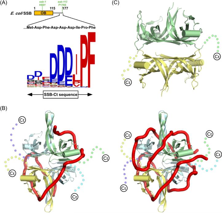
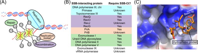
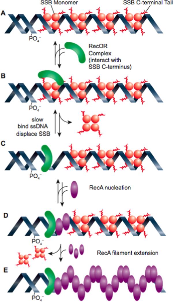
When duplex DNA is altered in almost any way (replicated, recombined, or repaired), single strands of DNA are usually intermediates, and single-stranded DNA binding (SSB) proteins are present. These proteins have often been described as inert, protective DNA coatings. Continuing research is demonstrating a far more complex role of SSB that includes the organization and/or mobilization of all aspects of DNA metabolism. Escherichia coli SSB is now known to interact with at least 14 other proteins that include key components of the elaborate systems involved in every aspect of DNA metabolism. Most, if not all, of these interactions are mediated by the amphipathic C-terminus of SSB. In this review, we summarize the extent of the eubacterial SSB interaction network, describe the energetics of interactions with SSB, and highlight the roles of SSB in the process of recombination. Similar themes to those highlighted in this review are evident in all biological systems.


Figures

Figure 1
Figure 1
(A) Schematic representation of SSB. The E. coli SSB OB domain (residues 1−115) is shown as a box with its structurally dynamic C-terminal tail (residues 116−177) as a line. The sequence of the E. coli SSB-Ct element is displayed with its conservation across 280 eubacterial species represented as a logo in which the height of the residue relates to its frequency at the given position. Logo residues are colored to indicate the hydrophobic (red), electronegative (blue), polar (black), or electropositive (green) nature of their side chains. (B) Ribbon diagram of the proposed structures of the E. coli (SSB)35 (left) and (SSB)65 (right) ssDNA binding models . Each monomer in the tetramer is separately colored and its C-terminus is shown schematically as a dashed line. ssDNA is shown as a red tube. (C) Ribbon diagram of the crystal structure of D. radiodurans SSB . OB folds are colored as for E. coli SSB, but with two OB folds in each monomer of the dimer. C-terminal tails are displayed as dots.
Figure 2
Figure 2
(A) Schematic representation of SSB interactions. SSB proteins (yellow) are depicted at tetramers with C-termini (Ct) interacting with ovals symbolizing proteins involved the major genome maintenance pathways of DNA replication (teal), recombination (purple), replication restart (orange), and repair (green). (B) List of proteins that are known to physically interact with SSB with their requirement for the SSB-Ct for interaction given. Citations for the interactions are given in the text. Highlighting colors indicate the major genome maintenance activities of the proteins (color coding as in (A)) and the sections in which each is described (except for Topoisomerase III, which is described in the recombination section with RecQ). (C) Binding site for the E. coli SSB C-terminus on Exonuclease I . Surface representation of Exonucelase I is stained in blue, red, and white to highlight positive, negative, and hydrophobic electrostatic features, respectively. The final four residues of the SSB C-terminus are shown in ball and stick form. Features shown to be critical for SSB binding by Exonuclease I are labeled.
Figure 3
Figure 3
Loading of RecA protein onto SSB-coated ssDNA by the RecOR proteins. The RecO protein, in a complex with RecR, first binds to the C-terminus of SSB. The RecOR complex with SSB is then rearranged to permit direct binding of RecOR to the ssDNA and displacement of an SSB tetramer. Once RecOR is loaded, RecA interacts with RecOR (perhaps in a way that alters the conformation of the RecA C-terminus so as to expose an intrinsic loading surface), and RecA nucleation occurs. This is followed by rapid and unassisted RecA filament extension. This figure is based on recent studies of the loading process ; .

References

    1. Watson JD, Crick FH. Genetical implications of the structure of deoxyribonucleic acid. Nature. 1953;171(4361):964. - PubMed
    1. Watson JD, Crick FH. Molecular structure of nucleic acids; a structure for deoxyribose nucleic acid. Nature. 1953;171(4356):737. - PubMed
    1. Varghese AJ. Photochemistry of nucleic acids and their constituents. Photophysiology. 1972;(7):207. - PubMed
    1. Hanawalt PC. The U.V. sensitivity of bacteria: its relation to the DNA replication cycle. Photochem Photobiol. 1966;5(1):1. - PubMed
    1. Hanawalt PC. Normal replication of DNA after repair replication in bacteria. Nature. 1967;214(5085):269. - PubMed

Publication types

MeSH terms

LinkOut - more resources