. 2008 Nov 18;47(46):12047-56.
doi: 10.1021/bi801604x. Epub 2008 Oct 21.

Oxidative protein folding in vitro: a study of the cooperation between quiescin-sulfhydryl oxidase and protein disulfide isomerase

Affiliations

Oxidative protein folding in vitro: a study of the cooperation between quiescin-sulfhydryl oxidase and protein disulfide isomerase

Pumtiwitt C Rancy et al. Biochemistry. .

Abstract

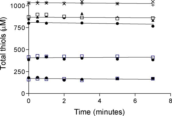
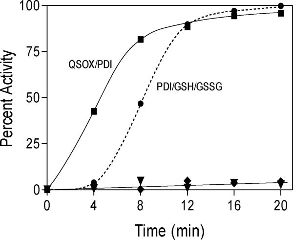
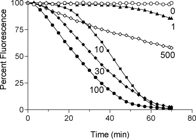
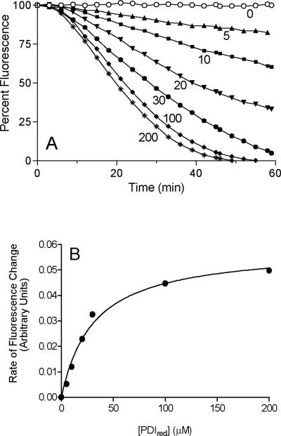
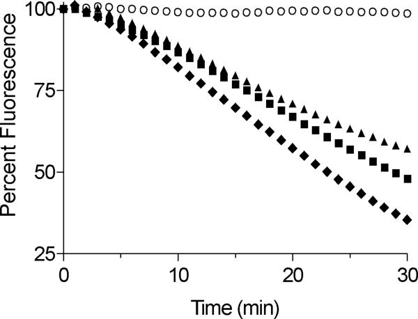
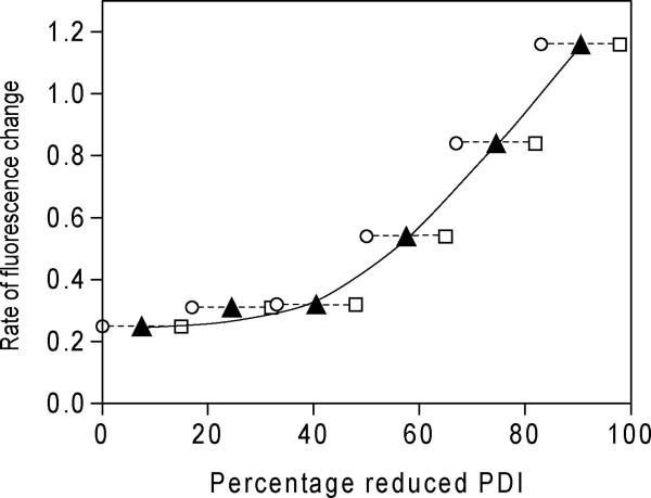
The flavin-dependent quiescin-sulfhydryl oxidase (QSOX) inserts disulfide bridges into unfolded reduced proteins with the reduction of molecular oxygen to form hydrogen peroxide. This work investigates how QSOX and protein disulfide isomerase (PDI) cooperate in vitro to generate native pairings in two unfolded reduced proteins: ribonuclease A (RNase, four disulfide bonds and 105 disulfide isomers of the fully oxidized protein) and avian riboflavin binding protein (RfBP, nine disulfide bonds and more than 34 million corresponding disulfide pairings). Experiments combining avian or human QSOX with up to 200 muM avian or human reduced PDI show that the isomerase is not a significant substrate of QSOX. Both reduced RNase and RfBP can be efficiently refolded in an aerobic solution containing micromolar concentrations of reduced PDI and nanomolar levels of QSOX without any added oxidized PDI or glutathione redox buffer. Refolding of RfBP is followed continuously using the complete quenching of the fluorescence of free riboflavin that occurs on binding to apo-RfBP. The rate of refolding is half-maximal at 30 muM reduced PDI when the reduced client protein (1 muM) is used in the presence of 30 nM QSOX. The use of high concentrations of PDI, in considerable excess over the folding protein client, reflects the concentration prevailing in the lumen of the endoplasmic reticulum and allows the redox poise of these in vitro experiments to be set with oxidized and reduced PDI. In the absence of either QSOX or redox buffer, the fastest refolding of RfBP is accomplished with excess reduced PDI and just enough oxidized PDI to generate nine disulfides in the protein client. These in vitro experiments are discussed in terms of current models for oxidative folding in the endoplasmic reticulum.


Figures

FIGURE 1
FIGURE 1
Oxidative folding models for Ero1 and QSOX. Panel A shows the PDI-first model of oxidative folding driven by Ero1p. QSOX oxidizes reduced unfolded proteins directly in panel B.
FIGURE 2
FIGURE 2
Reduced PDI as a substrate of QSOX. For each cognate and non-cognate PDI/QSOX pairing the oxidation of reduced PDI was evaluated by following the disappearance of CxxC thiols (using 50–200 μM PDI; 200–800 μM –SH groups; see Experimental Procedures) with 100 nM QSOX in 50 mM phosphate buffer, 1 mM EDTA, at pH 7.5 and 25 °C. Catalase (10 nM) was included in these incubations to discharge the hydrogen peroxide generated by QSOX. Aliquots were withdrawn at the indicated times for analysis of thiol content using DTNB (see Experimental Procedures). Open squares: avian QSOX and the indicated thiol titers of human PDI; filled circles: avian QSOX and avian PDI; filled triangles: human PDI and human QSOX. Crosses and checks indicate thiol consumption in an illustrative control reaction in the absence of QSOX using 250 μM of avian and human PDI respectively.
FIGURE 3
FIGURE 3
Oxidative refolding of reduced RNase. Reduced RNase 10 μM (80 μM –SH) in 50 mM Tris buffer, pH 7.5, 25 °C was incubated with 5 μM reduced PDI and 50 nM QSOX (squares); 50 nM QSOX alone (triangles); 5 μM reduced PDI and 1 mM GSH and 0.2 mM GSSG (circles); or 1 mM GSH and 0.2 mM GSSG alone (diamonds). At the times indicated samples were quenched with NEM and assayed for RNase using cCMP (see Experimental Procedures).
FIGURE 4
FIGURE 4
Stereoview of the crystal structure of riboflavin binding protein. The isoalloxazine ring of the bound riboflavin (gold) is sandwiched between Tyrosine 75 and Tryptophan 156 (grey). None of the 9 disulfide bonds come closer than 9 Å to the isoalloxazine ring. The N-terminus is at the bottom left, and the C-terminus is at the end of the helix at the bottom right of the structure.
FIGURE 5
FIGURE 5
QSOX cooperates with PDI to refold RfBP. Avian QSOX (0–500 nM) in 50 mM phosphate buffer (pH 7.5, 1 mM EDTA, 25 °C), was mixed with 0.8 μM riboflavin and 30 μM reduced human PDI (120 μM CxxC thiols; see Experimental Procedures). The reaction was initiated by the addition of reduced RfBP (1 μM; 18 μM cysteine thiols with a carry-over of ~ 50 mM guanidine hydrochloride). Binding of riboflavin was assessed by the decrease in riboflavin fluorescence emission.
FIGURE 6
FIGURE 6
The effect of increasing concentrations of reduced PDI on the refolding of RfBP driven by QSOX. Panel A: the conditions of Figure 5 were used except that a concentration of 30 nM QSOX was used and the reduced PDI concentration was varied from 0 to 200 μM. Panel B: the maximal rates of riboflavin rebinding attained for each curve in panel A show a hyperbolic dependence with half-saturation at 30 μM reduced PDI.
FIGURE 7
FIGURE 7
Comparison of the effectiveness of different oxidative refolding systems for reduced RfBP. Reduced RfBP was incubated with riboflavin (1 μM and 0.8 μM, respectively) in 50 mM phosphate buffer, pH 7.5, 25 °C in the presence of: a redox buffer of 5 mM GSH/1 mM GSSG (open circles); the redox buffer and 30 μM reduced PDI (triangles); redox buffer, reduced PDI and 30 nM QSOX (squares); or QSOX and reduced PDI alone (diamonds).
FIGURE 8
FIGURE 8
The influence of PDI redox poise on the refolding rate of reduced RfBP. Reduced RfBP (1 μM) was added to 0.8 μM riboflavin and 30 μM PDI at six redox ratios in the absence of QSOX or glutathione (see Experimental Procedures): the initial percentage of reduced PDI in the mixtures is depicted by the open circles. Reduced RfBP will transfer reducing equivalents to oxidized PDI as each experiment progresses. Open squares would represent stoichiometric transfer; see the Text. The solid triangles are the average between these two percentages.

References

    1. Pollard MG, Travers KJ, Weissman JS. Ero1p: a novel and ubiquitous protein with an essential role in oxidative protein folding in the endoplamic reticulum. Mol. Cell. 1998;1:171–182. - PubMed
    1. Frand AR, Kaiser CA. The ERO1 gene of yeast is required for oxidation of protein dithiols in the endoplasmic reticulum. Mol. Cell. 1998;1:161–170. - PubMed
    1. Hoober KL, Glynn NM, Burnside J, Coppock DL, Thorpe C. Homology between egg white sulfhydryl oxidase and quiescin Q6 defines a new class of flavin-linked sulfhydryl oxidases. J. Biol. Chem. 1999;274:31759–31762. - PubMed
    1. Benayoun B, Esnard-Fève A, Castella S, Courty Y, Esnard F. Rat seminal vesicle FAD-dependent sulfhydryl oxidase:biochemical characterization and molecular cloning of a member of the new sulfhydryl oxidase/quiescin Q6 gene family. J. Biol. Chem. 2001;276:13830–13837. - PubMed
    1. Dias-Gunasekara S, Gubbens J, van Lith M, Dunne C, Williams JA, Kataky R, Scoones D, Lapthorn A, Bulleid NJ, Benham AM. Tissue-specific expression and dimerization of the endoplasmic reticulum oxidoreductase Ero1beta. J. Biol. Chem. 2005;280:33066–33075. - PubMed

Publication types