

A basis function feature-based approach for skin lesion discrimination in dermatology dermoscopy images
- PMID: 18937777
- PMCID: PMC3185359
- DOI: 10.1111/j.1600-0846.2008.00307.x
A basis function feature-based approach for skin lesion discrimination in dermatology dermoscopy images
Abstract
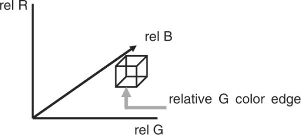
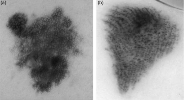
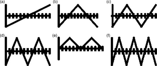
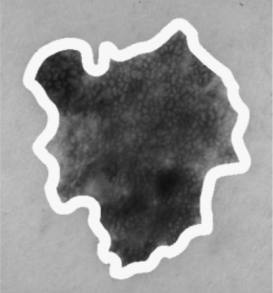
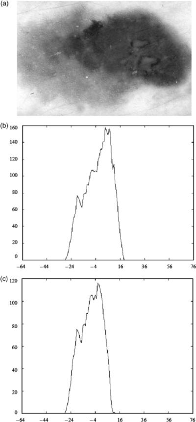
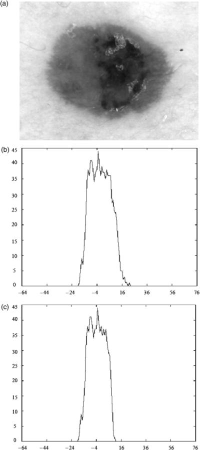
Background: Skin lesion color is an important feature for diagnosing malignant melanoma. New basis function correlation features are proposed for discriminating malignant melanoma lesions from benign lesions in dermoscopy images. The proposed features are computed based on correlating the luminance histogram of melanoma or benign labeled relative colors from a specified portion of the skin lesion with a set of basis functions. These features extend previously developed statistical and fuzzy logic-based relative color histogram analysis techniques for automated mapping of colors representative of melanoma and benign skin lesions from a training set of lesion images.
Methods: Using the statistical and fuzzy logic-based approaches for relative color mapping, melanoma and benign color features are computed over skin lesion region of interest, respectively. Luminance histograms are obtained from the melanoma and benign mapped colors within the lesion region of interest and are correlated with a set of basis functions to quantify the distribution of colors. The histogram analysis techniques and feature calculations are evaluated using a data set of 279 malignant melanomas and 442 benign dysplastic nevi images.
Results: Experimental test results showed that combining existing melanoma and benign color features with the proposed basis function features found from the melanoma mapped colors yielded average correct melanoma and benign lesion discrimination rates as high as 86.45% and 83.35%, respectively.
Conclusions: The basis function features provide an alternative approach to melanoma discrimination that quantifies the variation and distribution of colors characteristic of melanoma and benign skin lesions.
Figures
References
-
- Lightstone AC, Kopf AW, Garfinkel L. Diagnostic accuracy - a new approach to its evaluation. Arch Dermatol. 1965;91:497–502. - PubMed
-
- Grin C, Kopf AW, Welkovich B, Bar R, Levenstein M. Diagnostic accuracy in malignant melanoma. Arch Dermatol. 1990;126:763–766. - PubMed
-
- Lindelof B, Hedblad MA. Accuracy in the clinical diagnosis and pattern of malignant melanoma at a dermatological clinic. J Dermatol. 1994;21:461–464. - PubMed
-
- Friedman RJ, Rigel DS, Kopf AW. Early detection of malignant melanoma: the role of physician examination and self-examination of the skin. Ca-A Cancer J Clinicians. 1985;35:130–151. - PubMed
-
- Kopf A, Mintzis M, Bar R. Diagnostic accuracy in malignant melanoma. Arch Dermatol. 1975;111:1291–1292. - PubMed
Publication types
MeSH terms
Grants and funding
LinkOut - more resources
Full Text Sources
Medical
