Review
. 2008 Dec;68(3):293-308.
doi: 10.1016/j.bandc.2008.08.019. Epub 2008 Oct 19.

Development of eye-movement control

Affiliations
Review

Development of eye-movement control

Beatriz Luna et al. Brain Cogn. 2008 Dec.

Abstract

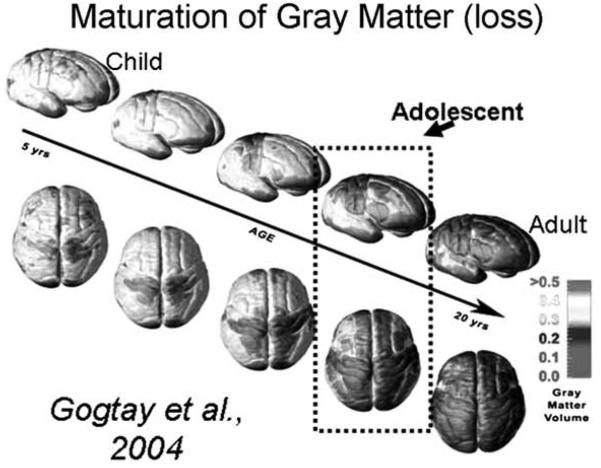
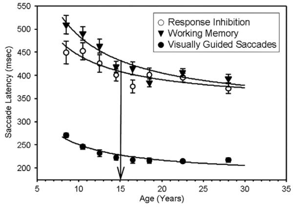
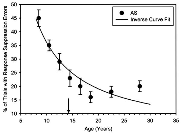
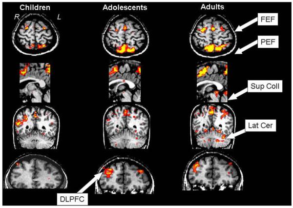
Cognitive control of behavior continues to improve through adolescence in parallel with important brain maturational processes including synaptic pruning and myelination, which allow for efficient neuronal computations and the functional integration of widely distributed circuitries supporting top-down control of behavior. This is also a time when psychiatric disorders, such as schizophrenia and mood disorders, emerge reflecting a particularly vulnerability to impairments in development during adolescence. Oculomotor studies provide a unique neuroscientific approach to make precise associations between cognitive control and brain circuitry during development that can inform us of impaired systems in psychopathology. In this review, we first describe the development of pursuit, fixation, and visually-guided saccadic eye movements, which collectively indicate early maturation of basic sensorimotor processes supporting reflexive, exogenously-driven eye movements. We then describe the literature on the development of the cognitive control of eye movements as reflected in the ability to inhibit a prepotent eye movement in the antisaccade task, as well as making an eye movement guided by on-line spatial information in working memory in the oculomotor delayed response task. Results indicate that the ability to make eye movements in a voluntary fashion driven by endogenous plans shows a protracted development into adolescence. Characterizing the transition through adolescence to adult-level cognitive control of behavior can inform models aimed at understanding the neurodevelopmental basis of psychiatric disorders.


Figures

Fig. 1
Fig. 1
View of cortical surface of the brain. Colors represent degree of thinning of gray matter. Blue indicates mature adult-levels have been reached. We have added a box around the brains that represent adolescence (figure from Gogtay et al. (2004). PNAS, 101, 8174–8179).
Fig. 2
Fig. 2
M ± 1 standard error of the M (SEM) of the latency to initiate a saccade in each task for each age group. Solid circles depict the latency to initiate a saccade to a visual stimulus during the visually guided saccade (VGS) task. Open circles depict the latency to initiate an eye movement to the opposite location of a visual target in the antisaccade (AS) task. Solid triangles depict the latency to initiate an eye movement to a remembered location in the oculomotor delayed response (ODR) task. Thick lines indicate the inverse curve fit on the M latency to initiate an eye-movement response in millisecond by age in years. Arrows depict the ages at which change-point analyses indicate adult levels of performance were reached (from Luna et al. (2004). Child Development, 75, 1357–1372).
Fig. 3
Fig. 3
Solid circles depict the M ± 1 standard error of the M (SEM) for the percent of trials with a response suppression failure in the antisaccade (AS) task. Thick lines depict the inverse curve fit on the response suppression failures by age in years. The arrow depicts the age at which change-point analyses indicate adult levels of performance were reached (from Luna et al. (2004). Child Development, 75, 1357–1372).
Fig. 4
Fig. 4
Mean group activity during a block antisaccade task for children, adolescents, and adults overlaid on top of the structure of a representative subject (from Luna et al. (2001). Neuroimage, 2001 13(5), 786–793).
Fig. 5
Fig. 5
Activation maps displayed on the partially inflated medial cortical surface of the right hemisphere for inhibitory errors in the AS task for children, adolescents, and adults. Results indicate similarities across age groups during the initial stage of error processing in the medFG/rACC. However, only adults show recruitment of dACC in later stages of error processing. Blue indicated deactivation. Red/Yellow indicated activation (adapted from Velanova et al. (2008). Cerebral Cortex, February 14 [Epub ahead of print]). (For interpretation of the references to color in this figure legend, the reader is referred to the web version of this paper.)
Fig. 6
Fig. 6
Mean ± 1 standard error of the mean for the accuracy to initiate a memory-guided saccade (solid circles) and the accuracy of the final gaze location (open circles) in the ODR task for each age group. Thick lines indicate the inverse curve fit for these data across the age-range studied. Arrows depict the age at which change-point analyses indicate adult levels of performance were reached (from Luna et al. (2004). Child Development, 75, 1357–1372).
Fig. 7
Fig. 7
Imaging results from both magnitude and extent of activation analyses. (A) Proportion of total number of voxels in each region of interest submitted to extent of activation analyses in all groups. (B) Each group image represents illustrative differences in both the magnitude and extent of activation in the group-averaged percent signal change functional maps. Children showed stronger activation bilaterally in the caudate nucleus, the thalamus, and anterior insula. Adolescents showed the strongest right DLPFC activation, and adults showed concentrated activation in left prefrontal and posterior parietal regions. (C) Group differences in the extent of activation as measured by the proportion of total active voxels in each region of interest for each age group. Despite the fact that the proportion of total voxels in the extent of activation analyses was consistent across the age groups, the groups showed large differences in the proportion of total active voxels across the regions of interest (from Scherf et al. (2006). Journal of Cognitive Neuroscience, 18(7), 1045–1058).
Fig. 8
Fig. 8
Mean (±1 SEM) of the proportion of trials with response inhibition failures in the antisaccade task (left graph) and absolute error of the initial memory-guided saccade in the ODR task (right) for the autism group (open circles) and the control group (solid circles). Shaded circles indicate the similarities in the rates of improvements between groups (from Luna et al. (2007). Biological Psychiatry, 61(4), 474–481).

References

    1. Amador N, Schlag-Rey M, Schlag J. Reward-predicting and reward-detecting neuronal activity in the primate supplementary eye field. Journal of Neurophysiology. 2000;84:2166–2170. - PubMed
    1. Amador N, Schlag-Rey M, Schlag J. Primate antisaccade. II. Supplementary eye field neuronal activity predicts correct performance. Journal of Neurophysiology. 2004;91:1672–1689. - PubMed
    1. Andersen SL. Stimulants and the developing brain. Trends in Pharmacological Sciences. 2005;26:237–243. - PubMed
    1. Angold A, Costello EJ, Worthman CM. Puberty and depression: The roles of age, pubertal status and pubertal timing. Psychological Medicine. 1998;28:51–61. - PubMed
    1. Aring E, Grönlund MA, Hellström A, Ygge J. Visual fixation development in children. Graefes Archives of Clinical and Experimental Opthalmology. 2007;245:1659–1665. - PubMed