Review
. 2009 Jan;53(1):10-8.
doi: 10.1016/j.jdermsci.2008.08.012. Epub 2008 Oct 19.

The hairless mouse in skin research

Affiliations
Review

The hairless mouse in skin research

Fernando Benavides et al. J Dermatol Sci. 2009 Jan.

Abstract

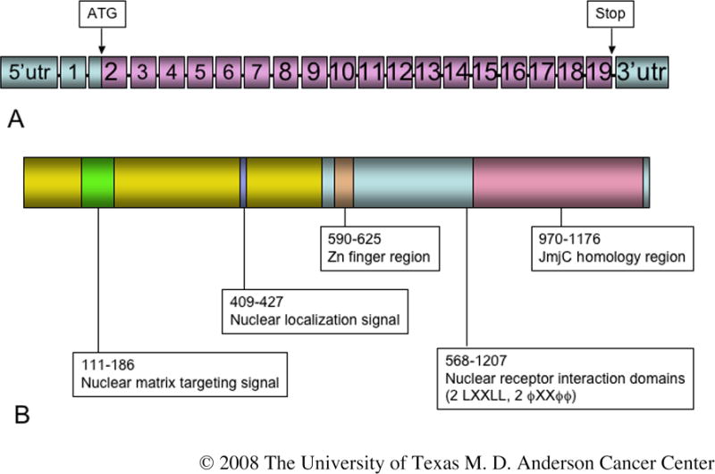
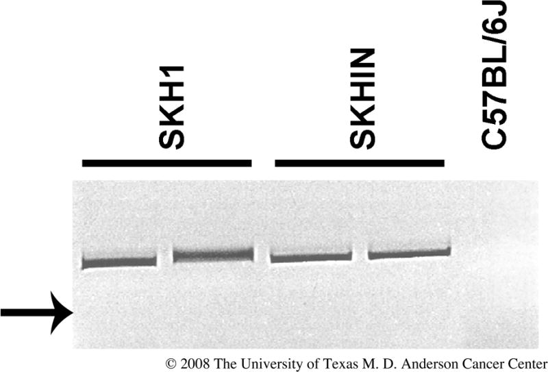
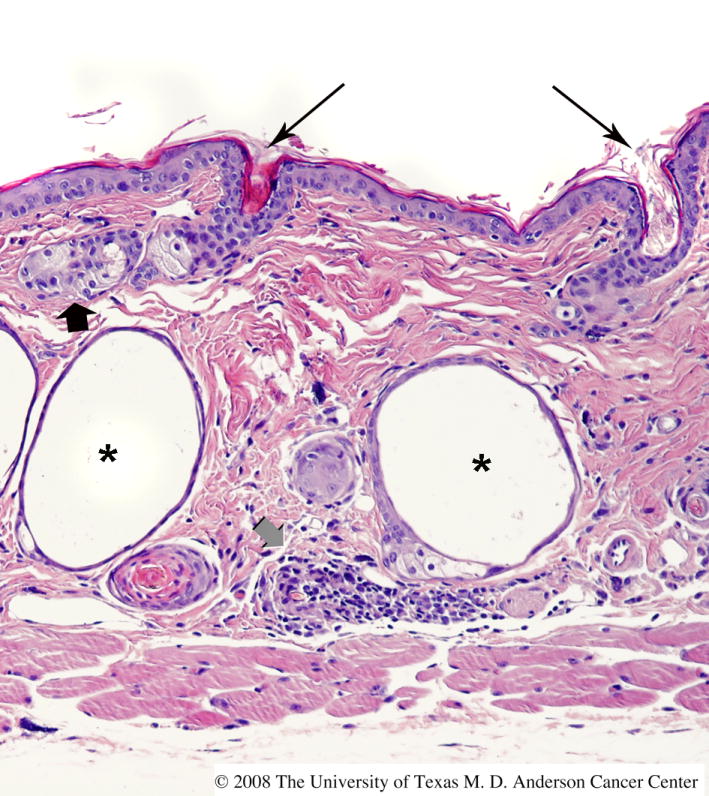
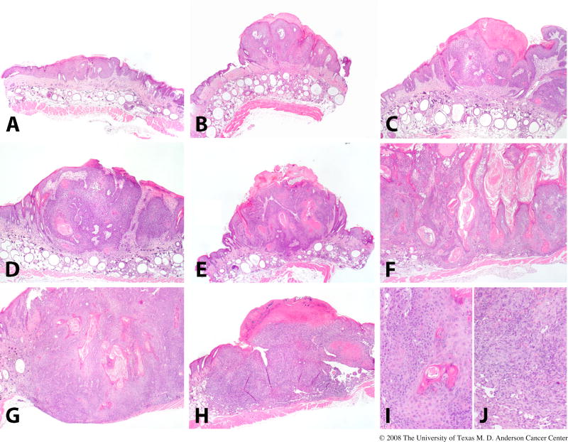
The hairless (Hr) gene encodes a transcriptional co-repressor highly expressed in the mammalian skin. In the mouse, several null and hypomorphic Hr alleles have been identified resulting in hairlessness in homozygous animals, characterized by alopecia developing after a single cycle of relatively normal hair growth. Mutations in the human ortholog have also been associated with congenital alopecia. Although a variety of hairless strains have been developed, outbred SKH1 mice are the most widely used in dermatologic research. These unpigmented and immunocompetent mice allow for ready manipulation of the skin, application of topical agents, and exposure to UVR, as well as easy visualization of the cutaneous response. Wound healing, acute photobiologic responses, and skin carcinogenesis have been extensively studied in SKH1 mice and are well characterized. In addition, tumors induced in these mice resemble, both at the morphologic and molecular levels, UVR-induced skin malignancies in man. Two limitations of the SKH1 mouse in dermatologic research are the relatively uncharacterized genetic background and its outbred status, which precludes inter-individual transplantation studies.


Conflict of interest statement

Conflict of Interest Statement None declared

Figures

Fig. 1
Fig. 1
The hairless gene and encoded protein. (A) The hairless gene contains 19 exons; translation begins in exon 2 at the ATG indicated. (B) The encoded protein is 1182 amino acids in length in which a number of functional domains have been identified, as indicated. Information from which this figure was compiled was obtained from a variety of sources [1,4,5, 14,88-90].
Fig. 2
Fig. 2
PCR identification of the hr allele. This shows the reverse image of an ethidium bromide-stained agarose gel of PCR products that identify the retroviral insertion in the Hr locus described for the hr allele. Primers employed were 5′-CAAGCCTTATTCGAACTAAC-3′ located within the retroviral insertion and 5′-AGATTTAACACAGGTGCTAG-3′ located in mouse genomic DNA. The presence of the 439 bp specific band in genomic DNA from SKH1 outbred and SKHIN inbred mice indicates that they carry the hr hypomorphic allele. The band was absent when C57BL/6 DNA was used in the reaction. We confirmed by direct sequencing that the PCR product contained retroviral sequences. The arrow indicates the position of the 400 basepair DNA size standard.
Fig. 3
Fig. 3
Histologic appearance of skin in mice homozygous for the hr allele. The skin of this FVB mouse homozygous for the hr allele exhibits characteristic features, including multiple dermal cysts (asterisks), utriculi connected to the skin surface (long black arrows), sebaceous gland hyperplasia (short black arrow), and dermal inflammation (short gray arrow).
Fig. 4
Fig. 4
Flow cytometry of peripheral blood leukocytes of the SKH1 mouse. Blood was collected by retroorbital bleeding, red blood cells were lysed, and the remaining leukocytes were incubated with anti-CD4 (A) or CD8 (B). Staining and flow cytometry were performed as previously described [49].
Fig 5
Fig 5
UVR-induced skin tumor development in the SKH1 mice. This figure illustrates the categories into which we classify skin lesions in SKH1 mice. Pre-malignant papillomas are shown in the upper panel, with grades 1, 2, 3, shown in (A), (B), and (C), respectively. Malignant micro-invasive SCC are shown in the middle panel, with grades 1, 2, 3, shown in (D), (E), and (F), respectively. A fully invasive SCC (G) and a spindle cell/anaplastic tumor (H) are shown in the lower panel. Closer views of the tumors in (G) and (H) are shown in (I) and (J). This detailed classification scheme has been useful in identifying small differences in skin tumor progression in several different protocols.

References

    1. Cachon-Gonzalez MB, Fenner S, Coffin JM, et al. Structure and expression of the hairless gene of mice. Proc Natl Acad Sci USA. 1994;91:7717–21. - PMC - PubMed
    1. Panteleyev AA, van der Veen C, Rosenbach T, et al. Towards defining the pathogenesis of the hairless phenotype. J Invest Dermatol. 1998;110:902–7. - PubMed
    1. Panteleyev AA, Botchkareva NV, Sundberg JP, et al. The role of the hairless (hr) gene in the regulation of hair follicle catagen transformation. Am J Pathol. 1999;155:159–71. - PMC - PubMed
    1. Djabali K, Aita VM, Christiano AM. Hairless is translocated to the nucleus via a novel bipartite nuclear localization signal and is associated with the nuclear matrix. J Cell Sci. 2001;114:367–76. - PubMed
    1. Potter GB, Beaudoin GM, 3rd, DeRenzo CL, et al. The hairless gene mutated in congenital hair loss disorders encodes a novel nuclear receptor corepressor. Genes Dev. 2001;15:2687–701. - PMC - PubMed

Publication types