

doi: 10.1021/nl801958b.
Epub 2008 Oct 22.
Nanocrystal core high-density lipoproteins: a multimodality contrast agent platform
Affiliations
- PMID: 18939808
- PMCID: PMC2629801
- DOI: 10.1021/nl801958b
Item in Clipboard
Nanocrystal core high-density lipoproteins: a multimodality contrast agent platform
Nano Lett.
2008 Nov.
Abstract
High density lipoprotein (HDL) is an important natural nanoparticle that may be modified for biomedical imaging purposes. Here we developed a novel technique to create unique multimodality HDL mimicking nanoparticles by incorporation of gold, iron oxide, or quantum dot nanocrystals for computed tomography, magnetic resonance, and fluorescence imaging, respectively. By including additional labels in the corona of the particles, they were made multifunctional. The characteristics of these nanoparticles, as well as their in vitro and in vivo behavior, revealed that they closely mimic native HDL.
Figures
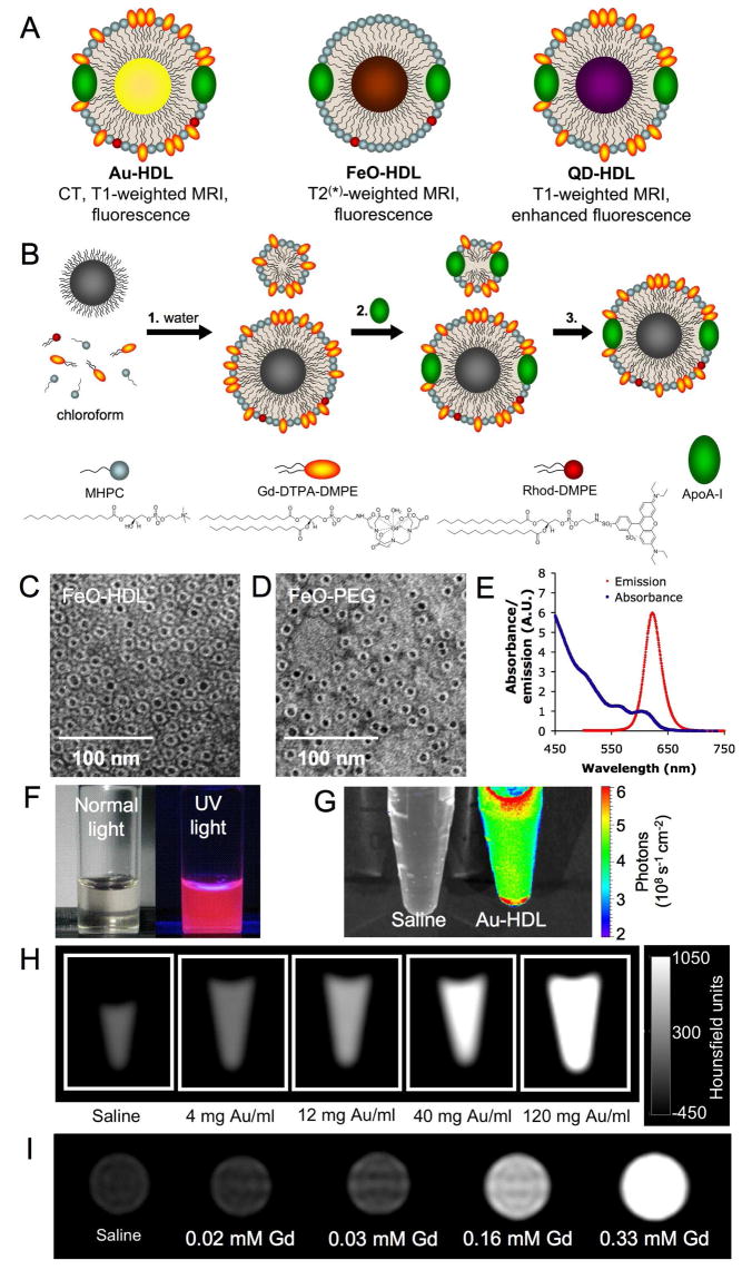
A schematic depiction of the different agents in this study. B summary of the synthesis procedure of the agents where 1, the phospholipids and nanocrystal in chloroform are added to water, 2, apoA-I is added and 3, the ‘empty’ particles are removed. C and D negative stain TEM images of FeO-HDL and FeO-PEG. E emission and absorption spectrum of the QD-HDL. F photograph of the QD-HDL in normal light (left) and under UV illumination (right). G, H, I phantoms of Au-HDL imaged using fluorescence, CT and MRI, respectively.
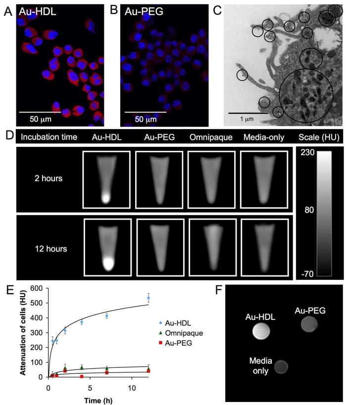
A confocal microscopy of cells incubated for 2 hours with Au-HDL or B Au-PEG, which appear red (rhodamine) and the nuclei are stained with DAPI (blue). C TEM image where black circles indicate areas of particle uptake. D CT images of the different cell pellets. E graph of the increase in CT attenuation of the cell pellets compared to media only incubated cells vs. time of incubation. F T1-weighted MR image of cell pellets.
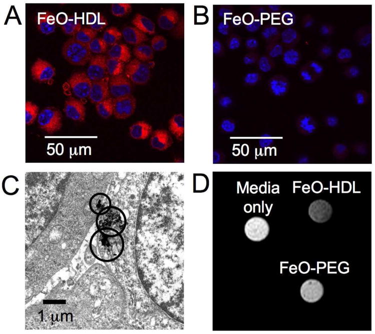
Confocal microscopy of cells incubated for 2 hours with A FeO-HDL and B FeO-PEG, where uptake is indicated by red (rhodamine) and the nuclei are stained with DAPI. C TEM of cells incubated with FeO-HDL, areas containing iron oxide are highlighted by circles. D T2-weighted MR image of cells incubated with FeO-HDL, FeO-PEG or media only for 4 hours.
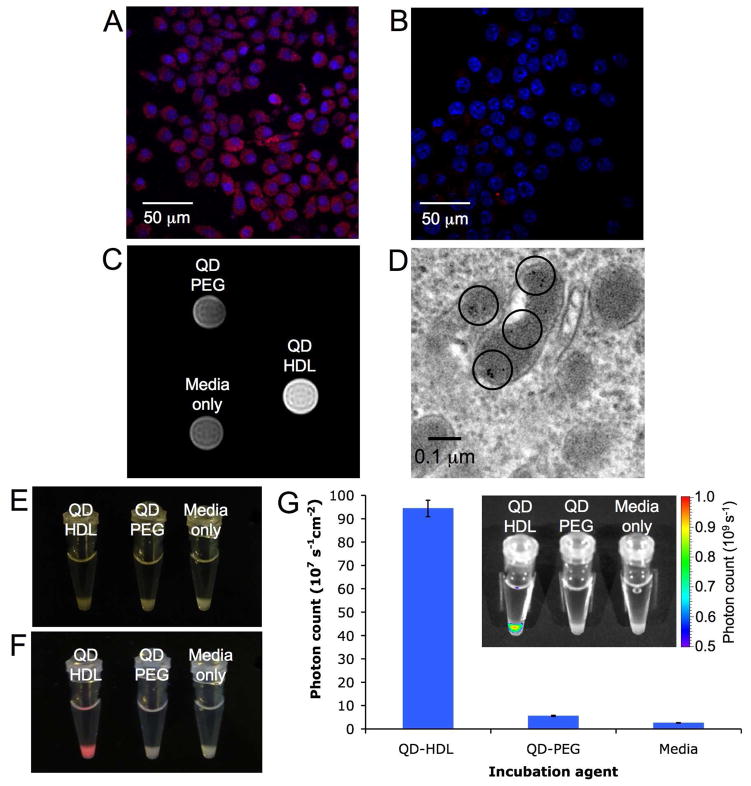
Confocal microscopy of macrophage cells incubated for 2 hours, where A QD-HDL and B QD-PEG uptake is indicated by red and the nuclei are blue. C T1-weighted MR image of pellets of cells incubated for 4 hrs. D TEM image of QD-HDL uptake, QDs are highlighted with black rings. Photographs of the cell pellets taken under E ambient light and F UV irradiation. G chart of fluorescent intensity of the cell pellets, with a typical fluorescent image inset.
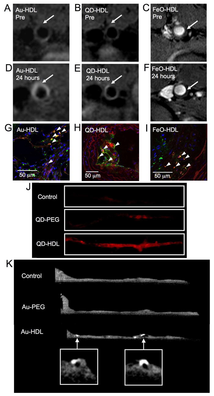
T1-weighted MR images of the aorta of apoE KO mice pre- (A, B) and 24 hours post-injection (D, E) with Au-HDL or QD-HDL. Arrows indicate areas enhanced in the post images. C, F T2*-weighted images of an apoE KO mouse pre- and 24 hours post-injection with FeO-HDL. G, H, I confocal microscopy images of aortic sections of mice injected with nanocrystal HDL. Red is nanocrystal HDL, macrophages are green and nuclei and blue. Yellow indicates co-localization of nanocrystal HDL with macrophages and is indicated by arrowheads. J fluorescence image of aortas of mice injected with QD-HDL, QD-PEG and saline. K ex vivo sagittal CT images of the aortas of mice injected with Au-HDL, Au-PEG and saline.
Comment in
- Nano Lett. 4:7.
References
-
- Mulder WJM, Cormode DP, Hak S, Lobatto ME, Silvera S, Fayad ZA. Nat Clin Pract Cardiovasc Med. 2008;5:S103–S111. - PubMed
-
- Kim D, Park S, Lee JH, Jeong YY, Jon S. J Am Chem Soc. 2007;129:7661–7665. - PubMed
-
- El-Sayed IH, Huang X, El-Sayed MA. Nano Lett. 2005;5:829–834. - PubMed
-
- Jaffer FA, Libby P, Weissleder R. Circulation. 2007;116:1052–1061. - PubMed
-
- de Vries IJM, Lesterhuis WJ, Barentsz JO, Verdijk P, van Krieken JH, Boerman OC, Oyen WJG, Bonenkamp JJ, Boezeman JB, Adema GJ, Bulte JWM, Scheenen TWJ, Punt CJA, Heerschap A, Figdor CG. Nature Biotechnology. 2005;23:1407–1413. - PubMed
Publication types
MeSH terms
Substances
Grants and funding
LinkOut - more resources
Full Text Sources
Other Literature Sources
