Comparative Study
. 2008 Oct 9;60(1):84-96.
doi: 10.1016/j.neuron.2008.09.027.

V3 spinal neurons establish a robust and balanced locomotor rhythm during walking

Affiliations
Comparative Study

V3 spinal neurons establish a robust and balanced locomotor rhythm during walking

Ying Zhang et al. Neuron. .

Abstract

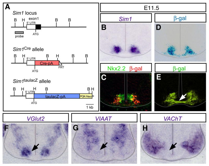
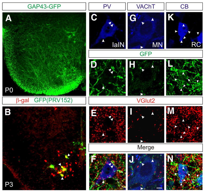
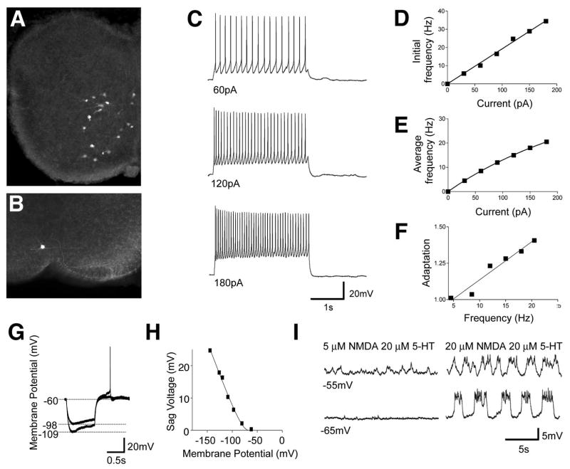
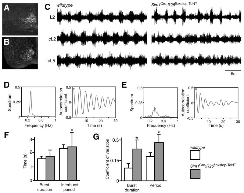
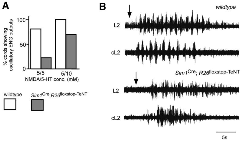
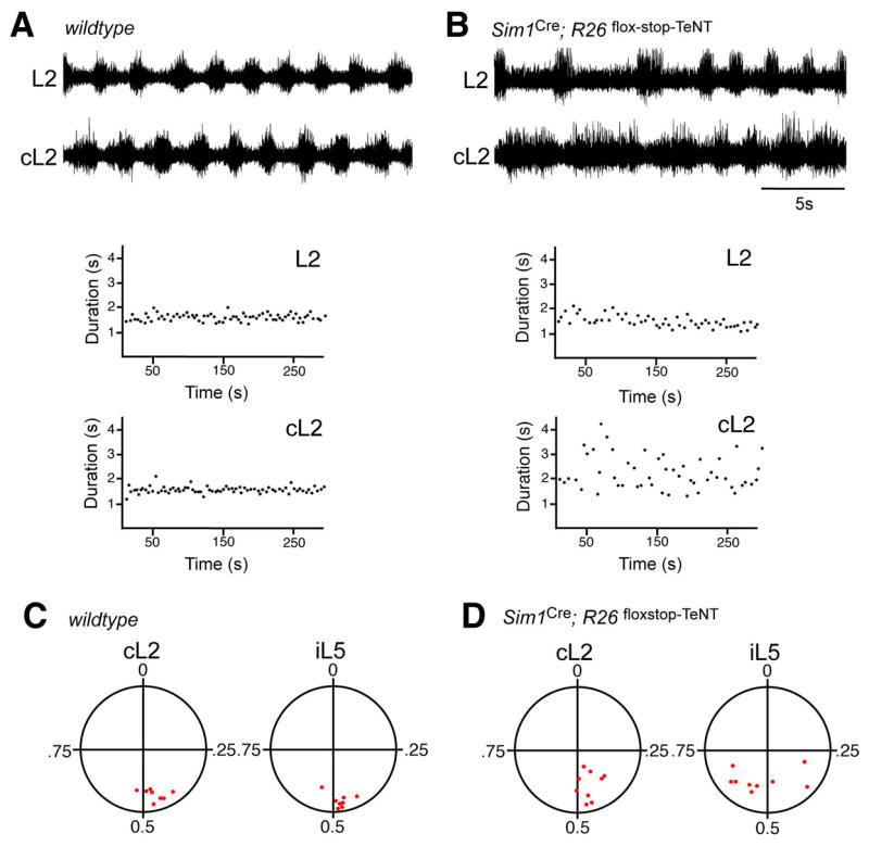
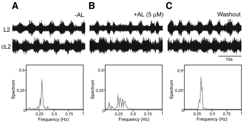
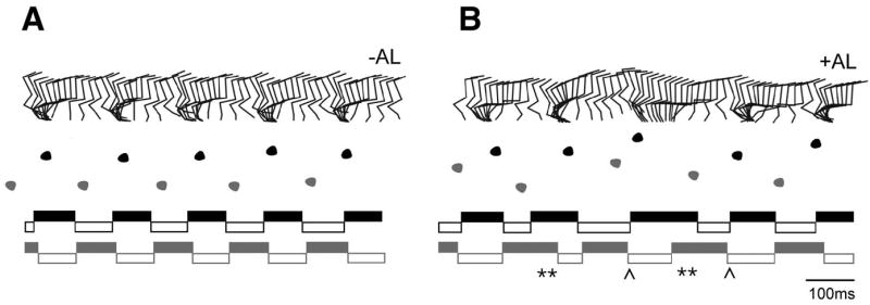
A robust and well-organized rhythm is a key feature of many neuronal networks, including those that regulate essential behaviors such as circadian rhythmogenesis, breathing, and locomotion. Here we show that excitatory V3-derived neurons are necessary for a robust and organized locomotor rhythm during walking. When V3-mediated neurotransmission is selectively blocked by the expression of the tetanus toxin light chain subunit (TeNT), the regularity and robustness of the locomotor rhythm is severely perturbed. A similar degeneration in the locomotor rhythm occurs when the excitability of V3-derived neurons is reduced acutely by ligand-induced activation of the allatostatin receptor. The V3-derived neurons additionally function to balance the locomotor output between both halves of the spinal cord, thereby ensuring a symmetrical pattern of locomotor activity during walking. We propose that the V3 neurons establish a regular and balanced motor rhythm by distributing excitatory drive between both halves of the spinal cord.


Figures

Figure 1
Figure 1. Generation and characterization of Sim1Cre and Sim1taulacZ mice
(A) Schematic diagrams of wild type Sim1 locus and the targeted Sim1 alleles. Gene cassettes encoding Cre recombinase and taulacZ were inserted into the first exon of the Sim1 gene to generate the Sim1Cre and Sim1taulacZ alleles, respectively. The FRT-flanked neomycin cassette in the Sim1Cre allele was removed by crossing Sim1Cre founder mice with an ACTB:Flpe transgenic line. (B-D) Analysis of Sim1Cre mediated recombination. Sim1Cre mice were crossed with Rosa26floxstop-lacZ (R26lacZ) reporter mice (Soriano, 1999). (B) The distribution of Sim1 transcripts at E11.5. (C) Immunohistochemistry showing that β-gal+ cells (red) arise from Nkx2.2-expressing p3 progenitors (green). (D) β-galactosidase activity in V3 neurons (D) is comparable to Sim1 expression (B). (E) Analysis of Sim1taulacz E11.5 spinal cord showing V3 neurons project axons across the ventral midline (arrow). (F-H) In situ hybridization of markers for glutamatergic (VGlut2), inhibitory (VIAAT) and cholinergic (VAChT) neurons at E11.5 showing VGlut2 is selectively expressed in the V3 domain (arrow).
Figure 2
Figure 2. V3-derived neurons are glutamatergic neurons that make synaptic contacts on motor neurons and locomotor-related interneurons
(A) Transverse upper lumbar spinal cord section in Sim1Cre; R26floxstop-GAP43-GFP mice at P0 reveals the presence of GFP-labeled V3-derived axons throughout the ventral spinal cord. (B) Transynaptic labeling of spinal cord interneurons in Sim1Cre; R26floxstop-lacZ mice by PRV152. Injections of PRV152 into hindlimb muscles shows that V3-derived neurons synapse with contralateral motor neurons. Note the co-localization of β-gal (red) and GFP in V3 commissural neurons that are transynaptically labeled with PRV152 (arrowheads). (C-N) In Sim1Cre; R26floxstop-GAP43-GFP mice, V3 axons (GFP, green) make glutamatergic contacts (red, arrowheads) onto Ia inhibitory interneurons (IaIN, C-F) that express parvalbumin (PV, blue), onto VAChT-immunolabeled (blue) motor neurons (G-J) and onto calbindin+ Renshaw cells (K-N). Images C-F and K-N are from P18 spinal cords whereas images G-J are from a P0 spinal cord.
Figure 3
Figure 3. Cellular properties of V3-derived neurons in lamina VIII
(A) Vibratome slice from P0 Sim1Cre; ZnG spinal cord showing V3-derived neurons expressing GFP. (B) An example of a whole cell patch-clamp recorded V3 neuron labeled with neurobiotin. (C and D) Representative response of a ventral V3-derived neuron to a series of 2s depolarizing currents (C). A linear relationship was found in the initial spike frequency as a function of the increasing input currents (D). (E) The relationship of the average spike frequency as a function of the input current was fitted by a non-linear regression function (Y=a*x/(1+x/b)). (F) A moderate but linear increase in the spike frequency adaptation was found along the increasing spike frequency (n==14 cells). The level of spike adaptation was determined by the average of the last three spike frequencies divided by the average of the first-three spike frequencies for varying 2 second current steps. Current steps of 20–210 pA were applied to each cell. (G-H) Small to moderate sag voltages and post rebound potentials were produced by a series of 1 s hyperpolarizing currents in lamina VII/VIII V3 neurons. The amplitude of the sag voltage was strongly voltage dependent (H). (I) Some V3-derived neurons display slow oscillations in membrane potential in the presence of 20 μM NMDA/20 μM 5-HT but not 5 μM NMDA/20 μM 5-HT.
Figure 4
Figure 4. Suppression of the synaptic transmission of V3-derived neurons disrupts the locomotor rhythm
(A-B) Comparison of β-gal staining in Sim1taulacZ E12.5 spinal cord (A) and GFP expression in Sim1Cre; R26floxstop-TeNT embryos (B) showing selective expression of the GFP-tetanus toxin light chain subunit fusion protein (GFP-TeNT) in V3-derived neurons. (C) Extracellular recordings from L2, contralateral L2 (cL2) and the cL5 ventral roots of wildtype (left) and Sim1Cre; R26floxstop-TeNT (right) P0 animals. (D and E) Recordings from the L2 ventral root in wildtype control cords exhibit a narrow peak for the power spectrum distribution of oscillatory frequency (D, left). Spinal cords from Sim1Cre;R26floxstop-TeNT animals exhibit a broad frequency band (E, left). Autocorrelation coefficient analysis on L2 ventral root recordings shows the oscillatory outputs from Sim1Cre; R26floxstop-TeNT spinal cords show a strong reduction in coherency (E, right) compared to wildtype animals (D, right). The average time constant decay for the autocorrelation plot was -0.067±0.001s and -0.117±0.004s for wildtype and Sim1Cre; R26floxstop-TeNT animals, respectively (p<0.05). Note that the autocorrelation is equal to 1 at time 0 (not shown). (F and G) Sim1Cre; R26floxstop-TeNT cords show increased variability in burst duration, interburst period, and step cycle period compared to cords isolated from control animals. Lines indicate standard deviation. The coefficient of variation (CV) of the burst width (left) and the oscillation period (right) of ventral root ENGs (E,F) were significantly greater (p<0.05) in Sim1Cre; R26floxstop-TeNT animals (gray) than in wildtypes (white). Asterisk indicates significant difference from the control.
Figure 5
Figure 5. Attenuation of neurotransmission in V3-derived neurons impairs fictive locomotion
(A) Sim1Cre; R26floxstop-TeNT spinal cords (gray bars) show impaired production of locomotor-like oscillations following the application of NMDA and 5-HT (5 μM/5 μM (left) and 5 μM/10 μM (right), respectively) when compared to wildtype animals (white bars). (B) Recordings of flexor–related L2 motor activity reveals a decrease in locomotor–like oscillatory outputs following electrical stimulation of L3 sensory roots in the Sim1Cre; R26floxstop-TeNT cord (lower traces) compared to the wildtype cord (upper traces). Most Sim1Cre; R26floxstop-TeNT cords displayed a highly degraded pattern of flexor motor activity.
Figure 6
Figure 6. Sim1Cre; R26floxstop-TeNT animals display asymmetrical patterns of left-right activity during drug-induced locomotion
(A,B) Example of ENG recordings made from left and right L2 ventral roots over a prolonged period (>10 min) of stable locomotor activity induced by NMDA (5 μM) and 5-HT (10 μM). Control wildtype cords (C) typically exhibit a stable pattern of locomotor activity marked by low variance in the cycle to cycle burst duration for each L2 ventral root. Sim1Cre; R26floxstop-TeNT spinal cords show an asymmetrical pattern of flexor-related motor activity between both halves of the spinal cord, together with the high cycle to cycle variability in the burst duration period (D). While the duration of flexor-related burst activity in the left and right halves of spinal cords are closely matched, the cL2 bursts in this Sim1Cre; R26floxstop-TeNT cord were prolonged compared to the other L2 ventral root. The lower panels show the burst duration for the flexor-related recording shown above over a 5 minute period. Note the increased variability and asymmetry in the step cycle period in the Sim1Cre; R26floxstop-TeNT cord (L2, 2.70±0.67 secs, range 1.62-4.30 secs; cL2 1.72±0.27 secs, range 1.37-2.67 secs) compared to the control cord (L2, 1.59±0.10 secs, range 1.24-1.92 secs; cL2, 1.53±0.14 secs, range 1.27-1.90 secs). (C, D) Circular plots (Zar, 1974) showing the phase coupling between right L2 (cL2) and left L5 (iL5) with respect to left L2 ventral roots over a 5 minute period of stable locomotor-like activity. Each point represents the calculated vector point for a single spinal cord. Points located near 0.5 represent alternation, while those near 1 represent coactivation. Note left-right alternation (cL2) is normal in the majority of the Sim1Cre; R26floxstop-TeNT cords.
Figure 7
Figure 7. Acute suppression of the V3 neuronal activity disturbed the locomotion activity
(A-C) Extracellular ventral root recordings from left and right L2 ventral roots of a P1 Sim1Cre; AlstR192 mouse before (A), during (B) and after (C, washout) application of 2μM allatostatin peptide. Activation of the allatostatin receptor in V3-derived neurons leads to a decrease in the rhythmicity of locomotor-like outputs during NMDA and 5-HT induced bouts of fictive locomotion. Power spectrum distribution of L2 ventral root oscillatory frequency before, during and after the application of the synthetic ligand (lower panels) show increased variance in the step cycle period when allatostatin is present.
Figure 8
Figure 8. Allatostatin-induced attenuation of V3 neuronal activity in adult Sim1Cre; AlstR192 mice
Kinematic analysis of Sim1Cre; AlstR192 mice before and after application of allatostatin ligand to the lumbar spinal cord. ACSF alone (left) and 1mM allatostatin in ACSF (right) were applied to the exposed spinal cord (L2-L4 segments) of a Sim1Cre; AlstR192 mouse. Upper panels: Stick diagrams illustrating the stance and swing movements of the left hindlimb. Allatostatin application causes a disordered gait as illustrated by the increased variability in timing and phasing of stepping movements. Middle panels: Foot placement analysis during the same walking sequence, which shows increased variability in the positioning of the hind paw following application of allatostatin. Note the meandering trajectory following application of allatostatin. Lower panels: Stance phase (solid bars) and swing phase (empty bars) for the left (L) and right (R) hindlimbs during the same bout of locomotion. Application of allatostatin causes a marked disruption in the duration and timing of the stance and swing phases. Periods of substantial overlap between the stance (extensor) phase of the left and right hindlimbs are indicated by a double asterisk. Skipping movements involving overlapping swing (flexor) phases for the left and right hindlimbs are indicated by a chevron.

Comment in

References

    1. Akay T, Fouad K, Pearson KG. New technique for drug application to the spinal cord of walking mice. J Neurosci Meth. 2008;171:39–47. - PubMed
    1. Al-Mosawie A, Wilson JM, Brownstone RM. Heterogeneity of V2-derived interneurons in the adult mouse spinal cord. Eur J Neurosci. 2007;26:3003–3015. - PubMed
    1. Alvarez FJ, Jonas PC, Sapir T, Hartley R, Berrocal MC, Geiman EJ, Todd AJ, Goulding M. Postnatal phenotype and localization of spinal cord V1 derived interneurons. J Comp Neurol. 2005;493:177–192. - PMC - PubMed
    1. Aton SJ, Herzog ED. Come together, right…now synchronization of rhythms in a mammalian circadian clock. Neuron. 48:531–534. - PMC - PubMed
    1. Baines RA, Uhler JP, Thompson A, Sweeney ST, Bate M. Altered electrical properties in Drosophila neurons developing without synaptic transmission. J Neurosci. 2001;21:1523–1531. - PMC - PubMed

Publication types

MeSH terms

Substances