. 2008 Oct 20;15(10):1091-103.
doi: 10.1016/j.chembiol.2008.09.008.

A full-length group 1 bacterial sigma factor adopts a compact structure incompatible with DNA binding

Affiliations

A full-length group 1 bacterial sigma factor adopts a compact structure incompatible with DNA binding

Edmund C Schwartz et al. Chem Biol. .

Abstract

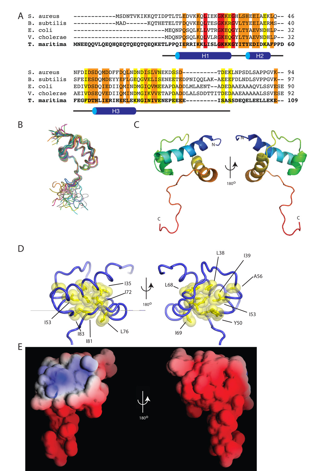
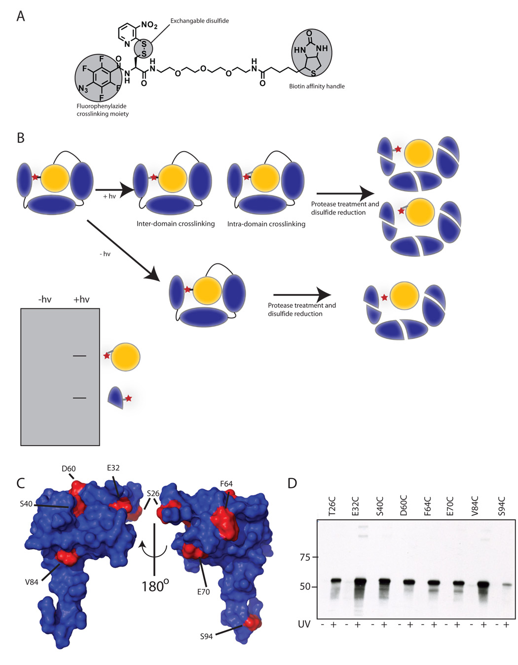
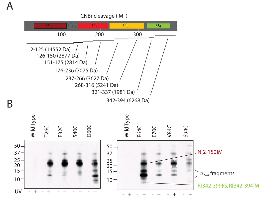
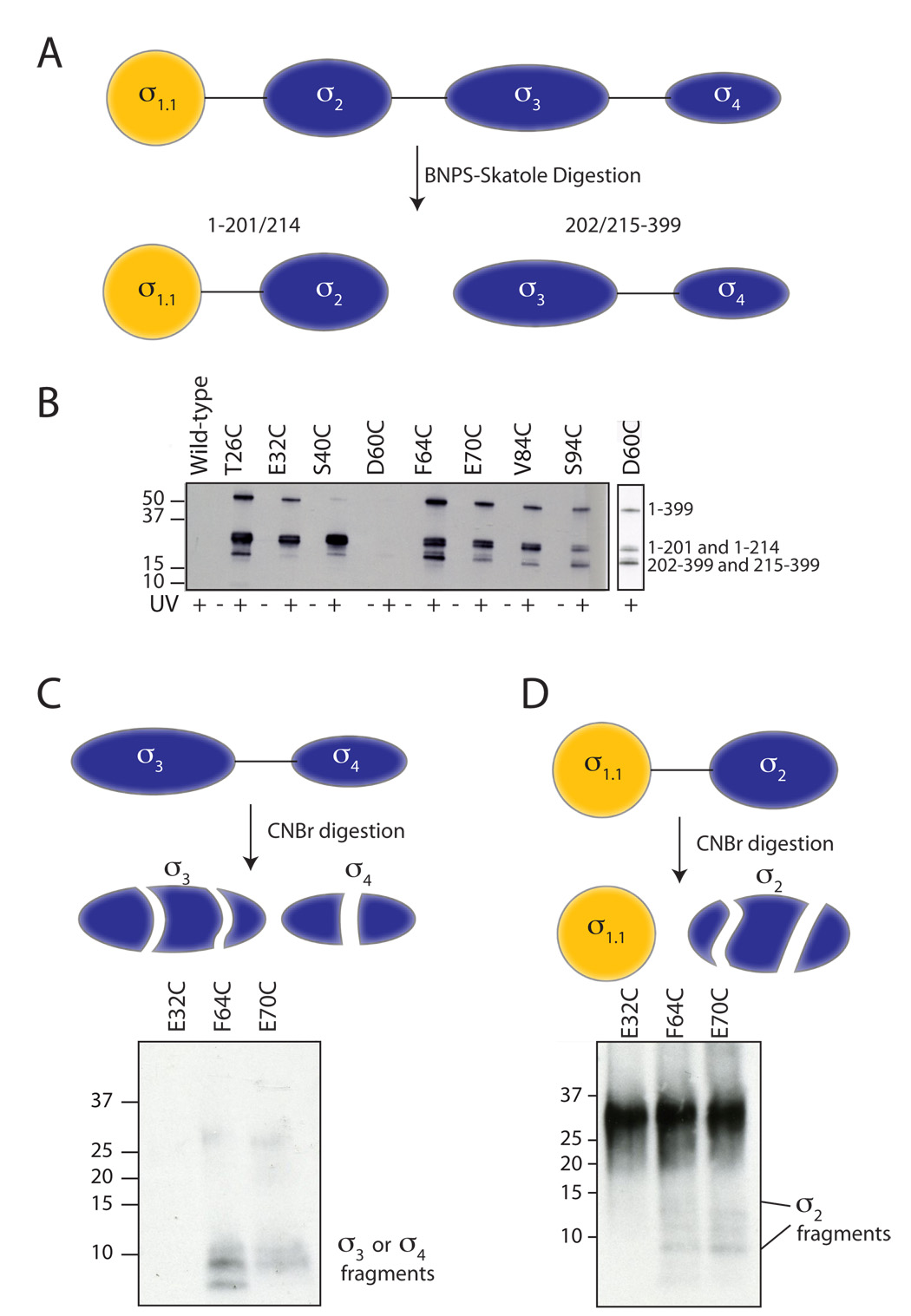
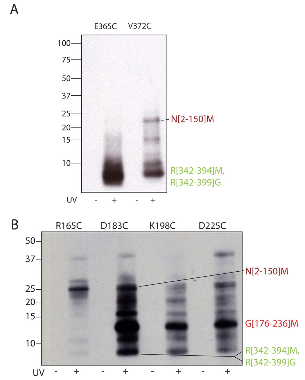
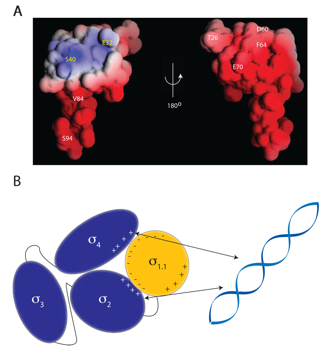
The sigma factors are the key regulators of bacterial transcription initiation. Through direct read-out of promoter DNA sequence, they recruit the core RNA polymerase to sites of initiation, thereby dictating the RNA polymerase promoter-specificity. The group 1 sigma factors, which direct the vast majority of transcription initiation during log phase growth and are essential for viability, are autoregulated by an N-terminal sequence known as sigma1.1. We report the solution structure of Thermotoga maritima sigmaA sigma1.1. We additionally demonstrate by using chemical crosslinking strategies that sigma1.1 is in close proximity to the promoter recognition domains of sigmaA. We therefore propose that sigma1.1 autoinhibits promoter DNA binding of free sigmaA by stabilizing a compact organization of the sigma factor domains that is unable to bind DNA.


Figures

Figure 1
Figure 1. The structure of σ1.1
(A) An alignment of σ1.1 from five different bacterial species generated using the ClustalW program. Highlighted in red are identical residues, in orange are conserved residues and in yellow are semi-conserved residues. The black line below the alignment represents the construct used for structural determination, with secondary structural elements (H1–H3) indicated. (B) A superimposition of the backbone of the 20 lowest energy structures of σ1.1 obtained from calculations using NMR-derived restraints. Each structure is individually colored. The N-termini are in the upper left and the C-termini are in the bottom half of the figure. (C) Cartoon representation of the secondary structure elements of σ1.1. The structure is rainbow spectrum colored from N-terminus (blue) to C-terminus (red). (D) Cartoon representation of σ1.1. Residues that form the hydrophobic core are shown in yellow. Images generated using MacPYMOL (Delano Scientific). (E) The electrostatic surface of σ1.1. Red is negative, blue is positive, white is neutral. Image generated using GRASP (Nicholls et al., 1991).
Figure 2
Figure 2. Crosslinking to detect intramolecular interactions
(A) Chemical structure of the photo-crosslinker used in this study. A synthetic scheme is provided in supplemental material. (B) The crosslinker (red star) is attached by disulfide exchange to a surface cysteine in the σ factor. Following photo-crosslinking and digestion, the disulfide is reduced, transferring the tag to regions in close proximity to σ1.1. The transferred-tag is detected by electrophoretic methods. (C) σ1.1 residues mutated for crosslinking studies. The surface of σ1.1 is rendered in blue with residues that were mutated to cysteine for attachment of the crosslinker highlighted in red. Images were generated using MacPYMOL (Delano Scientific). (D) Crosslinking can be induced by uv irradiation. Indicated σA mutants were labeled with crosslinker by disulfide exchange. Samples that had either been or had not been irradiated at 325 nm light were then reduced with DTT. Samples were then resolved by SDS-PAGE and analyzed by Western blot (Streptavidin-HRP).
Figure 3
Figure 3. Interdomain crosslinking from σ1.1
(A) Predicted CNBr digestion of Tm σA. The domain structure of σA (top) is shown with the predicted CNBr digestion fragments below. Residue numbers of cleavage products are shown with molecular weights in parenthesis. (B) Photo-crosslinking from σ1.1 analyzed by label transfer and CNBr digestion. Crosslinker was attached at the indicated residues within σ1.1 through a disulfide. Wild-type σA, which lacks a native cysteine, was included as a control. Irradiated (UV+) and non-irradiated (UV−) samples were digested with CNBr, separated by reducing SDS-PAGE (10–20% acrylamide, tris-tricine buffer system) and probed for biotin by Western blotting (HRP-streptavidin). Note, only the irradiated cysteine mutants retained the biotin. The identity of the labeled bands is shown at right. Bands that have been identified by mass spectrometry are labeled with residue numbers. A full account of the assignment protocol is given in supplemental methods.
Figure 4
Figure 4. BNPS-Skatole and two-stage digestion of crosslinked products
(A) BNPS-skatole cleaves after Trp residues and, as a consequence, conveniently cuts σA between σ2 and σ3. (B) Photo-crosslinking from σ1.1 analyzed by label transfer and BNPS digestion. Crosslinker was attached at the indicated residues within σ1.1 through a disulfide. Wild-type σA, which lacks a native cysteine, was included as a control. Irradiated (UV+) and non-irradiated (UV−) samples were digested with BNPS, separated by reducing SDS-PAGE (4–12% acrylamide) and probed for biotin by Western blotting (HRP-streptavidin). Note, the D60C sample did not label well, so a longer exposure is shown to the right. The identity of the labeled bands (shown at right) was determined by in-gel trypsinolysis followed by mass spectrometry (data not shown). (C,D) Two-step digestion protocol. Irradiated crosslinker samples of the indicated attachment sites were digested with BNPS-skatole, separated by SDS-PAGE (4–12% acrylamide). Following excision and extraction from the gel, the C-terminal BNPS-skatole fragment (panel C) and N-terminal BNPS-skatole fragment (panel D) were further digested with CNBr and analyzed by SDS-PAGE (10–20% acrylamide, tris-tricine buffer system) and Western blot (HRP-streptavidin).
Figure 5
Figure 5. Interdomain crosslinking from σ4 and σ2
Crosslinker was attached at the indicated residues within σ4 (A) and σ2 (B) through a disulfide. Irradiated (UV+) and non-irradiated (UV−) samples were digested with CNBr, separated by reducing SDS-PAGE (10–20% acrylamide, tris-tricine buffer system) and probed for biotin by Western blotting (HRP-streptavidin). The identity of the labeled bands is shown at right. The N[2–150]M, R[342–394]M and R[342–399]G bands were identified by mass spectrometry, the G[176–236]M band was identified as described in the text.
Figure 6
Figure 6. Proposed model of DNA binding inhibition by σ1.1
(A) Crosslinker attachment sites are indicated on the electrostatic surface map of σ1.1 generated using the GRASP program. Indicated in white are the sites that were shown to make interdomain crosslinks. Indicated in yellow are the sites from which no interdomain crosslinking was observed. (B) Schematic showing the compaction model of σA autoinhibition. The negative surface of σ1.1 is capable of forming crosslinks to the DNA binding domains σ2 and σ4. It is thus likely that σ1.1 organizes the σ factor into a compacted structure that is incapable of binding DNA. σ2 forms crosslinks to σ4, indicating that these two domains must also be in close proximity.

Similar articles

Cited by

References

    1. Barne KA, Bown JA, Busby SJ, Minchin SD. Region 2.5 of the Escherichia coli RNA polymerase sigma70 subunit is responsible for the recognition of the 'extended-10' motif at promoters. EMBO J. 1997;16:4034–4040. - PMC - PubMed
    1. Borukhov S, Nudler E. RNA polymerase holoenzyme: structure, function and biological implications. Curr. Opin. Microbiol. 2003;6:93–100. - PubMed
    1. Bowers CW, Dombroski AJ. A mutation in region 1.1 of σ70 affects promoter DNA binding by Escherichia coli RNA polymerase holoenzyme. EMBO J. 1999;18:709–716. - PMC - PubMed
    1. Browning DF, Busby SJ. The regulation of bacterial transcription initiation. Nat. Rev. Microbiol. 2004;2:57–65. - PubMed
    1. Burgess RR, Travers AA, Dunn JJ, Bautz EK. Factor stimulating transcription by RNA polymerase. Nature. 1969;221:43–46. - PubMed

Publication types

Associated data