Review
. 2008 Dec;14(6):571-83.
doi: 10.1177/1073858408320293. Epub 2008 Oct 20.

An emerging role for voltage-gated Na+ channels in cellular migration: regulation of central nervous system development and potentiation of invasive cancers

Affiliations
Review

An emerging role for voltage-gated Na+ channels in cellular migration: regulation of central nervous system development and potentiation of invasive cancers

William J Brackenbury et al. Neuroscientist. 2008 Dec.

Abstract

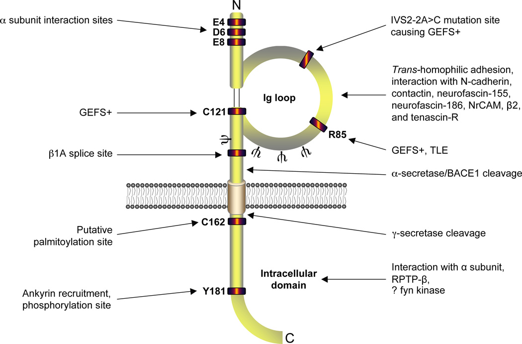
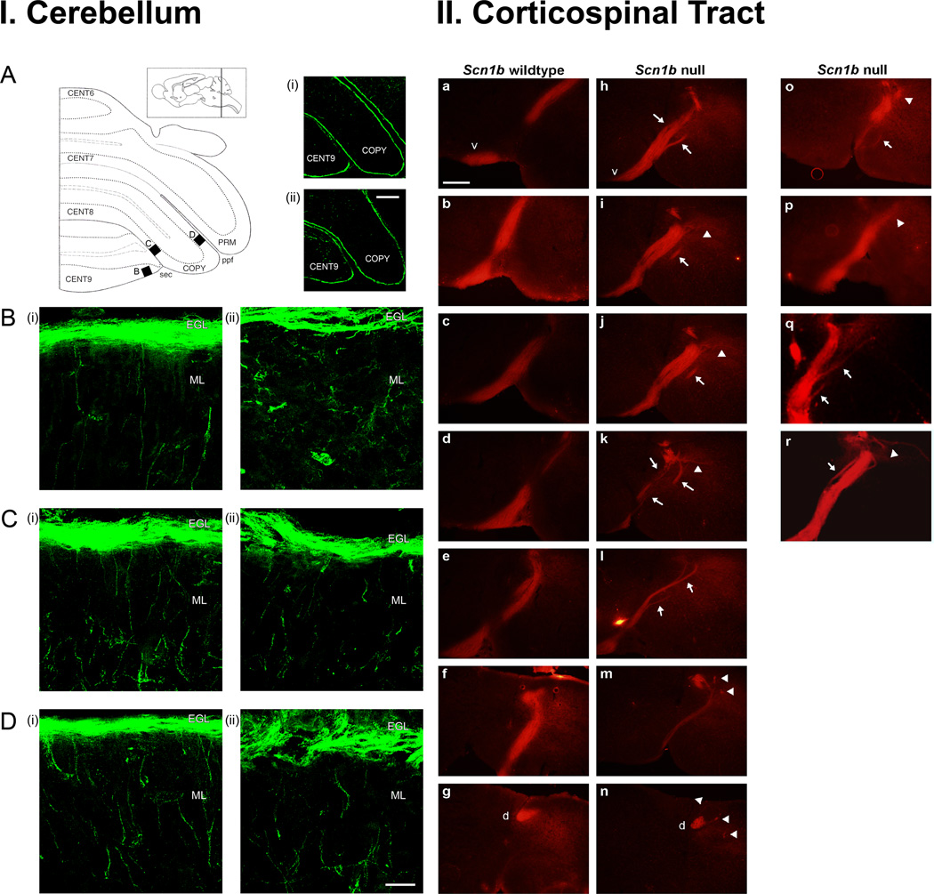
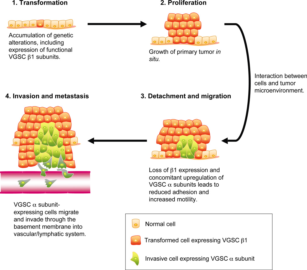
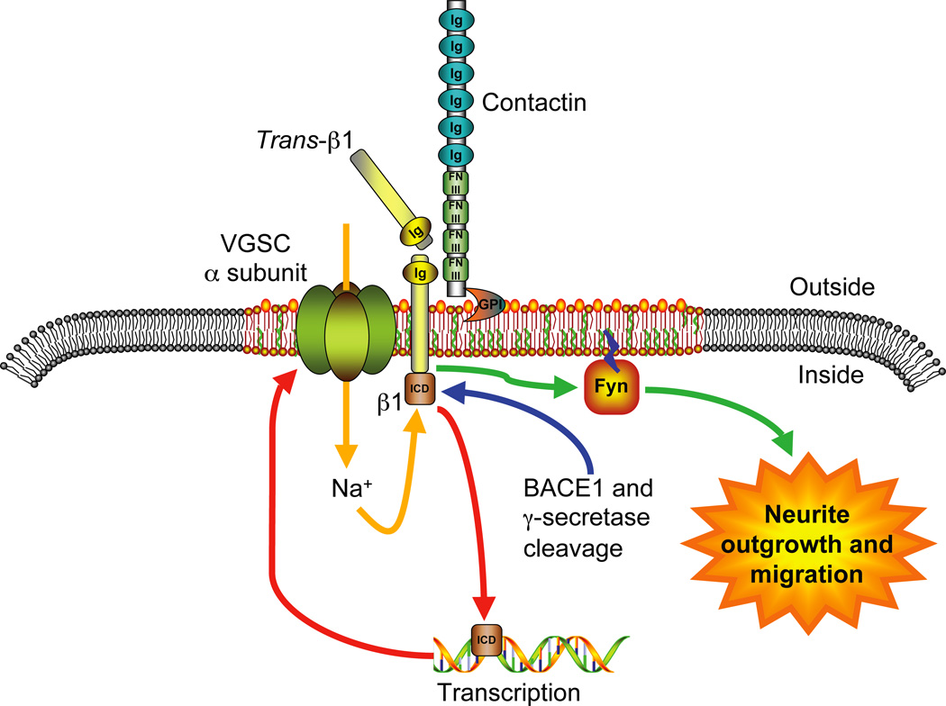
Voltage-gated Na(+) channels (VGSCs) exist as macromolecular complexes containing a pore-forming alpha subunit and one or more beta subunits. The VGSC alpha subunit gene family consists of 10 members, which have distinct tissue-specific and developmental expression profiles. So far, four beta subunits (beta1-beta4) and one splice variant of beta1 (beta1A, also called beta1B) have been identified. VGSC beta subunits are multifunctional, serving as modulators of channel activity, regulators of channel cell surface expression, and as members of the immunoglobulin superfamily, cell adhesion molecules (CAMs). beta subunits are substrates of beta-amyloid precursor protein-cleaving enzyme (BACE1) and gamma-secretase, yielding intracellular domains (ICDs) that may further modulate cellular activity via transcription. Recent evidence shows that beta1 regulates migration and pathfinding in the developing postnatal CNS in vivo. The alpha and beta subunits, together with other components of the VGSC signaling complex, may have dynamic interactive roles depending on cell/tissue type, developmental stage, and pathophysiology. In addition to excitable cells like nerve and muscle, VGSC alpha and beta subunits are functionally expressed in cells that are traditionally considered nonexcitable, including glia, vascular endothelial cells, and cancer cells. In particular, the alpha subunits are up-regulated in line with metastatic potential and are proposed to enhance cellular migration and invasion. In contrast to the alpha subunits, beta1 is more highly expressed in weakly metastatic cancer cells, and evidence suggests that its expression enhances cellular adhesion. Thus, novel roles are emerging for VGSC alpha and beta subunits in regulating migration during normal postnatal development of the CNS as well as during cancer metastasis.


Figures

Figure 1
Figure 1
Basic functional architecture of the β1 subunit. Amino acid residues responsible for interaction with α subunit (McCormick and others 1998; Spampanato and others 2004), generalized epilsepsy with febrile seizures plus (GEFS+) and temporal lobe epilepsy (TLE) (Audenaert and others 2003; Meadows and others 2002; Scheffer and others 2007; Wallace and others 2002), site of intron retention (Kazen-Gillespie and others 2000; Qin and others 2003), putative palmitoylation site (McEwen and others 2004), ankryin interaction site (Malhotra and others 2002) and tyrosine phosphorylation site (Malhotra and others 2004) are shown. Regions identified for N-glycosylation (ψ) (McCormick and others 1998), the Ig loop (Isom and Catterall 1996), BACE1 and α/γ-secretase cleavage (Wong and others 2005), RPTPβ interaction (Ratcliffe and others 2000), and possible fyn kinase interaction (Brackenbury and others 2008; Malhotra and others 2001) are also marked. It should be noted that the C-terminal domain of β1 is also critical for α-β subunit interactions (Spampanato and others 2004). Figure was produced using Science Slides 2006 software.
Figure 2
Figure 2
Scn1b null mice exhibit CNS pathfinding errors. I. β1 modulates axonal migration in the postnatal developing cerebellum in vivo. (A) Schematic outline of left side of cerebellum in the coronal plane. Black boxes B–D show locations of high-magnification images in (B) – (D). CENT6-9, central lobe, lobules 6–9; COPY, copula pyramidis; PRM, paramedian lobule; sec, secondary fissure; prepyramidal fissure. Inset, parasagittal diagram indicating rostrocaudal location of coronal section. Right-hand plates (i) and (ii): low-magnification images (10X) of TAG-1 immunolabeling (green) on coronal cerebellar sections from Scn1b wildtype, and Scn1b null P14 littermates, respectively. Scale bar: 100 µm. (B) – (D) High-magnification (100X) Z-series projections of the same (i) Scn1b wildtype, and (ii) Scn1b null cerebellar sections, at the locations defined in (A). Scale bar: 20 µm. Three mice of each genotype were examined, with similar results. II. Loss of β1 results in axonal pathfinding abnormalities in the corticospinal tract. (A) – (G) Consecutive coronal sections across the pyramidal decussation in a Scn1b wildtype brain. (H) – (N) Consecutive coronal sections across the pyramidal decussation in a Scn1b null brain. (O) – (R) Example sections from further Scn1b null mice. Arrows in (H) – (L) and (O) – (R): defasciculation across pyramidal decussation. Arrowheads in (I) – (K) and (O) – (R): mislocalization of axons lateral to dorsal column. Arrowheads in (M), (N): axons deviating from dorsal column after pyramidal decussation. v, ventral pyramid; d, dorsal column. Scale bar, 250 µm. Six Scn1b null mice were examined. All 6 showed similar CST abnormalities compared to 7 wildtype mice. Figure reproduced with permission (Brackenbury and others 2008).
Figure 3
Figure 3
Proposed model for VGSC α and β subunit involvement in metastatic cancer progression. β1 is expressed in transformed weakly metastatic cancer cells, contributing to their adhesiveness within the proliferating tumor in situ (Chioni and others 2006). In response to signaling interactions between cancer cells and the local tumor microenvironment, VGSC α subunit expression is proposed to be upregulated, and β1 expression downregulated (Brackenbury and Djamgoz 2007; Chioni and others 2006; Ding and others 2008; Ding and Djamgoz 2004; Onganer and Djamgoz 2007). Reduction of β1 is proposed to reduce the cells’ adhesiveness, increasing migration (Chioni and others 2006). The metastatic cells’ migration and invasion is further potentiated by VGSC α subunit activity (Brackenbury and others 2007; Brackenbury and Djamgoz 2006; Smith and others 1998). Figure was produced using Science Slides 2006 software.
Figure 4
Figure 4
A VGSC macromolecular signaling complex in migrating neurons. A proposed trans-adhesive interaction between β1 on an adjacent neuronal or glial cell and the VGSC macromolecular signaling complex on the cerebellar granule neuron, comprising the α subunit, β1 and contactin, initiates a signaling cascade through fyn kinase leading to neurite outgrowth and migration (Brackenbury and others 2008). In addition, cleavage of β1 by BACE1 and γ-secretase is proposed to release the β1 intracellular domain (ICD), which may enhance transcription of VGSC α subunit(s) (Kim and others 2007; Wong and others 2005). The system may be further fine-tuned by signaling mechanism(s) resulting from Na+ influx through the VGSC α subunit (e.g. Brackenbury and Djamgoz 2006). Figure was produced using Science Slides 2006 software.

References

    1. Abdul M, Hoosein N. Inhibition by anticonvulsants of prostate-specific antigen and interleukin-6 secretion by human prostate cancer cells. Anticancer Res. 2001;21(3B):2045–2048. - PubMed
    1. Abdul M, Hoosein N. Voltage-gated sodium ion channels in prostate cancer: expression and activity. Anticancer Res. 2002;22(3):1727–1730. - PubMed
    1. Abrahamsson PA. Neuroendocrine cells in tumour growth of the prostate. Endocr Relat Cancer. 1999;6(4):503–519. - PubMed
    1. Adachi K, Toyota M, Sasaki Y, Yamashita T, Ishida S, Ohe-Toyota M, et al. Identification of SCN3B as a novel p53-inducible proapoptotic gene. Oncogene. 2004;23(47):7791–7798. - PubMed
    1. Allen DH, Lepple-Wienhues A, Cahalan MD. Ion channel phenotype of melanoma cell lines. J Membr Biol. 1997;155(1):27–34. - PubMed

Publication types

Substances

LinkOut - more resources