. 2008 Nov 15;121(Pt 22):3747-56.
doi: 10.1242/jcs.029769. Epub 2008 Oct 21.

Signaling through urokinase and urokinase receptor in lung cancer cells requires interactions with beta1 integrins

Affiliations

Signaling through urokinase and urokinase receptor in lung cancer cells requires interactions with beta1 integrins

Chi-Hui Tang et al. J Cell Sci. .

Abstract

The urokinase receptor (uPAR) is upregulated upon tumor cell invasion and correlates with poor lung cancer survival. Although a cis-interaction with integrins has been ascribed to uPAR, whether this interaction alone is critical to urokinase (uPA)- and uPAR-dependent signaling and tumor promotion is unclear. Here we report the functional consequences of point mutations of uPAR (H249A-D262A) that eliminate beta1 integrin interactions but maintain uPA binding, vitronectin attachment and association with alphaV integrins, caveolin and epidermal growth factor receptor. Disruption of uPAR interactions with beta1 integrins recapitulated previously reported findings with beta1-integrin-derived peptides that attenuated matrix-dependent ERK activation, MMP expression and in vitro migration by human lung adenocarcinoma cell lines. The uPAR mutant cells acquired enhanced capacity to adhere to vitronectin via uPAR-alphaVbeta5-integrin, rather than through the uPAR-alpha3beta1-integrin complex and they were unable to initiate uPA signaling to activate ERK, Akt or Stat1. In an orthotopic lung cancer model, uPAR mutant cells exhibited reduced tumor size compared with cells expressing wild-type uPAR. Taken together, the results indicate that uPAR-beta1-integrin interactions are essential to signals induced by integrin matrix ligands or uPA that support lung cancer cell invasion in vitro and progression in vivo.


Figures

Fig. 1
Fig. 1
uPAR expression, uPA binding and membrane partners of cells expressing HD mutant uPAR. (A) FACS analysis of uPAR expression in H1299 cells. Control, uPAR-knockdown, WT and HD cells were harvested and incubated with antibodies against uPAR, followed by APC-conjugated secondary antibodies. (B) uPA 1-48-Fc binding to H1299 cells. uPAR-knockdown (shu), WT and HD cells were acid-washed to remove bound endogenous uPA, cells were incubated with 1-48-Fc (0–50 nM) at 4°C for 1 hour and the bound 1-48-Fc was detected by anti-Fc-APC. The mean fluorescence was plotted. Data represents mean ± s.d. from three independent experiments. (C) Co-immunoprecipitation of uPAR with caveolin, αV integrin, or EGFR. H1299 cells expressing WT and HD uPAR were lysed in 1% Triton X-100 lysis buffer and the lysates were immunoprecipitated with mAb to uPAR (R2). The lysates and immunoprecipitates were then separated by SDS-PAGE and blotted for uPAR (399R), caveolin pAb, αV integrin pAb or EGFR pAb. The experiment was performed at least three times with similar results.
Fig. 2
Fig. 2
Change of mechanism in vitronectin adhesion by H249A-D262A mutations on uPAR. (A) vitronectin adhesion of cells expressing HD uPAR does not require uPAR–α3β1-integrin association. H1299 and HEK293 cells expressing WT and HD uPAR were seeded on to vitronectin (2 μg/ml)-coated wells with or without 10 μg/ml mAb against integrin α3 (P1B5) or α5 (P1D6), nonimmune IgG (IgG) or 0.4 mM peptide α325 or its scrambled version, scα325. After incubation for 1 hour, cells were rinsed and adherent cells were stained with Giemsa. (B) Vitronectin adhesion of cells expressing HD uPAR is mediated by uPA–uPAR–αVβ5-integrin. uPAR-knockdown H1299 cells (shu), HEK293 non-transfected cells (ctl) and H1299 and HEK293 cells expressing WT and HD uPAR were seeded on vitronectin-coated wells with or without 10 μg/ml mAb to uPAR (ATN615, ATN617), integrin αVβ3 (LM609), αVβ5 (P1D6), 0.5 mM peptide RGD or control RAD, or 0.4 mM peptide β1P1 or its scrambled version, scβ1P1. Cells were incubated, rinsed and stained as above. The adhesion assays shown above are representative of at least three independent experiments.
Fig. 3
Fig. 3
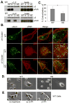
Loss of uPAR–β1-integrin association and change of cell morphology by H249A-D262A mutations on uPAR. (A) H, D or HD mutations in uPAR disrupt the formation of uPAR–β1-integrin complexes. H1299 cells expressing WT, H, D or HD uPAR were lysed in 1% Triton X-100 lysis buffer and the lysates were immunoprecipitated with monoclonal antibody to α3 integrin (P1B5) or α5 integrin (P1D6). The lysates and immunoprecipitates were then separated by SDS-PAGE and blotted for uPAR (R2) and integrin α3 or integrin α5. (B) uPAR co-clusters with α3 integrin but not α6 integrin. H1299 cells expressing WT or HD uPAR were plated on fibronectin and stained with mouse mAb to integrin α3 (P1B5) or rat mAb to integrin α6 (GoH3) on ice. The receptors were clustered with fluorophore-conjugated secondary antibody (Alexa Fluor 350 for α3 integrin or Alex Fluor 568 for α6 integrin) at 37°C or left nonclustered as controls. Cells were then fixed and stained with conjugated anti-uPAR antibody (Rhodamine-conjugated ATN615 for α3-integrin-clustered cells or ATN615 with Alexa Fluor 350 for α6-integrin-clustered cells). To facilitate visualization, images were converted to pseudo color using Zeiss LSM Image Browser software: integrin (green), uPAR (red) and sites of colocalization (yellow). The indicated areas in the merged images were magnified and shown on the right. The above experiments were performed three times with similar results. (C) Quantification of α3 integrin co-clustering with uPAR. Percentage colocalization (colocalized α3÷total α3×100) from three independent experiments was measured using Zeiss LSM Image Browser software: WT (n=21) and HD (n=24), Student’s t-test, *P<0.005. (D) Phase images of H1299 cells expressing WT or HD uPAR. WT cells show robust lamellipodia formation that is absent in HD cells. The indicated areas were magnified and shown on the right. (E) Phase images of H1299 cells expressing WT uPAR with or without treatment of uPAR–β1-integrin blocking peptide β1P1 or its scrambled control. β1P1-treated WT cells show no lamellipodia formation.
Fig. 4
Fig. 4
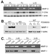
Matrix-mediated signaling requires uPAR–β1-integrin association. (A) Matrix-induced MMP9 expression and ERK activation are dependent on uPAR–β1-integrin association. H1299 cells expressing WT, H, D and HD uPAR were serum-starved and cultured on poly-L-lysine (PL), fibronectin (Fn) or laminin-5 (Ln5), for 20 minutes or 24 hours in serum-free DMEM. The conditioned media after 24 hours on matrices were analyzed by gelatin zymography. Clear zones of degradation are seen at 92 kDa indicated as MMP9. Lysates from cells cultured for 20 minutes on matrices were immunoblotted with anti-ERK-P and anti-ERK antibodies. (B) Matrix-induced MMP1 expression is dependent on uPAR–β1-integrin association. Cells expressing WT and HD uPAR were serum-starved and cultured on poly-L-lysine, fibronectin or laminin-5 for 48 hours in serum-free DMEM. The conditioned media were immunoblotted for MMP1. Some WT cells were pretreated with the MEK inhibitor PD98059 (10 μM). Total ERK was detected as a loading control. (C) uPAR–β1-integrin blocking peptides inhibit matrix-induced MMP expression. Serum-starved cells expressing WT uPAR were pretreated with 0.4 mM peptide α325, scrambled α325, β1P1 or scrambled β1P1 and then plated on fibronectin or laminin-5 for 24 hours (for MMP9) or 48 hours (for MMP1) in serum-free DMEM. The conditioned media were analyzed by gelatin zymography for MMP9 or immunoblotted for MMP1. The volume of conditioned medium loaded to the gel was normalized to total protein in the lysate. All data shown are representative of three experiments with similar results.
Fig. 5
Fig. 5
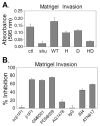
Both uPA-uPAR and uPAR association with β1 integrins are critical to lung cancer cell invasion. (A) Matrigel invasion of H1299 cells. Control, uPAR knockdown (shu), WT, H, D, or HD uPAR expressing H1299 cells were seeded into 24-well Transwell units (1×10⁵ cells/well in triplicate) and allowed to invade Matrigel for 24 hours. The cells that had passed through the filter and attached to the bottom of the filter coated with fibronectin were fixed and stained with Giemsa. The stained cells on the membrane were extracted with 10% acetic acid. Cell invasiveness was evaluated by measuring OD595 nm. (B) Effects of inhibitors on H1299 cell invasion. H1299 cells were pretreated with antibodies (10 μg/ml): 394, uPA activity neutralizing antibody; ATN617, uPAR mAb blocking uPA binding, or control IgG; peptides (100 μM): β1P1 or scβ1P1; inhibitors: GM6001 (0.1 μM), PD98059 (10 μM) or AG1478 (1 μM). The invasion assay was carried out as described above. The results are expressed as a percentage of inhibition observed in the no treatment control. All data are representative of three independent experiments carried out in triplicate.
Fig. 6
Fig. 6
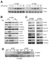
uPA signaling requires uPAR–β1-integrin interactions. (A) Time course of ERK activation in H1299 and H1264 cells. Cells were serum-starved for 24 hours, acid-washed to remove bound endogenous uPA and then incubated with pro-uPA (10 nM) in serum-free medium for different time points (minutes): 0, 0.5, 1, 5, 10, 30. The cells were lysed in RIPA buffer and immunoblotted with anti-phospho-ERK and anti-total-ERK antibodies, respectively. (B) uPA-induced signaling in H1299 cells. H1299 cells expressing WT or HD uPAR were serum-starved, acid washed and then incubated with pro-uPA (10 nM) in serum-free medium for 5 minutes to assess ERK activation and 30 minutes to assess activation of Akt, GSK-3β or Stat. The lysates were immunoblotted with anti-phospho- and anti-total-antibodies to ERK, Akt, GSK-3β, or Stat1 or Stat3. (C) Effects of inhibitors on uPA-induced signaling. H1299 and H1264 cells expressing endogenous uPAR were serum-starved, acid washed, pre-treated with uPAR–β1-integrin blocking peptide (β1P1) or scrambled peptide (scβ1P1) and then incubated with pro-uPA (10 nM) in serum-free medium. (D) EGF-induced ERK phosphorylation is not affected by H249A and/or D262A mutations in uPAR. H1299 cells expressing WT, H, D or HD uPAR were serum-starved and incubated with 20 ng/ml EGF for 10 minutes and cell lysates were assessed for ERK activation. All the experiments were performed at least three times with similar results.
Fig. 7
Fig. 7
H249A-D262A mutations on uPAR inhibit orthotopic lung tumor growth in vivo. (A) Tumor area is reduced in HD-mutant group and uPAR-knockdown group. WT or HD uPAR expressing H1299 cells or control (ctl) or uPAR knockdown (shu) H1299 cells were injected into the left lungs of nude mice (2×10⁵ in 20 μl of 10% Matrigel/mouse). 35 days later the lungs were harvested, embedded in OCT and serial sections were inspected for tumor size by examining green fluorescence. Average tumor area was measured using SimplePCI software and plotted: WT (n=20) and HD (n=22), Mann-Whitney U-test, **P<0.044; ctl (n=13) and uPAR knockdown (shu; n=14), Mann-Whitney U-test, *P<0.019. (B) Representative images of GFP-positive tumors. A view showing only a small part of the tumor mass from a lung injected with control cells and a view showing the whole tumor from injected uPAR-knockdown cells. The images were taken under ×10 magnification by fluorescence microscopy using a Spot Camera and imaging software.

Similar articles

Cited by

References

    1. Adachi T, Kar S, Wang M, Carr BI. Transient and sustained ERK phosphorylation and nuclear translocation in growth control. J Cell Physiol. 2002;192:151–159. - PubMed
    1. Aguirre Ghiso JA, Kovalski K, Ossowski L. Tumor dormancy induced by downregulation of urokinase receptor in human carcinoma involves integrin and MAPK signaling. J Cell Biol. 1999;147:89–104. - PMC - PubMed
    1. Ahmed N, Oliva K, Wang Y, Quinn M, Rice G. Downregulation of urokinase plasminogen activator receptor expression inhibits Erk signalling with concomitant suppression of invasiveness due to loss of uPAR-beta1 integrin complex in colon cancer cells. Br J Cancer. 2003;89:374–384. - PMC - PubMed
    1. Blasi F, Carmeliet P. uPAR: a versatile signalling orchestrator. Nat Rev Mol Cell Biol. 2002;3:932–943. - PubMed
    1. Carriero MV, Del Vecchio S, Capozzoli M, Franco P, Fontana L, Zannetti A, Botti G, D’Aiuto G, Salvatore M, Stoppelli MP. Urokinase receptor interacts with alpha(v)beta5 vitronectin receptor, promoting urokinase-dependent cell migration in breast cancer. Cancer Res. 1999;59:5307–5314. - PubMed

Publication types

Substances