. 2008 Oct 28;105(43):16555-60.
doi: 10.1073/pnas.0806401105. Epub 2008 Oct 21.

Role of HAMP domains in chemotaxis signaling by bacterial chemoreceptors

Affiliations

Role of HAMP domains in chemotaxis signaling by bacterial chemoreceptors

Cezar M Khursigara et al. Proc Natl Acad Sci U S A. .

Abstract

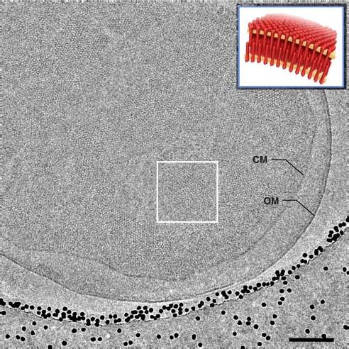
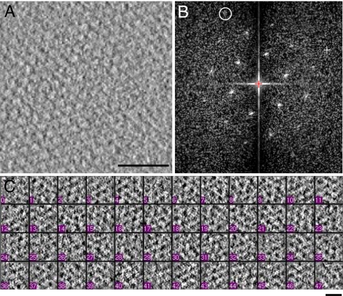
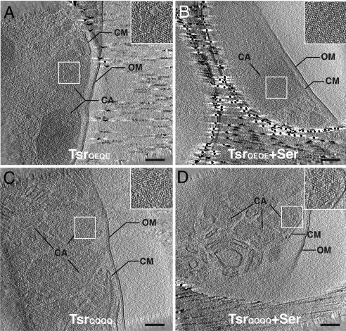
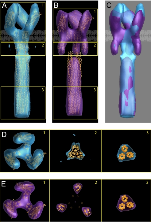
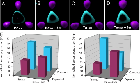
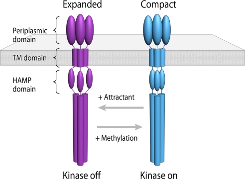
Bacterial chemoreceptors undergo conformational changes in response to variations in the concentration of extracellular ligands. These changes in chemoreceptor structure initiate a series of signaling events that ultimately result in regulation of rotation of the flagellar motor. Here we have used cryo-electron tomography combined with 3D averaging to determine the in situ structure of chemoreceptor assemblies in Escherichia coli cells that have been engineered to overproduce the serine chemoreceptor Tsr. We demonstrate that chemoreceptors are organized as trimers of receptor dimers and display two distinct conformations that differ principally in arrangement of the HAMP domains within each trimer. Ligand binding and methylation alter the distribution of chemoreceptors between the two conformations, with serine binding favoring the "expanded" conformation and chemoreceptor methylation favoring the "compact" conformation. The distinct positions of chemoreceptor HAMP domains within the context of a trimeric unit are thus likely to represent important aspects of chemoreceptor structural changes relevant to chemotaxis signaling. Based on these results, we propose that the compact and expanded conformations represent the "kinase-on" and "kinase-off" states of chemoreceptor trimers, respectively.


Conflict of interest statement

The authors declare no conflict of interest.

Figures

Fig. 1.
Fig. 1.
Cryo-electron microscopy of Tsr chemoreceptor assemblies in whole E. coli cells. Shown is a projection image recorded from a plunge-frozen E. coli cell engineered to overproduce TsrQEQE (in the absence of serine) by using low-dose cryo-electron microscopy. The projection image shows patches of chemoreceptor assemblies in the native cytoplasmic membrane (CM) contained within a cell with an intact outer membrane (OM). The cell is close to the edge of a hole in a holey carbon grid containing vitreous ice; the black dots are 15-nm gold particles added for use as fiducial markers in tomogram reconstruction. (Inset) A schematic perspective view of “zipper”-like chemoreceptor assemblies from a region such as that enclosed by the white box. (Scale bar: 100 nm.)
Fig. 2.
Fig. 2.
Cryo-electron tomographic analysis of crystalline Tsr assemblies. (A and B) A 4-nm-thick tomographic slice from a region of a reconstructed tomogram of whole cells containing receptor arrays, and its Fourier transform, respectively. The circled diffraction spot in B is at a resolution of ≈33 Å. (C) Representative examples of tomographic slices corresponding to local receptor clusters extracted from whole cell tomograms. [Scale bars: 50 nm (A) and 10 nm (C).]
Fig. 3.
Fig. 3.
Cryo-electron tomography of Tsr chemoreceptor assemblies in whole E. coli cells. Shown are tomographic slices (≈10 nm thick) generated from E. coli cells engineered to overproduce TsrQEQE in the absence (A) or presence (B) of serine (Ser), and TsrQQQQ in the in the absence (C) or presence (D) of serine. Chemoreceptor assemblies (CA) are observed as crystalline patches embedded in cytoplasmic membrane (CM). The crystalline patches are observed throughout the cells and are contained by the outer membrane (OM); an expanded view of the patches is shown in the Inset to each panel. (Scale bars: 100 nm.)
Fig. 4.
Fig. 4.
Identification of two trimeric Tsr conformations and interpretation of the density maps in terms of known structures of subdomains. Averaged density maps of TsrQEQE in both the compact (A) and expanded (B) conformations, with structural coordinates corresponding to chemoreceptor models fitted to a single trimer of receptor dimers. (C) Superposition of the two density maps highlighting overlap in the cytoplasmic domain and differences in the HAMP and periplasmic domains. A schematic view of the lipid bilayer is shown to highlight the locations of the putative transmembrane region in A–C. (D) Sections of the map from regions marked 1–3 in A correspond to the periplasmic domain, HAMP domain, and end of the cytoplasmic domain, respectively. Densities corresponding to each of these regions can be unambiguously assigned, although the map is not at a resolution where the rotational orientations of the individual domains can be determined. (E) Sections from the map shown in B displayed as described for D. The density in the cytoplasmic domain (section 3) was fit with a trimer obtained from three monomers derived from the structure of this region in the serine chemoreceptor Tsr reported by Kim et al. (11). Note that the packing of the trimer in our map is different from the organization of monomers reported in the crystals used to determine the structure (37) (PDB ID code 1QU7). Both conformations are interpreted by using the same packing arrangement in the cytoplasmic domain.
Fig. 5.
Fig. 5.
Effects of ligand binding and chemoreceptor methylation on the distribution of Tsr conformations. Tomographic volumes were generated for TsrQEQE and the fully methylated TsrQQQQ variants of Tsr in the absence and presence of the attractant serine (Ser), and the distribution of compact or expanded conformations of the HAMP domains was compared. Shown are sectional views from superimposed density maps of each Tsr variant, TsrQEQE (A), TsrQEQE+Ser (B), TsrQQQQ (C), and TsrQQQQ+Ser (D) demonstrating the compact (cyan) and expanded (magenta) conformation of the HAMP domain (as shown in Fig. 4, section 2). (E) Addition of the ligand serine to the TsrQEQE variant causes an increase in the proportion of receptors with an expanded conformation (magenta bars) of the HAMP domain and a concomitant decrease in the proportion of receptors in the compact conformation (cyan bars). Conversely, methylation of Tsr (TsrQQQQ) causes an increase in the proportion of the receptors in the compact state (compare TsrQEQE from E with TsrQQQQ from F). (F) Ligand binding to the TsrQQQQ variant causes a conformational shift similar to that seen in E for TsrQEQE.
Fig. 6.
Fig. 6.
A two-state model describing conformational signaling in chemoreceptor trimers. Trimeric chemoreceptors exist in an equilibrium between two conformations and adopt either expanded or compact arrangements of the HAMP signaling domain. Binding of the attractant serine initiates movement in the transmembrane helix (9), which, in turn, shifts the conformational equilibrium of the HAMP domain in favor of the expanded conformation (magenta). Based on the known effects of serine binding to reduce the activity of the CheA kinase, we propose that this expanded conformation of the HAMP domain corresponds to the “kinase-off” state. Conversely, an increase in chemoreceptor methylation shifts the equilibrium in favor of the compact HAMP conformation (cyan), corresponding to the “kinase-on” state.

Similar articles

Cited by

References

    1. Wadhams GH, Armitage JP. Making sense of it all: Bacterial chemotaxis. Nat Rev Mol Cell Biol. 2004;5:1024–1037. - PubMed
    1. Webre DJ, Wolanin PM, Stock JB. Modulated receptor interactions in bacterial transmembrane signaling. Trends Cell Biol. 2004;14:478–482. - PubMed
    1. Boldog T, Grimme S, Li M, Sligar SG, Hazelbauer GL. Nanodiscs separate chemoreceptor oligomeric states and reveal their signaling properties. Proc Natl Acad Sci USA. 2006;103:11509–11514. - PMC - PubMed
    1. Studdert CA, Parkinson JS. Insights into the organization and dynamics of bacterial chemoreceptor clusters through in vivo crosslinking studies. Proc Natl Acad Sci USA. 2005;102:15623–15628. - PMC - PubMed
    1. Studdert CA, Parkinson JS. In vivo crosslinking methods for analyzing the assembly and architecture of chemoreceptor arrays. Methods Enzymol. 2007;423:414–431. - PubMed

Publication types

LinkOut - more resources