Review
. 2009 Mar-Apr;44(2):115-27.
doi: 10.1093/alcalc/agn079. Epub 2008 Oct 21.

Mechanisms of neurodegeneration and regeneration in alcoholism

Affiliations
Review

Mechanisms of neurodegeneration and regeneration in alcoholism

Fulton T Crews et al. Alcohol Alcohol. 2009 Mar-Apr.

Abstract

Aims: This is a review of preclinical studies covering alcohol-induced brain neuronal death and loss of neurogenesis as well as abstinence-induced brain cell genesis, e.g. brain regeneration. Efforts are made to relate preclinical studies to human studies.

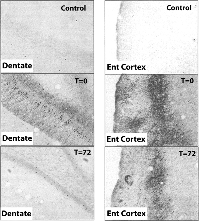
Methods: The studies described are preclinical rat experiments using a 4-day binge ethanol treatment known to induce physical dependence to ethanol. Neurodegeneration and cognitive deficits following binge treatment mimic the mild degeneration and cognitive deficits found in humans. Various histological methods are used to follow brain regional degeneration and regeneration.

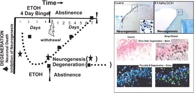
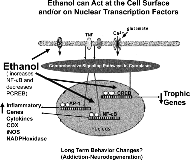
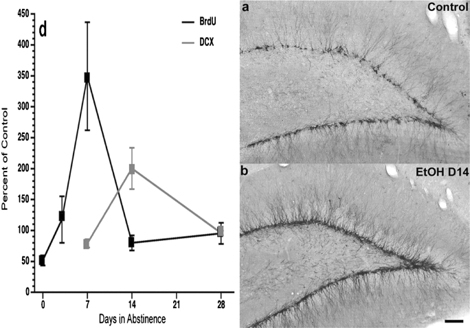
Results: Alcohol-induced degeneration occurs due to neuronal death during alcohol intoxication. Neuronal death is related to increases in oxidative stress in brain that coincide with the induction of proinflammatory cytokines and oxidative enzymes that insult brain. Degeneration is associated with increased NF-kappaB proinflammatory transcription and decreased CREB transcription. Corticolimbic brain regions are most sensitive to binge-induced degeneration and induce relearning deficits. Drugs that block oxidative stress and NF-kappaB transcription or increase CREB transcription block binge-induced neurodegeneration, inhibition of neurogenesis and proinflammatory enzyme induction. Regeneration of brain occurs during abstinence following binge ethanol treatment. Bursts of proliferating cells occur across multiple brain regions, with many new microglia across brain after months of abstinence and many new neurons in neurogenic hippocampal dentate gyrus. Brain regeneration may be important to sustain abstinence in humans.

Conclusions: Alcohol-induced neurodegeneration occurs primarily during intoxication and is related to increased oxidative stress and proinflammatory proteins that are neurotoxic. Abstinence after binge ethanol intoxication results in brain cell genesis that could contribute to the return of brain function and structure found in abstinent humans.


Figures

Fig. 1
Fig. 1
Ethanol-induced brain damage and inhibition of neurogenesis, and time course of degeneration. Shown on the left is the time course of neurodegeneration (dotted line) with greater damage and loss of neurogenesis as negative downward values. Neurodegeneration (squares, binge ethanol agyrophilic silver stain) and loss of neurogenesis (stars, BrdU+ir) are both plotted during ethanol treatment. Acute treatment with ethanol results in inhibition of neurogenesis and increased apoptotic cells (Obernier et al., ; He et al., 2005). Although little silver stain degeneration is found with acute ethanol treatments, silver stain takes 24– 48 h to develop suggesting that cell death may have been missed in acute studies. The 4-day binge shows progressively increased silver stain from Day 2 throughout the 4-day period. Abstinence following the binge results in a progressive loss of cell death silver stain markers with complete clearance after ∼1 week (Crews and Braun, 2003). See also Crews et al. (2004). Pictures right top: examples of ethanol inhibition of neurogenesis (top left: control, top right: ethanol; 5 gm/kg, i.g.) in adolescent animals. Shown are brain sections of frontal subventricular zone neural progenitor cells (BrdU + IR, black dots on section). Note how acute ethanol has eliminated these progenitors completely [no black dots—low first star in time course; adapted from Crews et al. (2006a)]. Pictures right, middle and bottom: degeneration. Brain sections identify 4-day binge ethanol-induced necrotic degeneration in hippocampus visualized by agyrophilic amino cupric silver stain (black—middle photo) or Fluoro-Jade B (green–-bottom). Note binge ethanol degeneration, but none in controls. [Adapted from Obernier et al. (2002a).]
Fig. 2
Fig. 2
Binge ethanol-induced brain damage. Shown are brain sections from hippocampal dentate gyrus (left 3 panels) and entorhinal cortex (right 3 panels). Agyrophilic silver stain identifies dying neurons as confirmed by multiple methods showing binge ethanol-induced neurodegeneration (Obernier et al., 2002a). Note little or no cell death in controls (top panels) with significant silver staining in 4-day binge ethanol-treated animals sacrificed just after the last ethanol dose (T = 0; middle 2 panels). The bottom panels show brain sections 72 h after the last ethanol dose. Note the loss of silver stain at 72 h of abstinence. Since silver staining of dying neurons occurs 1–3 days after insults (Switzer III, 2000), it is likely that damage occurs during binge intoxication with little additional damage during abstinence–withdrawal.
Fig. 3
Fig. 3
Reversal learning identifies persistent perseverative repetitive behaviors following binge ethanol administration. Morris water maze learning of platform location was identical in binge ethanol and control animals on 4–11 days of abstinence. Both readily learned the platform location. However, they differed in the reversal-learning task of the Morris water maze. Following the 7 days of place learning, animals are tested in a reversal-learning task. The submerged platform was placed in the quadrant opposite that in which it had been placed during the reference memory task (southwest quadrant). The animals were given four trials, each consisting of a 90-s ceiling and a 60-s intertrial interval. Once the animal reached the platform, it was allowed to remain on the platform for ∼10 s. If the animal failed to reach the platform within the trial ceiling, the experimenter gently guided the animal through the water and placed it on the platform where it would remain for 10 s. The animal was then removed to its home cage, which was warmed with a heating pad, to await the next trial. (A) ETOH animals required a significantly greater number of trials to reach the criterion than CON animals [t(14) = 2.376; *P < 0.05]. (B) Time line of binge treatment and behavioral testing: After a 7-day acquisition of the platform location, the submerged platform was moved to the opposite quadrant and animals were given four trials (12 days’ postbinge treatment). A vertical view of the track was taken by a CON and an ETOH rat during the first trial of the reversal-learning task. The open circle represents the location of the submerged platform the animals were trained to and the patterned circle represents the location of the platform during the reversal-learning task. Note the perseverative behavior shown by the ETOH animal with numerous re-entries into the original goal quadrant. The ETOH animal also failed to reach the new platform locations within the 90-s ceiling. [Adapted from Obernier et al., (2002b).]
Fig. 4
Fig. 4
Schematic representation of ethanol-induced changes in transcription. Ethanol can modulate multiple signaling pathways with convergence on multiple transcription factors, three of which are illustrated: ethanol increased proinflammatory gene expression, the convergence on increased NFκB and AP1 transcription initiating and sustaining proinflammatory cascades. CREB is the transcription factor that regulates the transcription of prosurvival target genes, such as bcl2 and brain-derived neurotrophic factor. Ethanol exposure leads to imbalance between procytokine-oxidative stress and pro-survival gene transcription, causing neuronal atrophy, shrinkage and degeneration and inhibiting neurogenesis. [See Crews et al. (2004) and Zou and Crews (2005) for details.]
Fig. 5
Fig. 5
Ethanol increases CREB and decreases NF-κB DNA binding. HEC slices were treated with various concentrations of ethanol for 24 h and then removed for processing of nuclear extracts. Nuclear extracts are processed for electrophoretic mobility shift assay (EMSA). EMSA involves an oligonucleotide probe specific for the DNA binding site of each transcription factor with gel separation of transcription factor-bound probe providing an index of transcription factor activation and binding to DNA. (Cont = control; E25 and E50, ethanol 25 and 50 mM, respectively.) Note the decreased CREB binding with increasing concentrations of ethanol and the increased NF-κB binding (band size) with ethanol addition to brain slices. [Adapted from Zou and Crews (2005).]
Fig. 6
Fig. 6
Bursts of neurogenesis during abstinence from binge ethanol treatment. Left: time course of changes in neurogenesis in hippocampal dentate gyrus during abstinence after the 4-day ethanol binge treatment (Nixon and Crews, 2004). BrdU labels newborn cells providing an index of progenitor proliferation. Each timepoint represents BrdU + immunohistochemistry, 4 h after BrdU dosing. Note the peak in proliferation (BrdU) at 1 week of abstinence. Doublecortin (DCX) is expressed in neural progenitors during differentiation into mature neurons (Brown et al., 2003). Note the peak on DCX expression at 14 days of abstinence. Images of DCX immunohistochemistry in control (a) and 14 days of abstinence after the binge (b) illustrate abstinence-induced neurogenesis. [Adapted from Nixon and Crews (2004).]
Fig. 7
Fig. 7
Temporal relationship of binge ethanol-induced neurodegeneration and abstinence-induced cell genesis. Neurodegeneration is represented as down on the y-axis (solid squares–dotted line). Regeneration is represented as up (cell genesis—solid stars, dashed line). The 4-day binge ethanol treatment has been extensively studied as a model of alcohol dependence and alcohol-induced neurodegeneration. Multiple markers of neuronal cell death, particularly agyrophilic silver stain, have characterized pyramidal dark cell degeneration that shows greater degeneration (down) over the 4-day binge. This neuronal cell death during intoxication corresponds with ethanol inhibition of dentate gyrus neurogenesis (solid stars below midline represent inhibition of neurogenesis). The dotted line represents the course of both markers of degeneration. Symptoms of physical withdrawal occur during the first 24 h (lightning bolt). The bursts of cell genesis (stars above midline) occur in the week following ethanol treatment. After 2 days of abstinence following the binge (T48), a cell genesis burst occurs in multiple brain regions shown to primarily differentiate into microglia at 1 and 2 months of abstinence (upper stripped arrow—sun symbol). At a later time, 7 days of abstinence, a hippocampal (T168) cell burst in dentate gyrus results in more new neurons 1 month later (lower stripped arrow—smiley face). Studies have shown that cells that survive 1 month tend to persist for long periods in the brain (Kempermann et al., 2004). These studies suggest that abstinence from ethanol has increased new microglia across broad regions of brain.
Fig. 8
Fig. 8
Regeneration of brain is related to increased CREB transcription, increased neurogenesis and cell genesis. The temporal relationship of binge ethanol-induced neurodegeneration and abstinence-induced cell genesis related to pCREB immunohistochemical staining in dentate gyrus of control, binge ethanol-treated animals (4-day Etoh-T0) and animals at 72 h of abstinence following the 4-day binge (4-day EtOH/72-h withdrawal). Control brain section—middle picture (A) shows pCREB + IR particularly high in the subgranular zone (SGZ) where neurogenesis occurs, but staining is throughout. After 4 days of binge ethanol (4-day Etoh-T0), pCREB staining is decreased when neurogenesis is inhibited and granule cells degenerate. However, after 72 h of abstinence with the first 24 including the excessive excitation of behavioral withdrawal result in a marked increase in pCREB staining (top photo—4-days EtOH/72-h withdrawal) that coincides with or precedes increased cell and neurogenesis and loss of degeneration markers. [pCREB immunohistochemistry adapted from Bison and Crews (2003).]

Similar articles

Cited by

References

    1. Altman J, Das GD. Autoradiographic and histological evidence of postnatal hippocampal neurogenesis in rats. J Comp Neurol. 1965;124:319–35. - PubMed
    1. Alvarez-Buylla A, Garcia-Verdugo JM. Neurogenesis in adult subventricular zone. J Neurosci. 2002;22:629–34. - PMC - PubMed
    1. Armstrong RJ, Barker RA. Neurodegeneration: a failure of neuroregeneration? Lancet. 2001;358:1174–6. - PubMed
    1. Banks WA. Blood–brain barrier transport of cytokines: a mechanism for neuropathology. Curr Pharm Des. 2005;11:973–84. - PubMed
    1. Baydas G, Tuzcu M. Protective effects of melatonin against ethanol-induced reactive gliosis in hippocampus and cortex of young and aged rats. Exp Neurol. 2005;194:175–81. - PubMed