. 2008 Nov 1;181(9):5948-55.
doi: 10.4049/jimmunol.181.9.5948.

IL-23 promotes maintenance but not commitment to the Th17 lineage

Affiliations

IL-23 promotes maintenance but not commitment to the Th17 lineage

Gretta L Stritesky et al. J Immunol. .

Abstract

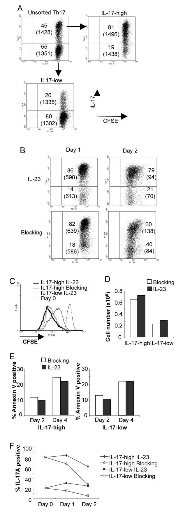
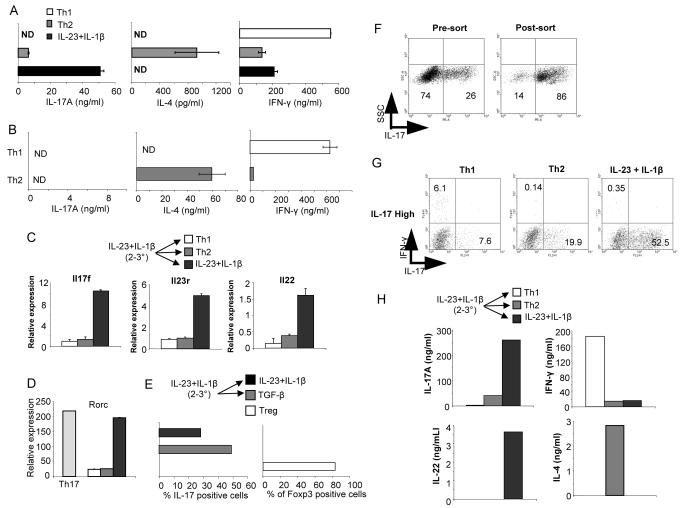
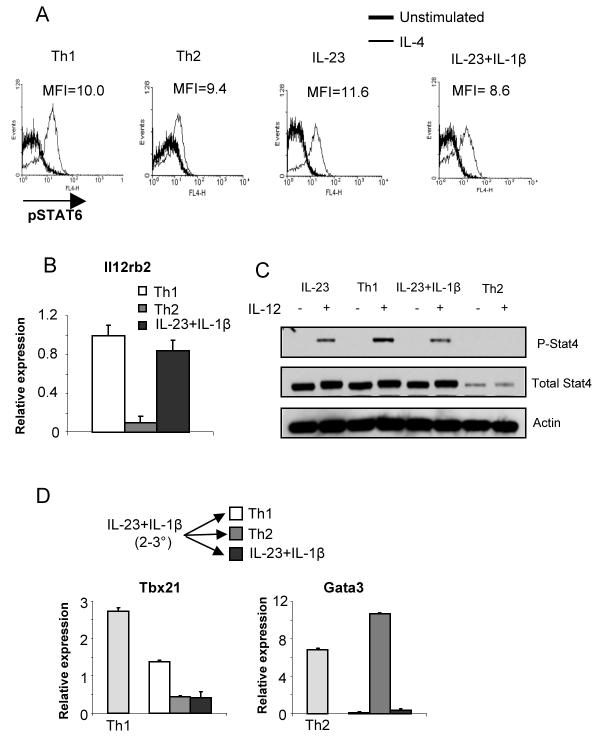
IL-23 plays a critical role establishing inflammatory immunity and enhancing IL-17 production in vivo. However, an understanding of how it performs those functions has been elusive. In this report, using an IL-17-capture technique, we demonstrate that IL-23 maintains the IL-17-secreting phenotype of purified IL-17(+) cells without affecting cell expansion or survival. IL-23 maintains the Th17 phenotype over multiple rounds of in vitro stimulation most efficiently in conjunction with IL-1beta. However, in contrast to Th1 and Th2 cells, the Th17 phenotype is not stable and when long-term IL-23-stimulated Th17 cultures are exposed to Th1- or Th2-inducing cytokines, the Th17 genetic program is repressed and cells that previously secreted IL-17 assume the cytokine secreting profile of other Th subsets. Thus, while IL-23 can maintain the Th17 phenotype, it does not promote commitment to an IL-17-secreting lineage.


Figures

FIGURE 1
FIGURE 1
Cytokine selection of IL-17+ Th17 cells. A, Naïve CD4+ T cells were activated, cultured in TGF-β+IL-6+IL-1β and blocking antibodies (anti-IFN-γ and anti-IL-4) for five days and activated before surface staining for IL-17 using cytokine capture. Cells were then sorted into IL-17-high and –low populations. B, Supernatants from IL-17-high and -low cells stimulated with anti-CD3 were tested for cytokines using ELISA. C, RNA was isolated from cells treated in (B) and gene expression was assessed using real-time PCR.
FIGURE 2
FIGURE 2
IL-23 maintains the IL-17 secreting phenotype without affecting cell expansion or survival. A, Naïve CD4+ T cells were activated, cultured in TGF-β+IL-6+IL-1β and blocking antibodies (anti-IFN-γ and anti-IL-4) for five days before sorting into IL-17-high and –low populations. Cells were then labeled with CFSE and for intracellular IL-17 following stimulation with PMA + ionomycin. Numbers indicate percent of cells in each quadrant and bracketed numbers indicate CFSE MFI. B, IL-17-high cells were cultured with IL-23 and blocking antibodies or blocking antibodies alone for the indicated times before cells were stimulated and stained for intracellular IL-17. Numbers indicate percent of cells in each quadrant and bracketed numbers indicate CFSE MFI. C, IL-17-high or -low CFSE-stained cells prepared as in B were cultured for two days with blocking antibodies in the presence or absence of IL-23 as indicated. CFSE staining is shown from freshly stained cells (Day 0) for comparison. D, IL-17-high and -low cells cultured as in B were counted after 48 hours. E, IL-17-high and -low cells were cultured as in B and were analyzed for Annexin V staining after two or four days of culture in the presence or absence of IL-23. F, IL-17-high and-low cells were stimulated with anti-CD3 and cultured with blocking antibodies with or without IL-23. Cells were stimulated with PMA+Ionomyocin for 4 hours and stained for intracellular IL-17.
FIGURE 3
FIGURE 3
IL-1β increases IL-23 stimulated maintenance of the Th17 phenotype. A, Naïve T cells were activated, primed with TGF-β+IL-6+IL-1β for the first round and cultured for 2 additional rounds of stimulation in the presence of blocking antibodies with or without IL-23. After each round of culture cells were stimulated and cell-free supernatants were tested for cytokine production using ELISA. B, Naïve T cells were activated and primed as in (A) and cultured for 2 additional rounds of stimulation with IL-23, IL-1β, or IL-23+IL-1β as indicated. Cytokine production was measured using ELISA.
FIGURE 4
FIGURE 4
IL-1b increases responsiveness of the Il17 locus. A, Naïve T cells were activated, primed with TGF-β+IL-6+IL-1β for the first round and cultured for 2 additional rounds of stimulation with IL-23, IL-1β, or IL-23+IL-1β as indicated. At the end of the first, second and third round of stimulation RNA was isolated from cells stimulated with anti-CD3 for 4 hours. Quantitative PCR of Il23r expression is shown as relative to Th1 cultures after the third round of stimulation. B, Cells cultured and stimulated as in (A) for three rounds were stimulated with IL-23 for 30 minutes before intracellular staining for phospho-Stat3. C, Naïve cells primed and cultured as in (A) were stimulated with IL-18 and IL-23 for 24 hours. Cell free supernatants were measured for IL-17 using ELISA.
FIGURE 5
FIGURE 5
IL-23 does not program commitment to the Th17 lineage. A, Naïve T cells were activated, primed with TGF-β+IL-6+IL-1β for the first round followed by 2 rounds of stimulation in IL-23 + IL-1β, were cultured for an additional round of stimulation in Th1 or Th2 conditions, or IL-23+IL-1β. Supernatants from anti-CD3 stimulated cells were tested for cytokine production using ELISA. B, Naïve CD4+ T cells were activated and cultured under Th1 or Th2 priming conditions for four rounds of stimulation. At the end of the fourth round of stimulation, supernatants from anti-CD3 stimulated cells were tested for cytokine production using ELISA. C, Cells stimulated and cultured as in (A) were stimulated with anti-CD3 for 4 hours and RNA was isolated for qPCR. D, Cells were stimulated and cultured for three rounds as in (A). After three days of culture in Th1, Th2 or IL-23+IL-1β conditions, RNA was isolated from control or switched cultures for qPCR. E, Naïve T cells were activated, primed with TGF-β+IL-6+IL-1β for the first round followed by 2 rounds of stimulation in IL-23 + IL-1β, were cultured for an additional round of stimulation in IL-23 + IL-1β or TGF-β + IL-2. The percentages of cells positive for Foxp3 or IL-17 intracellular staining are indicated with cells cultured for one week in TGF-β + IL-2 shown as a control for Foxp3 expression. F, Naïve CD4+ T cells were primed and cultured as in (A) and after the third round of culture, cells were enriched for IL-17-secreting cells by cytokine selection. Surface staining for IL-17 is shown pre- and post-sort. G, IL-17-high cells from (F) were cultured in Th1, Th2 or IL-23+IL-1β for an additional round of stimulation. Cells were stimulated for 4 hours and stained for intracellular IL-17 and IFN-γ. H, Supernatants from anti-CD3 stimulated cells cultured as in (G) were tested for cytokine production using ELISA.
FIGURE 6
FIGURE 6
Signals promoting Th1 or Th2 development are intact in Th17 cultures. A, Cells stimulated and cultured as in Figure 5A and 5B were incubated with IL-4 for 30 minutes and stained for phospho-Stat6. B, RNA from cells stimulated as in (A) for three rounds of stimulation was examined for relative levels of Il12rb2 expression. Levels are relative to three-round Th1 cultures. C, Cells stimulated and cultured as in (A) for 3 rounds of stimulation were then stimulated with IL-12 for 1 hour. Phospho-Stat4, total Stat4 and actin were detected by immunoblot. D, Cells were stimulated and cultured for three rounds as in (A). After three days of culture in Th1, Th2 or IL-23+IL-1β, RNA was isolated from control or switched cultures for qPCR to test for expression of the indicated genes. Expression is relative to the level of expression of each gene in IL-23 + IL-1β cultured cells before the fourth round of culture.

References

    1. Weaver CT, Hatton RD, Mangan PR, Harrington LE. IL-17 family cytokines and the expanding diversity of effector T cell lineages. Annu Rev Immunol. 2007;25:821–852. - PubMed
    1. Bettelli E, Korn T, Kuchroo VK. Th17: the third member of the effector T cell trilogy. Curr Opin Immunol. 2007;19:652–657. - PMC - PubMed
    1. Murphy KM, Reiner SL. The lineage decisions of helper T cells. Nat Rev Immunol. 2002;2:933–944. - PubMed
    1. Ivanov II, McKenzie BS, Zhou L, Tadokoro CE, Lepelley A, Lafaille JJ, Cua DJ, Littman DR. The orphan nuclear receptor RORgammat directs the differentiation program of proinflammatory IL-17+ T helper cells. Cell. 2006;126:1121–1133. - PubMed
    1. Yang XO, Pappu BP, Nurieva R, Akimzhanov A, Kang HS, Chung Y, Ma L, Shah B, Panopoulos AD, Schluns KS, Watowich SS, Tian Q, Jetten AM, Dong C. T helper 17 lineage differentiation is programmed by orphan nuclear receptors ROR alpha and ROR gamma. Immunity. 2008;28:29–39. - PMC - PubMed

Publication types

MeSH terms