. 2008 Nov 1;181(9):6435-46.
doi: 10.4049/jimmunol.181.9.6435.

Hepatitis C virus immune escape via exploitation of a hole in the T cell repertoire

Affiliations

Hepatitis C virus immune escape via exploitation of a hole in the T cell repertoire

Matthias Wölfl et al. J Immunol. .

Abstract

Hepatitis C virus (HCV) infection frequently persists despite eliciting substantial virus-specific immune responses. Thus, HCV infection provides a setting in which to investigate mechanisms of immune escape that allow for viral persistence. Viral amino acid substitutions resulting in decreased MHC binding or impaired Ag processing of T cell epitopes reduce Ag density on the cell surface, permitting evasion of T cell responses in chronic viral infection. Substitutions in viral epitopes that alter TCR contact residues frequently result in escape, but via unclear mechanisms because such substitutions do not reduce surface presentation of peptide-MHC complexes and would be expected to prime T cells with new specificities. We demonstrate that a known in vivo HCV mutation involving a TCR contact residue significantly diminishes T cell recognition and, in contrast to the original sequence, fails to effectively prime naive T cells. This mutant epitope thus escapes de novo immune recognition because there are few highly specific cognate TCR among the primary human T cell repertoire. This example is the first on viral immune escape via exploitation of a "hole" in the T cell repertoire, and may represent an important general mechanism of viral persistence.


Figures
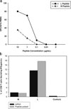
Figure 1
Figure 1
Decreased recognition of the methionine variant (Mp5) of the HLA A*0201 restricted T-cell epitope NS31406 is not due to loss of antigen processing and presentation or masking of low level responses. A. IFN-γ ELISpot was performed using PBMC obtained from Subject 28 one year following HCV infection when Mp5 was the dominant form of virus. Responses for PBMC incubated with the LP5 peptide (circles), which was present upon initial viremia, and Mp5 peptide (triangles), which was dominant six months following infection demonstrate decreased recognition of the Mp5 variant. B. Constructs containing either the Lp5 or Mp5 sequence were introduced via electroporation into EBV transformed cells from Subject 28. Immunogenicity of the construct-containing EBV cells (Black bars labeled mRNA) was assessed via intracellular cytokine staining (ICS) for IFN-γ using NS31406 specific-T cell lines generated from Subject 28. As a positive control, the same T cell line was tested for recognition of Lp5 and Mp5 peptides pulsed onto the surface of the autologous EBV transformed cells (Gray bars labeled peptide pulsed). There was no recognition of mock transfected EBV transformed cells (Black Control) or EBV transformed cells not pulsed with peptide (Grey Control). Recognition of the constructs as well as control constructs known not to represent processing mutations were similar to recognition when the peptides were pulsed onto the cell surface at a concentration of 0.001M, indicating that the Mp5 substitution did not prevent processing.
Figure 2
Figure 2
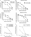
Prolonged CD8 T cell restimulation with Mp5 peptide failed to select a T cell line which recognized Mp5 better than Lp5A. PBMC were obtained from Subject 28 approximately six months after initial viremia when the Mp5 form of the virus was dominant. Those PBMC were stimulated in vitro with either the Lp5 (top panel) or Mp5 -peptide (lower panel) and supplemental interleukin-2 for two 10 day cycles and were subsequently tested for recognition of the Mp5 or Lp5-peptide. Independent of the peptide used for stimulation, both T cell lines recognized targets pulsed with the Lp5 peptide better than the Mp5 peptide in an ELISpot assay for IFN-γ. B. CD8 T cells isolated from Subject 28 after he developed Mp5 as the dominant form of the virus were restimulated with either Lp5 (top panel) or Mp5 pulsed (lower panel) DC followed by two 7 day rounds of stimulation with autologous PBMC pulsed with the same peptides as were pulsed on the DC. The response of both the Lp5 and Mp5 peptide stimulated CD8 T cells was subsequently assessed via IFN-γ ICS using LP5 (circles) or Mp5 (triangles) as antigens. Lp5 remained better recognized. C. PBMC form Subject 28 taken from the one-year time point were cultured in the presence of either Lp5 or Mp5 peptide. The precursor frequency of T cell specific for Lp5 or Mp5 was extremely low at this time point, however, after two rounds of stimulation with the peptide or the variant peptide, cells were tested for recognition of both peptides as demonstrated by IFN-gamma production and CD107a degranulation, in serial peptide dilutions in an intracellular cytokine assay. D. PBMC from an additional subject were used to generate an Lp5 specific T cell line as described in 2c. The T cell line recognized Lp5 peptide better than the Mp5 peptide, as measured by IFN-gamma production and degranulation.
Figure 3
Figure 3
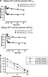
TCR from Subject 28 have decreased avidity for the Mp5/HLA-A*0201 complex. A. Binding of TCR from Subject 28 to Lp5 and Mp5 on HLA A*0201 was assessed by dissociation rates using the T cell lines described in Figure 2. Tetramer off-rate experiments were performed by staining Lp5- and Mp5- peptide primed T cell lines with Lp5- or Mp5- tetramer, washing, then incubating the cells with excess HLA-A2 antibody that prevented rebinding of any tetramer that had fallen off. An aliquot of each sample was removed following 0, 1, 2, 5, 7, 10, 20, 40, and 60 minutes of incubation and the MFI of Lp5- and Mp5- tetramer bound to the TCR for each line was recorded. The data were used to determine the time it took for half the tetramer to dissociate (t½) and demonstrated higher avidity of both Lp5 and Mp5 primed T cell lines for the Lp5-tetramer than the Mp5-tetramer. B. Serial dilution of tetramers were performed using half-log dilutions of tetramer and again demonstrated higher avidity of both Lp5 and Mp5 primed T cell lines for the Lp5-tetramer than the Mp5-tetramer.
Figure 4
Figure 4
Mp5 ineffectively primes naïve T cells. CD45RO−/CD8+ (naïve) T cells from HCV-negative individuals were primed under optimal conditions for generation of an antigen specific response with Lp5 or Mp5 and the generation of antigen specific responses assessed by tetramer staining. Priming was performed with five different donors in multiple experiments and in parallel groups for Lp5 and Mp5. An Lp5-specific response was obtained in 36 out of 38 lines and the majority of responses were vigorous. In contrast, a total of 14 out of 38 lines generated using Mp5 had a specific response for that variant as detected by tetramer staining, but nearly all were very low level responses and at the limit of detection in the assay. The Lp5 and Mp5 specific responses were compared via GEE analysis and the Lp5-specific responses were significantly more frequent and of higher magnitude than Mp5-specific responses. (p<.0001)
Figure 5
Figure 5
Responses to Lp5 in HCV naïve individuals derive from the naïve T cell population. A. Immunophenotyping of CD45RA+CCR7+ T-cells sorted cells showed a high purity of CD45RA+CCR7+CD28+CD62L+ naïve T cells (left panel), whereas a CD45RA-CCR7- control population was a CD45RO+ memory subpopulation (right panel). B. CD45RA+CCR7+ and CD45RA−CCR7− sorted T cells were expanded and restimulated for 2 cycles. Only the naïve CD45RA+CCR7+ population yielded a response to the Lp5 HCV epitope, as measured by magnitude (top) or percent positive (bottom). C. CD8+ cells were isolated by negative selection and then positively selected for CD62L or CD45RO expression. No Lp5-specific response was obtained from the CD45RO+ memory population, whereas a statistically significant number of wells were positive in the CD62L+ naïve group. D. Cells isolated by CD45RO depletion followed by CD8 positive selection were compared with CD8 cells obtained from PBMC by negative selection, depleted of CD45RO+ cells, then positively selected for CD62L expression. Following expansion and restimulation for 2 cycles, similar numbers of tetramer+ wells were detected from both populations containing naïve T cells.
Figure 6
Figure 6
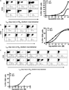
Mp5 primes naïve T cells that are primarily cross-reactive. In the right lower quadrant for each T cell line, numbers separated by a slash indicate the percent of T cells specific for NS31406/MFI. The percent of tetramer bound cells is shown using Lp5 (top row) or Mp5 (bottom row). Representative ICS data are shown for one line to the right of the FACS plots in each panel. A. Priming naïve cells with Lp5 peptide resulted in Lp5-specific responses that were cross-reactive for the Mp5-epitope as shown for three representative lines. The percent of T cells specific for Lp5 was usually higher and the MFI was always higher for Lp5 than Mp5 tetramer. Although cross-reactive, better IFN-γ production was observed in response to Lp5 versus Mp5B. For the T cells lines generated with Mp5 peptide, parallel staining with either Mp5 or Lp5/A*0201-MHC tetramers demonstrated that 8 of these lines were cross-reactive to the Lp5 epitope, displaying a similar or higher MFI, when stained with the Lp5 multimer, as shown for three representative lines. The percent of T cells specific for Lp5 and Mp5 and the MFI were usually equivalent. Responses vigorous enough to assess for cytokine production were very rare, but better or equivalent IFN-γ production was observed in response to Mp5 as compared to Lp5 for the lines that recognized both. C. Six of the 14 lines generated by Mp5 priming demonstrated a higher MFI after staining with the Mp5-multimer than the Lp5-multimer and one a much larger percent of Mp5 specific cells (25 vs. 0.1%), suggesting they were more specific for Mp5 and where assessed, they also produced more IFN-γ in response to Mp5.
Figure 7
Figure 7
Mp5 acts as a partial agonist and not as an antagonist. A. Mp5 fails to boost an Lp5-specific response. Using naïve T cells that had been primed with Lp5 -pulsed DC and expanded with Lp5-pulsed PBMC, a third round of stimulation was performed using either Lp5 (LLL) or Mp5 (LLM) and compared to a third round of culture with media alone (LL-). Both Lp5 and Mp5 bearing tetramers were used to assess the percent of cells specific for each antigen with no significant differences in recognition pattern. The percent of CD8+/Lp5 tetramer positive T cells did not differ significantly between LLM and LL-, demonstrating that Mp5 expanded specific T cells no better than media alone. B. The Mp5 peptide did not boost the Lp5-specific response, but does not have an inhibitory effect relative to a control peptide of comparable HLA-A*0201 binding either. A T-cell line was generated by stimulating naïve T-cells with the Lp5 peptide and the percent of Lp5 variant-specific T cell is shown in the FACS plot labeled (i) as 0.98%. This line was divided and restimulated with equal numbers of autologous PBMC pulsed with one of four peptide combinations; Lp5 only (ii), Mp5 only (iii), an equal mixture of Lp5 and control peptide (iv) or an equal mixture of Lp5 and Mp5 (v). Evaluation by tetramer staining 1 week after stimulation showed that Mp5 peptide did not boost or inhibit the Lp5-specific response relative to control peptide (Lp5 specific T cells 4.94% versus 4.85%).
Figure 8
Figure 8
Priming naïve cells with Lp5 peptide resulted in Lp5-specific responses that were very weakly cross-reactive for the Mp5-epitope. Data are shown for three representative lines in Figure 6a. Although cross-reactive, better IFN-γ (Figure 6a) and TNF-α production, degranulation (CD107a), and upregulation of activation markers (CD137, CD25) was observed in response to the Lp5 as compared to Mp5.

References

    1. Weiss RA, McMichael AJ. Social and environmental risk factors in the emergence of infectious diseases. Nat.Med. 2004;10:S70–S76. - PMC - PubMed
    1. Cox AL, Mosbruger T, Lauer GM, Pardoll D, Thomas DL, Ray SC. Comprehensive analyses of CD8+ T cell responses during longitudinal study of acute human hepatitis C. Hepatology. 2005;42:104–112. - PMC - PubMed
    1. Lechner F, Gruener NH, Urbani S, Uggeri J, Santantonio T, Kammer AR, Cerny A, Phillips R, Ferrari C, Pape GR, Klenerman P. CD8+ T lymphocyte responses are induced during acute hepatitis C virus infection but are not sustained. Eur.J.Immunol. 2000;30:2479–2487. - PubMed
    1. Cucchiarini M, Kammer AR, Grabscheid B, Diepolder HM, Gerlach TJ, Gruner N, Santantonio T, Reichen J, Pape GR, Cerny A. Vigorous peripheral blood cytotoxic T cell response during the acute phase of hepatitis C virus infection. Cell Immunol. 2000;203:111–123. - PubMed
    1. Rehermann B, Chang KM, McHutchison JG, Kokka R, Houghton M, Chisari FV. Quantitative analysis of the peripheral blood cytotoxic T lymphocyte response in patients with chronic hepatitis C virus infection. J.Clin.Invest. 1996;98:1432–1440. - PMC - PubMed

Publication types

MeSH terms