

Preliminary in vivo atherosclerotic carotid plaque characterization using the accumulated axial strain and relative lateral shift strain indices
- PMID: 18941278
- PMCID: PMC2891504
- DOI: 10.1088/0031-9155/53/22/008
Preliminary in vivo atherosclerotic carotid plaque characterization using the accumulated axial strain and relative lateral shift strain indices
Abstract
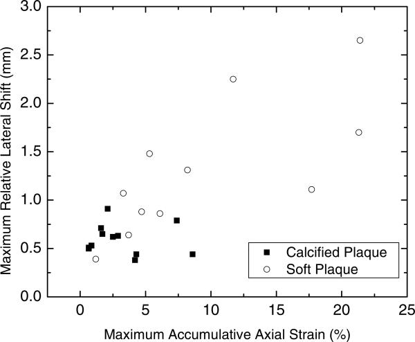
In this paper, we explore two parameters or strain indices related to plaque deformation during the cardiac cycle, namely, the maximum accumulated axial strain in plaque and the relative lateral shifts between plaque and vessel wall under in vivo clinical ultrasound imaging conditions for possible identification of vulnerable plaque. These strain indices enable differentiation between calcified and lipidic plaque tissue utilizing a new perspective based on the stiffness and mobility of the plaque. In addition, they also provide the ability to distinguish between softer plaques that undergo large deformations during the cardiac cycle when compared to stiffer plaque tissue. Soft plaques that undergo large deformations over the cardiac cycle are more prone to rupture and to release micro-emboli into the cerebral bloodstream. The ability to identify vulnerable plaque, prone to rupture, would significantly enhance the clinical utility of this method for screening patients. We present preliminary in vivo results obtained from ultrasound radio frequency data collected over 16 atherosclerotic plaque patients before these patients undergo a carotid endarterectomy procedure. Our preliminary in vivo results indicate that the maximum accumulated axial strain over a cardiac cycle and the maximum relative lateral shift or displacement of the plaque are useful strain indices that provide differentiation between soft and calcified plaques.
Figures


References
-
- AbuRahma AF, Wulu JT, Jr, Crotty B. Carotid plaque ultrasonic heterogeneity and severity of stenosis. Stroke. 2002;33:1772–5. - PubMed
-
- Baldewsing RA, Schaar JA, Mastik F, Oomens CW, van der Steen AF. Assessment of vulnerable plaque composition by matching the deformation of a parametric plaque model to measured plaque deformation. IEEE Trans. Med. Imaging. 2005;24:514–28. - PubMed
-
- Bertrand M, Meunier M, Doucet M, Ferland G. Ultrasonic biomechanical strain gauge based on speckle tracking. IEEE Ultrasonics Symp. 1989:859–64.
-
- Bridal SL, Fornes P, Bruneval P, Berger G. Correlation of ultrasonic attenuation (30 to 50 MHz) and constituents of atherosclerotic plaque. Ultrasound Med. Biol. 1997;23:691–703. - PubMed
-
- Cespedes EI. PhD Dissertation. University of Houston; Texas: 1993. Elastography: imaging of biological tissue elasticity.
Publication types
MeSH terms
Grants and funding
LinkOut - more resources
Full Text Sources
Medical