Review
. 2008;84(5):147-54.
doi: 10.2183/pjab.84.147.

Tetrodotoxin: a brief history

Affiliations
Review

Tetrodotoxin: a brief history

Toshio Narahashi. Proc Jpn Acad Ser B Phys Biol Sci. 2008.

Abstract

Tetrodotoxin (TTX), contained in puffer, has become an extremely popular chemical tool in the physiological and pharmacological laboratories since our discovery of its channel blocking action in the early 1960s. This brief review describes the history of discovery of TTX action on sodium channels, and represents a story primarily of my own work. TTX inhibits voltage-gated sodium channels in a highly potent and selective manner without effects on any other receptor and ion channel systems. TTX blocks the sodium channel only from outside of the nerve membrane, and is due to binding to the selectivity filter resulting in prevention of sodium ion flow. It does not impairs the channel gating mechanism. More recently, the TTX-resistant sodium channels have been discovered in the nervous system and received much attention because of their role in pain sensation. TTX is now known to be produced not by puffer but by bacteria, and reaches various species of animals via food chain.(Communicated by Masanori OTSUKA, M.J.A.).


Figures

Fig. 1.
Fig. 1.
Tetrodotoxin (TTX) block of muscle action potential without effect on delayed rectification. Intracellular microelectrode recording from a frog nerve-sartorius muscle preparation. (A) Normal muscle action potential evoked by nerve stimulation. (B) Responses to direct subthreshold depolarization and hyperpolarization in normal muscle. (C) Action potential generated by direct suprathreshold depolarization and hyperpolarization in normal muscle. (D) After application of 300 nM TTX, nerve stimulation failed to evoke muscle action potential. (E) In TTX, direct depolarization and hyperpolarization failed to evoke muscle action potential. (F) In TTX, stronger direct depolarization and hyperpolarization still failed to evoke muscle action potential revealing the presence of delayed rectification, indicative of potassium channel activation. 2)
Fig. 2.
Fig. 2.
Structures of tetrodotoxin (A) 15) and saxitoxin (B). 16)
Fig. 3.
Fig. 3.
Tetrodotoxin (94 nM) blocks action potentials and sodium channel currents without effect of potassium channel currents. Lobster giant axon under sucrose-gap voltage clamp. (A) Before and (B) during application of TTX. Holding potential was –120 mV including hyperpolarization caused by sucrose-gap conditions. Downward transient currents represent inward sodium currents, and upward steady-state currents represent outward potassium currents. Because of sucrose-gap hyperpolarization, a large depolarizing current was needed to evoke an action potential in A, and a large depolarizing current did not produce the action potential in the presence of TTX in B. 3)
Fig. 4.
Fig. 4.
Tetrodotoxin (TTX) blocks sodium channels from outside the nerve membrane in the cationic form, whereas local anesthetic molecules penetrate the nerve membrane in the uncharged form (B) and block both sodium and potassium channels from inside the nerve membrane in the cationic form. 42)
Fig. 5.
Fig. 5.
Tetrodotoxin (TTX) block of single sodium channel currents. (A) Single channel currents recorded from an outside-out membrane patch isolated from a neuroblastoma cell (N1E-115) in response to depolarization from a holding potential of –90 mV to –30 mV as shown at the bottom. (B) After application of 3 nM TTX to the external membrane surface. (C) Open time distributions before and after exposure to TTX. (D) Amplitude histograms before and after TTX. Temperature 10°C. TTX did not change the open channel characteristics but decreased the number of open channels to approximately half as 3 nM TTX was close to its IC50. 53), 54)
Fig. 6.
Fig. 6.
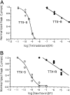
Dose-response relationships for TTX and STX block. Dorsal root ganglion cells expressing TTX-S (n=3) or TTX-R (n=3) currents were exposed to increasing concentrations of TTX or STX and pulsed once per minute to +10 mV to determine peak current amplitude. Steady-state peak current amplitudes reached at each concentration were normalized to control toxin-free amplitudes and plotted against toxin concentration. (A) TTX dose-response curve, with IC50 values of 0.3 nM (TTX-S) and 100 µM (TTX-R). (B) STX dose-response curve, with IC50 values of 0.5 nM (TTX-S) and 10 µM (TTX-R). 62)

References

    1. Noguchi, T. and Ebesu, J.S.M. (2001) J. Toxicol.-Toxin Rev. 20, 1–10
    1. Narahashi, T., Deguchi, T., Urakawa, N. and Ohkubo, Y. (1960) Amer. J. Physiol. 198, 934–938 - PubMed
    1. Narahashi, T., Moore, J.W. and Scott, W.R. (1964) J. Gen. Physiol. 47, 965–974 - PMC - PubMed
    1. Nastuk, W.L. and Hodgkin, A.L. (1950) J. Cell. Comp. Physiol. 35, 39–74
    1. Urakawa, N., Narahashi, T., Deguchi, T. and Ohkubo, Y. (1960) Amer. J. Physiol. 198, 938–942 - PubMed