Review
. 2008;84(7):246-63.
doi: 10.2183/pjab.84.246.

Glycine cleavage system: reaction mechanism, physiological significance, and hyperglycinemia

Affiliations
Review

Glycine cleavage system: reaction mechanism, physiological significance, and hyperglycinemia

Goro Kikuchi et al. Proc Jpn Acad Ser B Phys Biol Sci. 2008.

Abstract

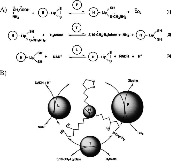
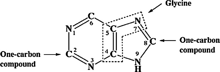
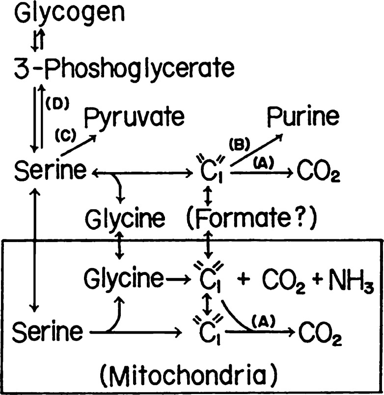
The glycine cleavage system catalyzes the following reversible reaction: Glycine + H(4)folate + NAD(+) <==> 5,10-methylene-H(4)folate + CO(2) + NH(3) + NADH + H(+)The glycine cleavage system is widely distributed in animals, plants and bacteria and consists of three intrinsic and one common components: those are i) P-protein, a pyridoxal phosphate-containing protein, ii) T-protein, a protein required for the tetrahydrofolate-dependent reaction, iii) H-protein, a protein that carries the aminomethyl intermediate and then hydrogen through the prosthetic lipoyl moiety, and iv) L-protein, a common lipoamide dehydrogenase. In animals and plants, the proteins form an enzyme complex loosely associating with the mitochondrial inner membrane. In the enzymatic reaction, H-protein converts P-protein, which is by itself a potential alpha-amino acid decarboxylase, to an active enzyme, and also forms a complex with T-protein. In both glycine cleavage and synthesis, aminomethyl moiety bound to lipoic acid of H-protein represents the intermediate that is degraded to or can be formed from N(5),N(10)-methylene-H(4)folate and ammonia by the action of T-protein. N(5),N(10)-Methylene-H(4)folate is used for the biosynthesis of various cellular substances such as purines, thymidylate and methionine that is the major methyl group donor through S-adenosyl-methionine. This accounts for the physiological importance of the glycine cleavage system as the most prominent pathway in serine and glycine catabolism in various vertebrates including humans. Nonketotic hyperglycinemia, a congenital metabolic disorder in human infants, results from defective glycine cleavage activity. The majority of patients with nonketotic hyperglycinemia had lesions in the P-protein gene, whereas some had mutant T-protein genes. The only patient classified into the degenerative type of nonketotic hyperglycinemia had an H-protein devoid of the prosthetic lipoyl residue. The crystallography of normal T-protein as well as biochemical characterization of recombinants of the normal and mutant T-proteins confirmed why the mutant T-proteins had lost enzyme activity. Putative mechanisms of cellular injuries including those in the central nervous system of patients with nonketotic hyperglycinemia are discussed.


Figures

Fig. 1
Fig. 1
A) Mechanism of the glycine cleavage reaction. P, H, T and L in the circles represent respective proteins. Lip, H4folate, 5,10-CH2-H4folate represent lipoyl moiety, tetrahydrofolate, and N5,N10-methylene-H4folate, respectively. B) Schematic presentation of the glycine cleavage reaction.
Fig. 2
Fig. 2
Stereo ribbon diagrams of the peaH (blue) and humanT (red) complex model. Cα of Gly299 of humanT (red) and Glu42 of peaH (blue) are presented as spheres with labels. The putative residues of humanT interacting with aminomethyl lipoate arm are also presented as spheres with labels (red). The aminomethyl lipoate lysine (Lys63 of peaH) and 5-CH3-H4folate are represented in ball-and-stick format with bonds colored in yellow. Asp39 and Lys226 of humanT corresponding to the residues involved in the intramolecular cross-linking in the E.coliT-E.coliH complex are represented in ball-and-stick format with bonds colored and labeled in green. The β-strands of peaH (β5 and β6) that form one side of the aminomethyl lipoate-binding cleft are labeled. The lower panel view is obtained by a 90° rotation of the upper one.
Fig. 3
Fig. 3
Glycine metabolism by rat liver mitochondria. Reaction mixtures contained, in a final volume of 2.8 ml, indicated amounts of 14C-glycine (0.01mCi/mmole) and 1 ml of mitochondrial suspension containing 10mg of protein. Reactions were carried out for 60 min at 37 °C. G stands for glycine.
Fig. 4
Fig. 4
Comparison of 14CO2 formation from 14C-serine and 14C-glycine catalyzed by rat liver mitochondria. Positions of 14C carbon in glycine and serine are shown in the figure. Reaction conditions were similar to those in Fig. 3 except for that various amounts of 14C-glycine (0.01mCi/mmole) or 14C-DL-serine (0.01mCi/mmole) were employed. G and S stand for glycine and DL-serine, respectively. The amounts of serine in the figure represent the amounts of L-serine.
Fig. 5
Fig. 5
Chemical structure of purine. Whole structure of glycine provides C4, C5 and N7 and N10-formyl-H4folate and N5,N10-methylene-H4folate provide C2 and C8.
Fig. 6
Fig. 6
Pathways for glycine and serine catabolism in vertebrate livers under physiological conditions. Route (A) is considerably limited in uricotelic animals. Route (B) is prevailing in uricotelic animals. Routes (C) and (D) in the soluble fractions are relatively minor. “C1” denotes N5,N10-methylene-H4folate and other one-carbon compounds.
Fig. 7
Fig. 7
Immunoblot of P-protein using liver homogenates of patients with nonketotic and ketotic hyperglycinemia. A) Lane 1, purified chicken P-protein (100 ng of protein); lane 2, mitochondrial extract from control human liver (100 μg); lanes 3 and 4, control human liver homogenates (300 μg); lanes 5 and 6, liver homogenates from patients with nonketotic hyperglycinemia (neonatal-onset type)(300 μg); lane 7, liver homogenate from a patient with propionic academia (one type of ketotic hyperglycinemias) (300 μg). B) Lanes 1 and 2, a control liver homogenate (100 and 200 μg of proteins); lanes 3, 4 and 5, liver homogenates from a patient with a late-onset type of nonketotic hyperglycinemia (50, 100 and 200 μg of proteins). Anti-chicken P-protein antibody was used in (A) and (B). C) Immunoblot of H-protein using anti-rat H-protein antibody. Lane 1, purified human H-protein (40 ng); lane 2, human liver mitochondrial extract (100 μg); lanes 3 and 4, control human liver homogenates; lanes 5, liver homogenate of the patient with propionic academia; lanes 6 and 7, liver homogenates of the patients with neonatal-onset type of nonketotic hyperglycinemia. Six μg of proteins were loaded onto lanes 3 to 7. Note that specimens loaded onto lanes 5, 6 and 7 in (A) correspond to those onto lanes 6, 7 and 5 in (C).
Fig. 8
Fig. 8
Nonketotic-hyperglycinemia-related mutation sites mapped on the overall topology of humanT. The mutant residues are depicted in a ball-and-stick representation with atoms colored in red. Residue numbers are labeled.
Fig. 9
Fig. 9
Metabolic map of H4folate derivatives.

Similar articles

Cited by

References

    1. Sagers, R. D. and Gunsalus, I. C. (1961) Intermediary metabolism of Diplococcus glycinophilus. I. Glycine cleavage and one carbon interconversions. J. Bacteriol. 81, 541–549 - PMC - PubMed
    1. Richert, D. A., Amberg, R. and Wilson, M. (1962) Metabolism of glycine by avian liver. J. Biol. Chem. 237, 99–103 - PubMed
    1. Kawasaki, H., Sato, T. and Kikuchi, G. (1966) A new reaction for glycine biosynthesis. Biochem. Biophys. Res. Commun. 23, 227–233 - PubMed
    1. Sato, T., Kochi, H., Motokawa, Y., Kawasaki, H. and Kikuchi, G. (1969) Glycine metabolism by rat liver mitochondria. I. Synthesis of two molecules of glycine from one molecule each of serine, bicarbonate and ammonia. J. Biochem. 65, 63–70 - PubMed
    1. Motokawa, Y. and Kikuchi, G. (1969) Glycine metabolism by rat liver mitochondria. II. Methylene tetrahydrofolate as the direct one carbon donor in the reaction of glycine synthesis. J. Biochem. 65, 71–75 - PubMed