. 2008;3(10):e3439.
doi: 10.1371/journal.pone.0003439. Epub 2008 Oct 21.

Diaphanous-related formin 2 and profilin I are required for gastrulation cell movements

Affiliations

Diaphanous-related formin 2 and profilin I are required for gastrulation cell movements

Shih-Lei Lai et al. PLoS One. 2008.

Abstract

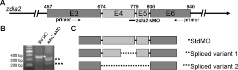
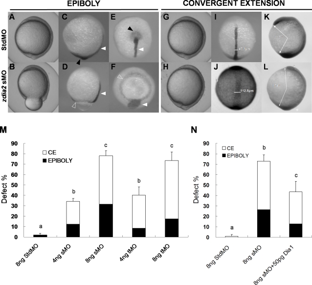
Intensive cellular movements occur during gastrulation. These cellular movements rely heavily on dynamic actin assembly. Rho with its associated proteins, including the Rho-activated formin, Diaphanous, are key regulators of actin assembly in cellular protrusion and migration. However, the function of Diaphanous in gastrulation cell movements remains unclear. To study the role of Diaphanous in gastrulation, we isolated a partial zebrafish diaphanous-related formin 2 (zdia2) clone with its N-terminal regulatory domains. The GTPase binding domain of zDia2 is highly conserved compared to its mammalian homologues. Using a yeast two-hybrid assay, we showed that zDia2 interacts with constitutively-active RhoA and Cdc42. The zdia2 mRNAs were ubiquitously expressed during early embryonic development in zebrafish as determined by RT-PCR and whole-mount in situ hybridization analyses. Knockdown of zdia2 by antisense morpholino oligonucleotides (MOs) blocked epiboly formation and convergent extension in a dose-dependent manner, whereas ectopic expression of a human mdia gene partially rescued these defects. Time-lapse recording further showed that bleb-like cellular processes of blastoderm marginal deep marginal cells and pseudopod-/filopod-like processes of prechordal plate cells and lateral cells were abolished in the zdia2 morphants. Furthermore, zDia2 acts cell-autonomously since transplanted zdia2-knockdown cells exhibited low protrusive activity with aberrant migration in wild type host embryos. Lastly, co-injection of antisense MOs of zdia2 and zebrafish profilin I (zpfn 1), but not zebrafish profilin II, resulted in a synergistic inhibition of gastrulation cell movements. These results suggest that zDia2 in conjunction with zPfn 1 are required for gastrulation cell movements in zebrafish.


Conflict of interest statement

Competing Interests: The authors have declared that no competing interests exist.

Figures

Figure 1
Figure 1. Sequence analysis of the zebrafish diaphanous 2 gene (zdia2).
(A) Nucleotide sequence of zdia2. A 5′-half zebrafish dia2 sequence was cloned by 5′RACE. It contained 376-bp 5′UTR and 1497-bp coding regions (the start codon is boxed). A putative Rho-binding domain predicted by HMMPFAM is underlined. Exon 4, a putative deleting region by a zdia2 splicing blocking MO, is shown with a gray background. (B) Amino acid sequence alignment of the cloned zDia2 with corresponding regions of human and mouse diaphanous-related formins 1, 2, and 3. Identical and conserved amino acids are shown with dark and gray backgrounds, respectively. (C) Phylogenic analysis of Dia members in chordates. The horizontal length is proportional to the estimated time from divergence of the gene from the related family member. h, human; m, mouse; z, zebrafish.
Figure 2
Figure 2. Ubiquitous expression of zdia2 during early embryonic development.
(A) Expression of a 578-bp fragment of zdia2 at different embryonic stages was examined by RT-PCR, and the transcription of a 717-bp β-actin fragment was used as an internal control. (B-I) WISH of zdia2 showed ubiquitous expression in the entire embryo during early stages, but the transcripts had enriched in the head region at 24 hpf. The riboprobe was synthesized from a 1487-bp fragment beginning at the start codon of zdia2.
Figure 3
Figure 3. The zDia2 N-terminal half domain specifically interacted with constitutively active RhoA and Cdc42 in the yeast two-hybrid assay.
Yeasts were transformed with designated constructs and tested for interaction by growth on selective medium lacking histidine with the addition of 4 mM 3AT. AD, GAL4 activation domain; BD, GAL4 DNA-binding domain.
Figure 4
Figure 4. The zdia2 splice-blocking morpholino oligonucleotide (sMO) caused splicing aberrance of zdia2 exons 4 and 5.
(A) Diagram of a partial pre-mRNA map of zdia2. The exons are shown in boxes, labeled with the corresponding exon number in E3 through E6, and the introns are represented by solid lines. A zdia2 sMO target site is at the end of E4 to the beginning of following intron, which is indicated underneath as a black bar. The zdia2 sMO would presumably interfere with the splicing of E4 shown in light grey. (B) The zdia2 sMO induced splicing aberrance as evidenced in the RT-PCR analysis using a primer pair as indicated in (A) by arrows. Two spliced variants were found in zdia2 sMO-treated embryos. The first variant (**) was 324 bp and the second variant (***) was 231 bp as compared to the 357-bp original transcript (*). (C) The spliced variants were sequenced (see Fig. S1) and shown as maps. The zdia2 sMO-affected exons are shown in light grey and the deleted regions are depicted in dotted lines.
Figure 5
Figure 5. Knockdown of zdia2 by MO interferes with gastrulation cell movements in a dose-dependent manner.
Embryos injected with 8 ng of standard control (std) MO (the upper row) and zdia2 sMO (the bottom row) were examined and photographed under a stereomicroscope at 10 hpf. Embryos were then fixed, stained by a no tail riboprobe (C, D, K and L, side view; E and F, vegetal view; I and J, dorsal view) or in combination with a goosecoid riboprobe and photographed (K and L, side view). Tail buds and notochords are respectively indicated by black and white arrowheads (C–F). The zdia2 sMO caused incomplete epiboly formation with a ring structure near the vegetal pole as indicated by the ntl staining (D and F, open arrowheads). Widths of the notochords were measured and labeled in I and J. Lengths of the body axis were also estimated by the angle (labeled) between the prechordal plate and tail bud as described in the text (K and L). (M) Different MOs at 4 or 8 ng as designated were injected, and the embryos were examined at 10 hpf. Each treatment was repeated at least three times and analyzed as described, and only upper error bars of standard deviations are shown. (N) Embryos were injected with stdMO, zdia2 sMO or zdia2 sMO with 50 pg mdia mRNA as indicated. Epiboly and convergent extension defects were determined, analyzed, and presented as described in (M). Different letters on top of each column indicate a significant difference between treatments (P<0.05).
Figure 6
Figure 6. Knockdown of zdia2 inhibits epiboly cell migration by inhibiting cell protrusion formation.
Embryos injected with the standard control MO (stdMO) or the zdia2 splice-blocking MO (sMO) were dechorionated, immobilized, monitored, and recorded on 20-minutes continuous time-lapse movies (See supplement movie S1 and S2 for the stdMO and sMO-injected embryo, respectively). Photographs from each recording at 5-minutes intervals are shown in (A) with the recording time given in the lower right corner. All photographs are positioned with animal pole up. In the magnified pictures, marginal deep cells of the stdMO-injected embryo (B) exhibited clear protrusive activity with two protruded cell processes indicated by arrowheads, but no such protrusions were evident in the zdia2 sMO-treated embryo (C). Bars indicate 100 µm. (D) Cell protrusions formation at the leading edge of marginal deep cells in both stdMO- and zdia2 sMO-injected embryos were quantified separately by counting protrusions formed within the first 50 frames form 3 different movies, respectively. Results were then analyzed and only upper error bars of standard deviations are shown. Different letters on top of each column indicate a significant difference between treatments (P<0.05).
Figure 7
Figure 7. Knockdown of zdia2 inhibits cell protrusion formation of prechordal plate (primordial) cells and lateral (mesendodermal) cells.
(A) Embryos injected with the standard control MO (stdMO) or the zdia2 splice-blocking MO (sMO) were dechorionated, immobilized, monitored, and recorded on 10-minutes continuous time-lapse movies. (B) Protrusions formation of cells on focus in both stdMO- and zdia2 sMO-injected embryos were quantified separately by counting protrusions formed within the first 10 minutes form at least 3 different movies, respectively. Results were then analyzed and only upper error bars of standard deviations are shown. Different letters on top of each column indicate a significant difference between treatments (P<0.05).
Figure 8
Figure 8. Knockdown of zdia2 inhibit actin condensation at the front edge of blastoderm.
Embryos injected with 8 ng stdMO and zdia2 sMO were fixed at the germ-ring stage and photographed under confocal microscope after rhodamine phalloidin staining. Filamentous actins (arrowheads) were enriched at the front edge of the epiboly marginal deep cells of the stdMO-injected embryo (A), but not in the zdia2 sMO treated one (B).
Figure 9
Figure 9. zdia2 function is required cell-autonomously for the cell protrusions and migration.
Rhodamine-labeled cells were transplanted from embryos injected with 8 ng StdMO (A) and 8 ng zdia2 sMO (B) with Q-rhodamine, respectively, to wild type embryos. (C) Curvillinear velocity (Vcl) and strait line velocity (Vsl) were calculated and quantified (p<0.05).
Figure 10
Figure 10. zDia2 coordinates with profilin I in the regulation of gastrulation cell movements.
(A) The stdMO, zdia2 sMO, pfn I tMO, or pfn II tMO were injected at 8 ng per embryo as indicated, while 4 ng zdia2 sMO plus 4 ng pfn I MO (MOs(pfn I+zdia2)) or pfn II MO (MOs(pfn II+zdia2)) were co-injected as shown. Each treatment was repeated at least three times and the results were then analyzed by student t test. The defects are categorized as in Fig. 6, and only upper error bars are shown. (B) Defect rates of embryos injected with 8 ng stdMO, pfn I MO, pfn I mRNA at 100 pg and 8 ng pfn I MO plus 100 pg pfn I mRNA were quantified as designated, and the embryos were examined by the same criterion as mentioned previously at 10 hpf. The results were analyzed by student t test., and only upper error bars are shown. Different letters on top of each column indicate a significant difference between treatments (P<0.05).
Figure 11
Figure 11. Rho mediates actin remodeling during gastrulation in zebrafish.
Rho acts through its downstream effector, Rho-associated kinase (ROCK), which presumably phosphorylates regulatory myosin light chains (MRLCs) and enforces actomyosin contractility. In conjunction, zDia2 or other formins may be activated by Rho and/or Cdc42 to accelerate actin-nucleation through the cooperation with profilin I at the front edge of migrating cells for the control of cellular migration during gastrulation in the zebrafish.

Similar articles

Cited by

References

    1. Solnica-Krezel L. Conserved patterns of cell movements during vertebrate gastrulation. Curr Biol. 2005;15:R213–R228. - PubMed
    1. Schier AF. Axis formation and patterning in zebrafish. Curr Opin Genet Dev. 2001;11:393–404. - PubMed
    1. Solnica-Krezel L. Gastrulation in zebrafish – all just about adhesion? Curr Opin Genet Dev. 2006;16:433–441. - PubMed
    1. Burridge K, Wennerberg K. Rho and Rac take center stage. Cell. 2004;116:167–179. - PubMed
    1. Hall A, Nobes CD. Rho GTPases: molecular switches that control the organization and dynamics of the actin cytoskeleton. Philos Trans R Soc Lond B Biol Sci. 2000;355:965–970. - PMC - PubMed

Publication types