. 2008;2(10):e325.
doi: 10.1371/journal.pntd.0000325. Epub 2008 Oct 22.

Mycolactone diffuses from Mycobacterium ulcerans-infected tissues and targets mononuclear cells in peripheral blood and lymphoid organs

Affiliations

Mycolactone diffuses from Mycobacterium ulcerans-infected tissues and targets mononuclear cells in peripheral blood and lymphoid organs

Hui Hong et al. PLoS Negl Trop Dis. 2008.

Abstract

Background: Buruli ulcer (BU) is a progressive disease of subcutaneous tissues caused by Mycobacterium ulcerans. The pathology of BU lesions is associated with the local production of a diffusible substance, mycolactone, with cytocidal and immunosuppressive properties. The defective inflammatory responses in BU lesions reflect these biological properties of the toxin. However, whether mycolactone diffuses from infected tissues and suppresses IFN-gamma responses in BU patients remains unclear.

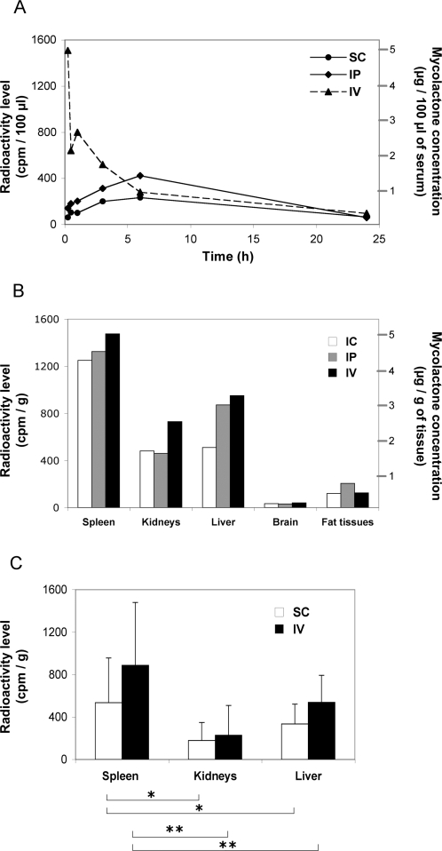
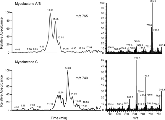
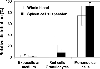
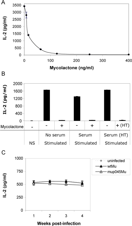
Methodology/principal findings: Here we have investigated the pharmacodistribution of mycolactone following injection in animal models by tracing a radiolabeled form of the toxin, and by directly quantifying mycolactone in lipid extracts from internal organs and cell subpopulations. We show that subcutaneously delivered mycolactone diffused into mouse peripheral blood and accumulated in internal organs with a particular tropism for the spleen. When mice were infected subcutaneously with M. ulcerans, this led to a comparable pattern of distribution of mycolactone. No evidence that mycolactone circulated in blood serum during infection could be demonstrated. However, structurally intact toxin was identified in the mononuclear cells of blood, lymph nodes and spleen several weeks before ulcerative lesions appear. Importantly, diffusion of mycolactone into the blood of M. ulcerans-infected mice coincided with alterations in the functions of circulating lymphocytes.

Conclusion: In addition to providing the first evidence that mycolactone diffuses beyond the site of M. ulcerans infection, our results support the hypothesis that the toxin exerts immunosuppressive effects at the systemic level. Furthermore, they suggest that assays based on mycolactone detection in circulating blood cells may be considered for diagnostic tests of early disease.


Conflict of interest statement

The authors have declared that no competing interests exist.

Figures

Figure 1
Figure 1. Pharmacokinetics of mycolactone in vivo.
Panel A shows the kinetics of mycolactone concentration in the circulating blood of mice following an intravenous (IV), intraperitoneal (IP) or subcutaneous (SC) injection of 300 µg of 14C-labeled mycolactone. Panel B shows radioactivity levels in various internal organs 24 h post injection of mycolactone via these three administration routes. The left axis indicates cpm levels and the right one the corresponding mycolactone concentrations, for a 300 cpm/µg activity. Data were acquired on pools of blood samples or homogenized organs (n = 3). Panel C illustrates the preferential distribution of mycolactone in the spleen, versus kidney and liver, following injection via the IV (n = 5) or the SC (n = 5) route in three independent experiments. Radioactivity levels in each organ were compared with the Friedman Test (Nonparametric Repeated Measures ANOVA) and Dunn's multiple comparison post-test (*: p<0,05; **: p<0,01).
Figure 2
Figure 2. Structural integrity of mycolactone following diffusion into internal organs.
The ion trace for m/z 765 (Mycolactone A/B) and m/z 749 (Mycolactone C), and the corresponding MS/MS spectra are shown for the spleen lipid extract of a mouse injected with mycolactone via the sc route. Data are representative of two independent experiments.
Figure 3
Figure 3. Mycolactone targets mononuclear cells in blood and spleen cell suspensions.
The distribution of mycolactone in the gradient density fractions of whole blood or spleen cell suspensions is shown. Mycolactone was added to whole blood (20 µg/ml) or spleen cell suspensions (20 µg/spleen) and incubated for 4 h or 1 h, respectively. The amount of mycolactone distributing in each gradient fractions was then determined by ESI-LC-MS quantitative analysis of their acetone-soluble lipid extracts. Data are mean percentages and SD of duplicate measurements, and are representative of two independent experiments.
Figure 4
Figure 4. M. ulcerans–produced mycolactone diffuses in internal organs of experimentally infected mice.
The distribution of mycolactone is shown in internal organs of mice ten weeks post sc infection with 104 M. ulcerans bacilli. The amount of mycolactone was determined by ESI-LC-MS analysis of acetone-soluble lipid extracts prepared from pools of 5 homogenized organs. Data correspond to the calculated amount of total mycolactones (A/B and C forms) per organ, nd: not detected.
Figure 5
Figure 5. Presence of mycolactone in serum during M. ulcerans infection.
A) The dose-dependent immunosuppressive activity of mycolactone on the IL-2 production of Jurkat T cells is shown. IL-2 concentration was measured in culture supernatants of Jurkat T cells incubated with mycolactone A/B for 6 h prior to stimulation with PMA/ionomycin for 16 h. B) The effect of control mouse sera on stimulation-induced production of IL-2 by Jurkat T cells is shown, by comparison to cells incubated in the absence of serum. The graph shows that this inhibitory effect can be neutralized by heating the control mouse sera at 90°C for 10 min (HT), prior to addition onto Jurkat T cells. Controls include cells in the absence of stimulation (NS), and cells activated in the presence of sera spiked with mycolactone (400 ng/ml), then heat-treated. C) Immunosuppressive activity of sera harvested from mice infected with wild-type M. ulcerans (wtMu) or the mycolactone deficient mutant mup045Mu, as compared to sera from uninfected animals. Data are mean and SD of duplicate measurements of IL-2 production by Jurkat T cells activated in the presence of mouse sera (n = 4), and are representative of two independent experiments.
Figure 6
Figure 6. Mycolactone is present in the mononuclear cells of M. ulcerans-infected animals.
C57BL/6 mice (n = 3) were infected by footpad injection of 104 wtMu or mup045Mu bacilli. The distribution of mycolactone in PBMCs and in the mononuclear cell fraction of draining lymph nodes (DLNs), inguinal lymph nodes (ILNs) and spleens (Spleen) is shown 6 weeks post infection. Mononuclear cells were isolated from pooled samples of whole blood (1 ml), pooled DLNs (n = 3), or from the ILNs and spleens of 3 individual mice. Acetone-soluble lipid were then extracted from cell pellets and mycolactone concentrations determined by quantitative LC/MS-MS analysis. Means and SD are shown for ILNs and spleens. C) IL-2 production after whole blood stimulation with anti-CD3 and -CD28 antibodies for 24 h. Data are mean and SD of IL-2 concentrations, as measured in duplicate for pooled blood samples (n = 4) after 2, 4 and 6 weeks of infection, and are representative of three independent experiments. Differences in IL-2 concentration between groups were analyzed by one-way ANOVA (*: p<0,05; **: p<0,01).

References

    1. Guarner J, Bartlett J, Whitney EAS, Raghunathan PL, Stienstra Y, et al. Histopathologic features of Mycobacterium ulcerans infection. Emerg Infect Dis. 2003;9:651–656. - PMC - PubMed
    1. George KM, Chatterjee D, Gunawardana G, Welty D, Hayman J, et al. Mycolactone: a polyketide toxin from Mycobacterium ulcerans required for virulence. Science. 1999;283:854–857. - PubMed
    1. Hong H, Demangel C, Pidot SJ, Leadlay PF, Stinear T. Mycolactones: immunosuppressive and cytotoxic polyketides produced by aquatic mycobacteria. Nat Prod Rep. 2008;25:447–454. - PMC - PubMed
    1. Adusumilli S, Mve-Obiang A, Sparer T, Meyers W, Hayman J, et al. Mycobacterium ulcerans toxic macrolide, mycolactone modulates the host immune response and cellular location of M. ulcerans in vitro and in vivo. Cell Microbiol. 2005;7:1295–1304. - PubMed
    1. Coutanceau E, Marsollier L, Brosch R, Perret E, Goossens P, et al. Modulation of the host immune response by a transient intracellular stage of Mycobacterium ulcerans: the contribution of endogenous mycolactone toxin. Cell Microbiol. 2005;7:1187–1196. - PubMed

Publication types