

Diverse expression patterns of LIM-homeodomain transcription factors (LIM-HDs) in mammalian inner ear development
- PMID: 18942141
- PMCID: PMC2860607
- DOI: 10.1002/dvdy.21735
Diverse expression patterns of LIM-homeodomain transcription factors (LIM-HDs) in mammalian inner ear development
Abstract
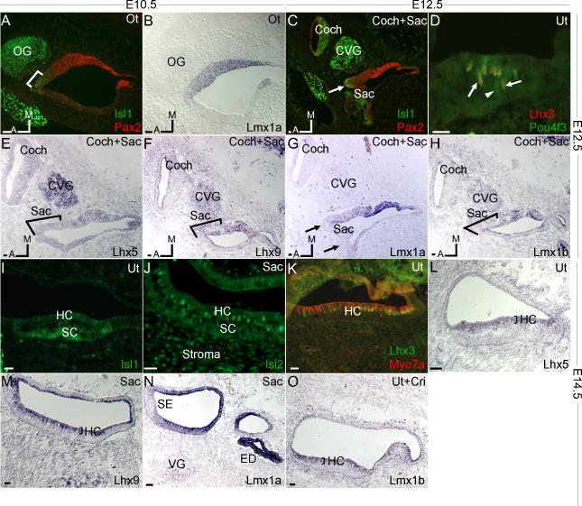
LIM-homeodomain transcription factors (LIM-HDs) are essential in tissue patterning and differentiation. But their expression patterns in the inner ear are largely unknown. Here we report on a study of twelve LIM-HDs, by their tempo-spatial patterns that imply distinct yet overlapping roles, in the developing mouse inner ear. Expression of Lmx1a and Isl1 begins in the otocyst stage, with Lmx1a exclusively in the non-sensory and Isl1 in the prosensory epithelia. The second wave of expression at E12.5 includes Lhx3, 5, 9, Isl2, and Lmx1b in the differentiating sensory epithelia with cellular specificities. With the exception of Lmx1a and Lhx3, all LIM-HDs are expressed in ganglion neurons. Expression of multiple LIM-HDs within a cell type suggests their redundant function.
Copyright (c) 2008 Wiley-Liss, Inc.
Figures


Similar articles
-
Expression of Islet1 marks the sensory and neuronal lineages in the mammalian inner ear.J Comp Neurol. 2004 Sep 27;477(4):412-21. doi: 10.1002/cne.20257. J Comp Neurol. 2004. PMID: 15329890 Free PMC article.
-
Comparative expression analysis of POU4F1, POU4F2 and ISL1 in developing mouse cochleovestibular ganglion neurons.Gene Expr Patterns. 2014 May;15(1):31-7. doi: 10.1016/j.gep.2014.03.001. Epub 2014 Apr 4. Gene Expr Patterns. 2014. PMID: 24709358 Free PMC article.
-
Islet-1 expression in the developing chicken inner ear.J Comp Neurol. 2004 Sep 6;477(1):1-10. doi: 10.1002/cne.20190. J Comp Neurol. 2004. PMID: 15281076
-
Cell fate determination, neuronal maintenance and disease state: The emerging role of transcription factors Lmx1a and Lmx1b.FEBS Lett. 2015 Dec 21;589(24 Pt A):3727-38. doi: 10.1016/j.febslet.2015.10.020. Epub 2015 Oct 23. FEBS Lett. 2015. PMID: 26526610 Review.
-
LIM Homeodomain (LIM-HD) Genes and Their Co-Regulators in Developing Reproductive System and Disorders of Sex Development.Sex Dev. 2022;16(2-3):147-161. doi: 10.1159/000518323. Epub 2021 Sep 10. Sex Dev. 2022. PMID: 34518474 Review.
Cited by
-
Deterioration of the Medial Olivocochlear Efferent System Accelerates Age-Related Hearing Loss in Pax2-Isl1 Transgenic Mice.Mol Neurobiol. 2016 May;53(4):2368-83. doi: 10.1007/s12035-015-9215-1. Epub 2015 May 20. Mol Neurobiol. 2016. PMID: 25990412
-
Early development of the cochlea of the common marmoset, a non-human primate model.Neural Dev. 2022 May 7;17(1):6. doi: 10.1186/s13064-022-00162-8. Neural Dev. 2022. PMID: 35524278 Free PMC article.
-
Pax2-Islet1 Transgenic Mice Are Hyperactive and Have Altered Cerebellar Foliation.Mol Neurobiol. 2017 Mar;54(2):1352-1368. doi: 10.1007/s12035-016-9716-6. Epub 2016 Feb 3. Mol Neurobiol. 2017. PMID: 26843111 Free PMC article.
-
Lmx1a is essential for marginal cell differentiation and stria vascularis formation.Front Cell Dev Biol. 2025 Mar 5;13:1537505. doi: 10.3389/fcell.2025.1537505. eCollection 2025. Front Cell Dev Biol. 2025. PMID: 40109362 Free PMC article.
-
Analysis of FGF20-regulated genes in organ of Corti progenitors by translating ribosome affinity purification.Dev Dyn. 2020 Oct;249(10):1217-1242. doi: 10.1002/dvdy.211. Epub 2020 Jul 10. Dev Dyn. 2020. PMID: 32492250 Free PMC article.
References
-
- Allan DW, Thor S. Together at last: bHLH and LIM-HD regulators cooperate to specify motor neurons. Neuron. 2003;38:675–677. - PubMed
-
- Andersson E, Tryggvason U, Deng Q, Friling S, Alekseenko Z, Robert B, Perlmann T, Ericson J. Identification of intrinsic determinants of midbrain dopamine neurons. Cell. 2006:393–405. - PubMed
-
- Barald KF, Kelley MW. From placode to polarization: new tunes in inner ear development. Development. 2004;131:4119–4130. - PubMed
-
- Bermingham NA, Hassan BA, Price SD, Vollrath MA, Ben-Arie N, Eatock RA, Bellen HJ, Lysakowski A, Zoghbi HY. Math1: an essential gene for the generation of inner ear hair cells. Science. 1999;284:1837–1841. - PubMed
-
- Bryant J, Goodyear RJ, Richardson GP. Sensory organ development in the inner ear: molecular and cellular mechanisms. Br Med Bull. 2002;63:39–57. - PubMed
Publication types
MeSH terms
Substances
Grants and funding
LinkOut - more resources
Full Text Sources
Other Literature Sources
Molecular Biology Databases
