. 2008 Nov;237(11):3115-27.
doi: 10.1002/dvdy.21762.

Signaling pathways regulating the expression of Prx1 and Prx2 in the chick mandibular mesenchyme

Affiliations

Signaling pathways regulating the expression of Prx1 and Prx2 in the chick mandibular mesenchyme

Aikaterini-El Doufexi et al. Dev Dyn. 2008 Nov.

Abstract

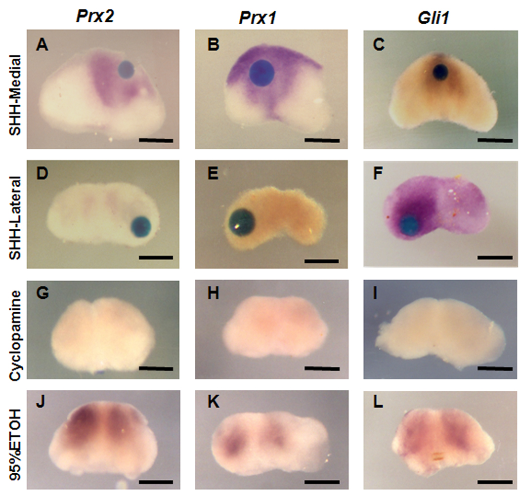
Prx1 and Prx2 are members of the aristaless-related homeobox genes shown to play redundant but essential roles in morphogenesis of the mandibular processes. To gain insight into the signaling pathways that regulate expression of Prx genes in the mandibular mesenchyme, we used the chick as a model system. We examined the patterns of gene expression in the face and the roles of signals derived from the epithelium on the expression of Prx genes in the mandibular mesenchyme. Our results demonstrated stage-dependent roles of mandibular epithelium on the expression of Prx in the mandibular mesenchyme and provide evidence for positive roles of members of the fibroblast and hedgehog families derived from mandibular epithelium on the expression of Prx genes in the mandibular mesenchyme. Our studies suggest that endothelin-1 signaling derived from the mesenchyme is involved in restricting the expression of Prx2 to the medial mandibular mesenchyme.


Figures

Figure 1
Figure 1. Prx expression in chick facial processes
Whole-mount in situ hybridization for Prx2 and Prx1 gene expression. Frontal views (A, C, E, G, I, K) and lateral views (B, D, F, H, J, L) of Prx2 (A–F) and Prx1 (G–L) expression in chick facial processes at E3 (A, B, G, H), E4 (C, D, I, J) and E5 (E, F, K, L). Note that at all stages Prx1 expression in the mandibular mesenchyme is extended into the lateral region as compared to Prx2. Note the higher levels of Prx2 expression in the frontonasal mass compared to Prx1 (indicated by arrows). Also note the higher levels of Prx1 expression in the 2nd and 3rd branchial arches compared to Prx2. Scale Bar: 250µm
Figure 2
Figure 2. The stage-dependent roles of mandibular epithelium on the expression of Prx in the mandibular mesenchyme
Whole-mount in situ hybridization for Prx2 (A–F) and Prx1 (G–K) in organ-cultured mandibular explants grown in the absence (A, C, E, G, I, K) and presence (B, D, F, H, J, L) of mandibular epithelium for 18 hrs. Removal of the epithelium at E3 resulted in complete loss of Prx2 (A) and Prx1 (G) expression compared to explants cultured in presence of mandibular epithelium (B and H). At E4, removal of the epithelium resulted in significant downregulation of Prx2 (C) and Prx1 (I) expression in medial mandibular mesenchyme compared to explants cultured with the overlying epithelium (D, J). Note the low but detectable expression of Prx2 (C) and Prx1 (I) in the mesenchyme around the midline in E4 cultures grown without the epithelium. At E5, removal of the epithelium did not affect the expression of Prx2 and Prx1 in the isolated mesenchyme grown without (E, K) and with epithelium (F, L). Scale Bar: 250µm
Figure 3
Figure 3. Roles of BMP signaling on the expression of Prx in mandibular mesenchyme
(A–I) Whole-mount in situ hybridization for Prx2 (A, D, G, J, M), Prx1 (B, E, H, K, N) and Msx1 (C, F, I, L, O) in organ-cultured mandible at E4 grown without epithelium and beads soaked in 0.3 mg/ml BMP7 (A–F) and 0.1% BSA (G–I) on the medial (A–C, H, I) and lateral (D–F, G, I) mesenchyme for 18 hrs. (A–C) Medially placed BMP7 beads were unable to restore expression of Prx2 (A) and Prx1 (B) in the medial mandibular mesenchyme. (C) Medially placed BMP7 bead restored the expression of Msx1 in the medial mesenchyme. (D–F) Laterally placed BMP7 did not induce ectopic expression of Prx2 (D), Prx1 (E) in the lateral mandibular mesenchyme. (F) Laterally placed BMP7 bead induced the ectopic expression of Msx1 in the lateral mesenchyme. (G–I) BSA beads placed on the lateral (G, I) or medial (H, I) mandibular mesenchyme did not induce/restore expression of Prx2 (G), Prx1 (H) and Msx1 (I) in the isolated mandibular mesenchyme. (J–O) Whole-mount in situ hybridization for Prx2 (J, M), Prx1 (K, N), and Msx1 (L, O) in organ-cultured mandible at E4 grown with epithelium and beads soaked in Noggin (J–L) and BSA (M–O) on the medial region for 18 hrs. Note the unchanged patterns of expression of Prx2 (J) and Prx1 (K) around the noggin beads as compared to the sides without the beads. (L) Note the lack of expression of Msx1 around the Noggin bead (indicated by dashed circle). Beads soaked in BSA did not affect the expression of Prx2 (M), Prx1 (N) and Msx1 (O) around the beads. Scale Bar: 250µm
Figure 4
Figure 4. Effects of beads soaked in FGF on the expression of Prx in the mandibular mesenchyme
Whole-mount in situ hybridization for Prx2 (A, D, G), Prx1 (B, E, H), and Barx1 (C, F, I) in organ-cultured mandibles at E4 grown without epithelium and beads soaked in 1 mg/ml of FGF8 (A–F) and 0.1% BSA (G–I) on the medial (A–C, H, J) and lateral (D–F, G) mesenchyme for 18 hrs. Medially placed FGF8 beads restored the expression of Prx2 (A), Prx1 (B) and induced ectopic expression of Barx1 (C) in the medial mandibular mesenchyme. Laterally placed FGF8 beads induced ectopic expression of Prx2 (D), Prx1 (E) and restored the expression of Barx1 (F) in the lateral mandibular mesenchyme. BSA beads did not restore expression of Prx2 (G), Prx1 (H) and Barx1 (I). Scale Bar: 250µm
Figure 5
Figure 5. Effects of inhibition of FGF signaling on the expression of Prx in the mandibular mesenchyme
Whole-mount in situ hybridization for Prx2 (A–D), Prx1 (E–H), and Barx1 (I–L) in organ-cultured mandible at E3 (A, C, E, G, I, K) and E4 (B, D, F, H, J, L) grown with epithelium and media containing 25 µM of Su5402 (A, B, E, F, I, J) and 0.04% DMSO (C, D, G, H, K, L) for 18 hrs. Note the lack of expression of Prx2 (A, B), and Prx1 (E, F) in explants grown in Su5402 at E3 and E4 as compared to explants grown in DMSO (C, D, G, H). Note that Su5402 inhibited Barx1 expression at E3 but not E4. Scale Bar: 250µm
Figure 6
Figure 6. Roles of Hh signaling on the expression of Prx in mandibular mesenchyme
(A–F) Whole-mount in situ hybridization for Prx2 (A, D, G, J), Prx1 (B, E, H, K), and Gli1 (C, F, I, L) in organ-cultured mandible at E4 grown without epithelium and with beads soaked in 1 mg/ml Shh placed in the medial (A–C) or lateral (D–F) mesenchyme for 18 hrs. (A–C) Medially placed Shh beads restored the expression of Prx2 (A), Prx1 (B) and Gli1 (C) in the medial mandibular mesenchyme. (D–F) Laterally placed Shh did not induce ectopic expression of Prx2 (D) and Prx1 (E) in the lateral mandibular mesenchyme. (F) Laterally placed Shh bead induced the ectopic expression of Gli1 in the lateral mesenchyme. (G–L) Whole-mount in situ hybridization for Prx2 (G, J), Prx1 (H, K), and Gli1 (I, L) in organ-cultured mandible at E4 grown with epithelium and media containing 10 µM of Cyclopamine (G–I) and vehicle (J–L) for 18 hrs. Note the absence of expression of Prx2 (G), Prx1 (K) and Gli1 (I) in cultures grown with Cyclopamine as compared to presence of Prx2 (J), Prx1 (H) and Gli1 (L) expression in cultures grown with vehicle. Scale Bar: 250µm.
Figure 7
Figure 7. Effects of ET-1 on the expression of Prx gene expression in mandibular mesenchymal explants
(A–F) Whole-mount in situ hybridization for Prx2 (A, D), Prx1 (B, E), and dHand (C, F) in organ-cultured mandibles at E4 grown without epithelium in media containing 0.3 µM of ET-1(A–C) or vehicle (D–F) for 18 hrs. Note the absence of Prx2 (A) and Prx1 (B) expression in explants grown in media containing ET-1 and water (D, E). Note the expression of dHand in cultures grown in media with ET-1 (C) and the absence of dHand expression in cultures grown with water (F). Scale Bar: 250µm
Figure 8
Figure 8. Effects of inhibition of ET-1 signaling on the expression of Prx in the mandibular mesenchyme
Whole-mount in situ hybridization for Prx2 (A–F), Prx1 (G–L), and dHand (M–R) in organ-cultured mandibles at E3 (A, D, G, J, M, P), E4 (B, E, H, K, N, Q) and E5 (C, F, I, L, O, R) grown with epithelium and media containing 10 µM of BQ123 (A–C, G–I, M–O) and water (D–F, J–L, P–R). Treatment of mandibular explants with BQ123 did not affect the expression of Prx2 (A, B, C) and Prx1 (G, H, I) at E3 (A, G), E4 (B, H) and E5 (C, I). Note the higher levels of Prx2 expression in explants treated with BQ123 (A) as compared to explants grown with water (D) at E3. Also note the higher and expanded domain of Prx2 expression in E4 cultures treated with BQ123 (B) compared to the explants cultured in control media (E). At E5 treatment with BQ123 did not affect the domain and level of expression of Prx2 (C) as compared to control (F). (G–I). Expression of Prx1 in explants grown in the presence of BQ123 at E3 (G), E4 (H), and E5 (I) is comparable to those grown in control media (J–L). (M) At E3 treatment of mandibular explants with BQ123 resulted in complete loss of dHand expression as compared to explants treated with water (P). At E4, in explants grown with BQ123 dHand expression is lost except for a small domain around the midline (N) as compared to control treatment (Q). At E5 treatment with BQ123 did not affect the expression of dHand (O) as compared to control treatment (R). Scale Bar: 250µm

References

    1. Abu-Issa R, Smyth G, Smoak I, Yamamura K, Meyers EN. Fgf8 is required for pharyngeal arch and cardiovascular development in the mouse. Development. 2002;129:4613–4625. - PubMed
    1. Barlow AJ, Bogardi JP, Ladher R, Francis-West PH. Expression of chick Barx-1 and its differential regulation by FGF-8 and BMP signaling in the maxillary primordia. Dev Dyn. 1999;214:291–302. - PubMed
    1. Barlow AJ, Francis-West PH. Ectopic application of recombinant BMP-2 and BMP-4 can change patterning of developing chick facial primordia. Development. 1997;124:391–398. - PubMed
    1. Brito JM, Teillet MA, Le Douarin NM. An early role for sonic hedgehog from foregut endoderm in jaw development: ensuring neural crest cell survival. Proc Natl Acad Sci U S A. 2006;103:11607–11612. - PMC - PubMed
    1. Buxton P, Davey MG, Paton IR, Morrice DR, Francis-West PH, Burt DW, Tickle C. Craniofacial development in the talpid3 chicken mutant. Differentiation. 2004;72:348–362. - PubMed

Publication types

LinkOut - more resources