Comparative Study
. 2009 Mar;15(3):341-52.
doi: 10.1002/ibd.20753.

Distinct cytokine patterns identified from multiplex profiles of murine DSS and TNBS-induced colitis

Affiliations
Comparative Study

Distinct cytokine patterns identified from multiplex profiles of murine DSS and TNBS-induced colitis

Philip Alex et al. Inflamm Bowel Dis. 2009 Mar.

Abstract

Background: The cytokine network in inflammatory bowel disease (IBD) is a complex, dynamic system that plays an important role in regulating mucosal innate and adaptive immune responses. While several studies have been done to evaluate immunomodulatory profiles in murine IBD, they have been limited to a relatively small number of cytokines that do not take into account its dependency of the interplay of multiple factors, and therefore the diagnostic potential of their cytokine profiles have been inconclusive.

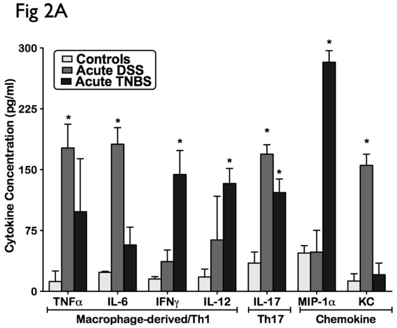
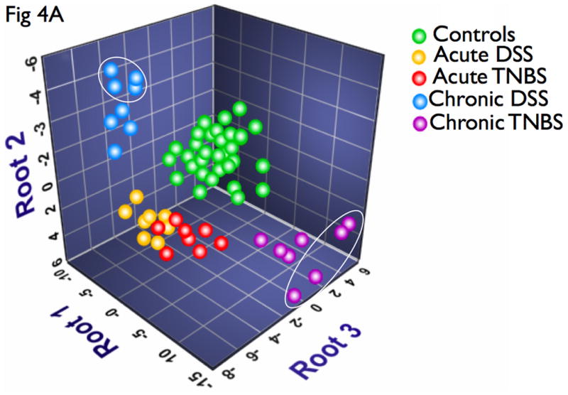
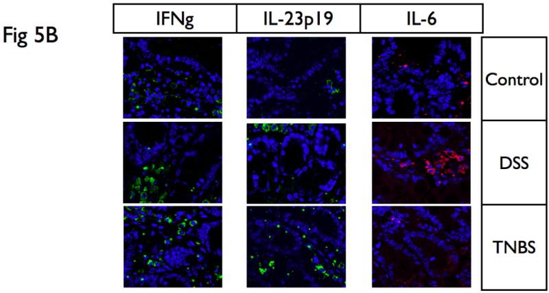
Methods: A novel approach of comprehensive serum multiplex cytokine profiling with biometric immunosandwich ELISA's was used to describe the modulation of 16 Th1, Th2, Th17 cytokines and chemokines in both acute and chronic murine models of DSS and TNBS-induced colitis. Advanced multivariate discriminant functional analyses (DFA) was used to identify statistically interrelated sets of variables with the most significant power to discriminate among the groups. Profiles of multiple cytokines seen systemically were also validated locally in colonic mucosa using Western blot analysis and fluorescent immunohistochemistry.

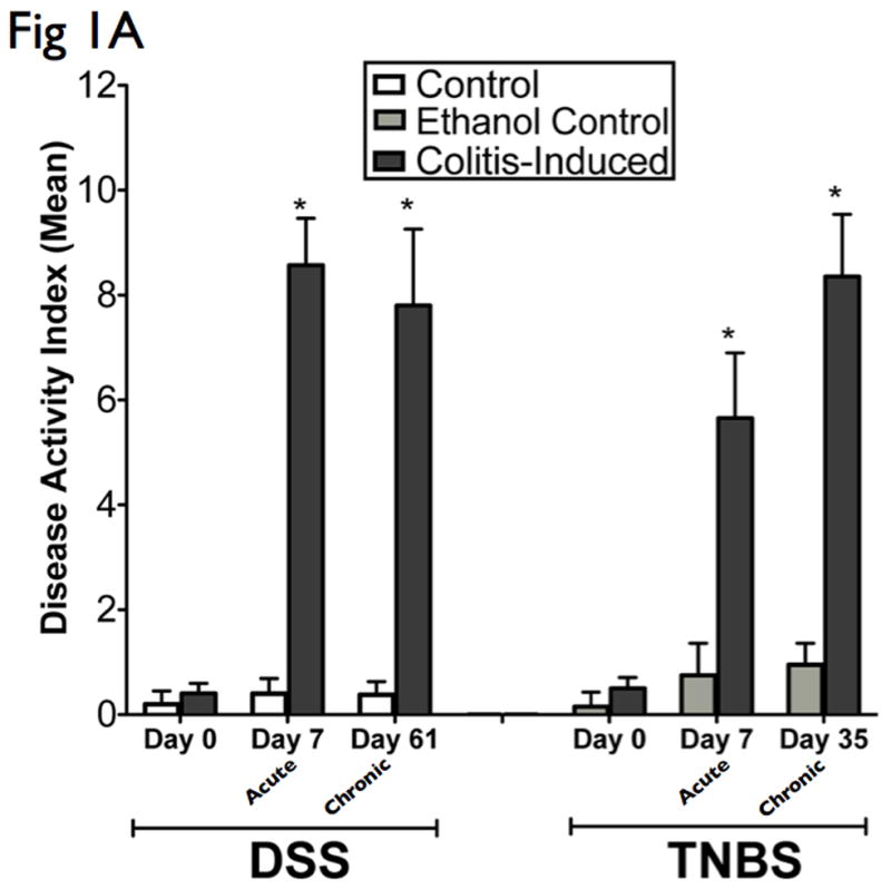
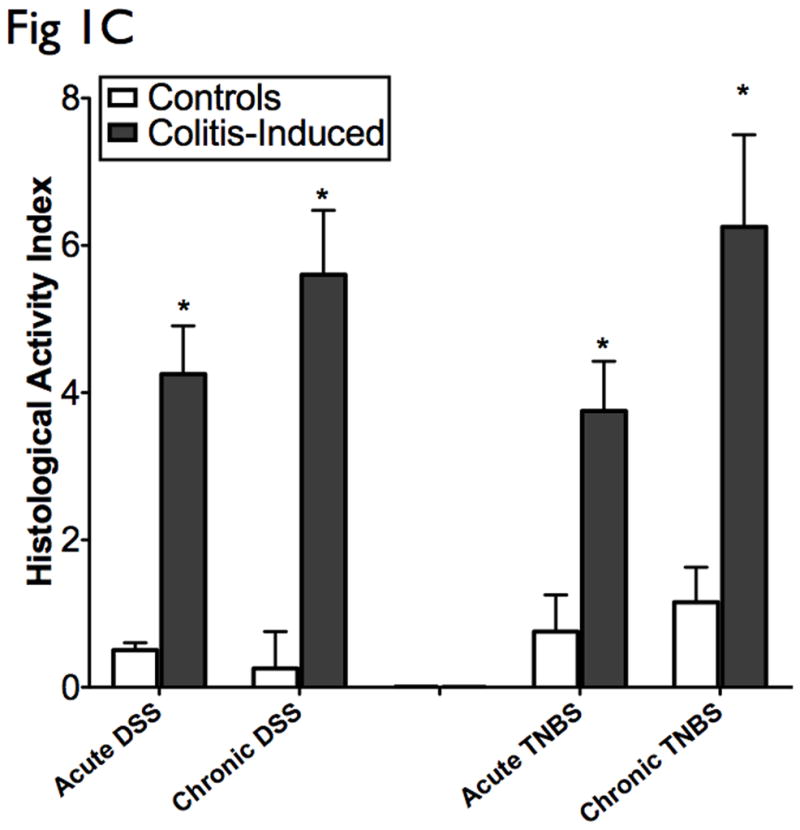
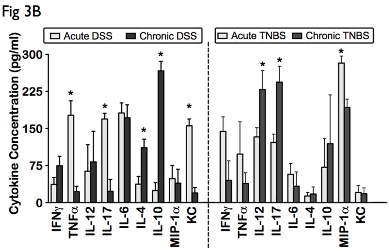
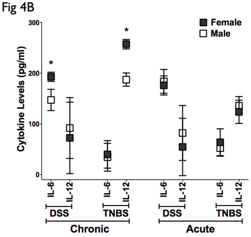
Results: Distinctive disease-specific cytokine profiles were identified with significant correlations to disease activity and duration of disease. TNBS colitis exhibits heightened Th1-Th17 response (increased IL-12 and IL-17) as the disease becomes chronic. In contrast, DSS colitis switches from a Th1-Th17-mediated acute inflammation (increased TNF-alpha, IL6, IL-17, and KC) to a predominant Th2-mediated inflammatory response (increase in IL-4 and IL-10 and concomitant decrease in TNF-alpha, IL6, IL-17, and KC) in the chronic state. Moreover, DFA identified discriminatory cytokine profiles that can be sufficiently used to distinguish unaffected controls from diseases, and one disease type from another. IL-6 and IL-12 stratified gender-associated disease activity in chronic colitis.

Conclusions: Our studies provide insight into disease immunopathogenesis and illustrate the significant potential of utilizing multiplex cytokine profiles and bioinformatics as diagnostic tools in IBD.


Figures

Figure 1
Figure 1. Administration of DSS and TNBS established acute and chronic experimental colitis
The results presented are representative of 8 mice from each group. (A) Clinical assessment of DSS and TNBS-induced colitis. DAI was scored from each mice for weight loss, stool consistency, and bleeding. (B) Histological analysis of acute and chronic DSS and TNBS colitis by H&E-stained colonic sections. (Structures: e, epithelial disruption; i, inflammatory infiltrate; m, lamina muscularis mucosae; s, submucosal edema; t, muscular thickening in lamina muscularis propria; magnification=20×). (C) For detailed histological analysis, colonic sections of each animal were scored in a blinded fashion as described in the Materials and Methods section. The resulting scores showed a significant increase in HAI after induction with colitis. (D) Myeloperoxidase activity in colonic mucosal scrapings were determined as described in the Materials and Methods section. Both acute and chronic DSS & TNBS had significant MPO activity compared with that in control animals. Mice induced with colitis demonstrated significant elevations in DAI, HAI and MPO, suggesting the effective establishment of acute and chronic experimental colitis.
Figure 2
Figure 2. Distinct cellular cytotoxic and chemotactic patterns identified in acute experimental colitis
Levels of 16 cytokines were measured simultaneously using a biometric multiplex assay from serum of mice induced with acute DSS and TNBS-induced colitis. (A) Cytokine pattern in acute colitis is represented by a macrophage-derived (TNFa, IL-6, IL-12 IFNg), strong chemotactic pattern (MIP-1a, KC), and a polarized Th1-Th17 panel (IL-17, TNFa, IFNg). (B) No significant differences were observed in levels of humoral cytokines, and other cellular cytokines and chemokines. This suggest that the cytokine profile of DSS and TNBS acute colitis is consistent with an acute inflammatory response.
Figure 3
Figure 3. Distinct differential immune response patterns identified in chronic experimental colitis
Levels of 16 cytokines were measured simultaneously using a biometric multiplex assay from serum of mice induced with chronic DSS and TNBS-induced colitis. (A) Cytokine profiles in chronic colitis is represented by an enhanced pro-humoral cytokine profile in chronic DSS (IL-6, IFN-γ, and IL-4, IL-10), and a distinct cytotoxic and chemotactic cytokine profile in chronic TNBS (IL-12, IL-17, and MIP-1α). (B) Acute inflammation in DSS colitis converts to a predominant Th2-mediated inflammatory response in the chronic state (lower levels of TNFα, IL-17 and KC, and elevated levels of IL-4, and IL-10), but chronic TNBS colitis is characterized by an enhanced Th1-Th17 immune response (higher IL-12 and IL-17 levels). (C) Levels of other cytokines, including chemokines did not change significantly between the models.
Figure 4
Figure 4. Multivariate analysis identifies novel subsets of discriminatory cytokines that can distinguish disease types and severity, and further identifiy disease-specific sex-stratification patterns
DFA was used to identify subset of cytokines whose expression values can be linearly combined in an equation, denoted a root, whose overall value is distinct for a given characterized group. (A) A graphical representation of the discriminatory potential of DFA. The variables identified by the DFA is plotted in three dimensions to visually represent the relative differences in cytokines among the distinct populations. Each ball represents an animal, and several clusters were mapped and had distinct relevance to the subgroups in context. One subgroup of the chronic DSS and TNBS colitis mapped further away from controls than the main group, marked by ovals, all of which were females. (B) Cytokine analysis of both the chronic DSS and TNBS colitis groups identified elevations in IL-6 and IL-12 in the females relative to males respectively, but not in acute colitis. There were no significant changes in CAI, HAI, and MPO between the genders (data not shown). These results suggest that the DFA is useful in evaluating relative disease activity, as more severe disease maps distal to unaffected controls.
Figure 5
Figure 5. Distinct cytokine profiles in tissues reflect and validate systemic cytokine levels
(A) Proteins were extracted from mucosa that was scraped from freshly excised colon, and samples were analyzed with SDS-PAGE Western-blots using primary antibodies for IL-6, IL-23p19, IL-12p40, IL-17, and IFNγ. While DSS colitis had significantly higher IL-6 protein expression in the colon than TNBS colitis, TNBS colitis had significantly higher IL-12p40, IL-23p19 and IL-17 protein expression in the colon than DSS colitis. In addition, acute TNBS colitis had higher IFNγ than acute DSS colitis. (B) Immunofluorescence analysis was performed on paraffin embedded tissue sections of DSS and TNBS acute colitis. IL-6 was significantly upregulated in DSS acute colitis, predominantly associated with lamina propria infiltrating mononuclear cells, while IFNγ and IL-23p19 were highly expressed in TNBS acute colitis. These suggest that cytokine profiles in tissues reflect that present within systemic levels. Actin was used as a loading control. The results presented are representative of 3 independent experiments.

References

    1. Podolsky DK. Inflammatory bowel disease. N Engl J Med. 2002;347:417–429. - PubMed
    1. Xavier RJ, Podolsky DK. Unravelling the pathogenesis of inflammatory bowel disease. Nature. 2007;448:427–434. - PubMed
    1. Cobrin GM, Abreu MT. Defects in mucosal immunity leading to Crohn’s disease. Immunol Rev. 2005;206:277–295. - PubMed
    1. Elson CO, Cong Y, McCracken VJ, Dimmitt RA, Lorenz RG, Weaver CT. Experimental models of inflammatory bowel disease reveal innate, adaptive, and regulatory mechanisms of host dialogue with the microbiota. Immunol Rev. 2005;206:260–276. - PubMed
    1. Pizarro TT, Cominelli F. Cytokine therapy for Crohn’s disease: advances in translational research. Annu Rev Med. 2007;58:433–444. - PubMed

Publication types

MeSH terms