. 2008 Dec 12;283(50):35042-52.
doi: 10.1074/jbc.M806603200. Epub 2008 Oct 22.

A pair of circularly permutated PDZ domains control RseP, the S2P family intramembrane protease of Escherichia coli

Affiliations

A pair of circularly permutated PDZ domains control RseP, the S2P family intramembrane protease of Escherichia coli

Kenji Inaba et al. J Biol Chem. .

Abstract

The sigma(E) pathway of extracytoplasmic stress responses in Escherichia coli is activated through sequential cleavages of the anti-sigma(E) protein, RseA, by membrane proteases DegS and RseP. Without the first cleavage by DegS, RseP is unable to cleave full-length RseA. We previously showed that a PDZ-like domain in the RseP periplasmic region is essential for this negative regulation of RseP. We now isolated additional deregulated RseP mutants. Many of the mutations affected a periplasmic region that is N-terminal to the previously defined PDZ domain. We expressed these regions and determined their crystal structures. Consistent with a recent prediction, our results indicate that RseP has tandem, circularly permutated PDZ domains (PDZ-N and PDZ-C). Strikingly, almost all the strong mutations have been mapped around the ligand binding cleft region in PDZ-N. These results together with those of an in vitro reaction reproducing the two-step RseA cleavage suggest that the proteolytic function of RseP is controlled by ligand binding to PDZ-N.


Figures

FIGURE 1.
FIGURE 1.
Sequence features, structures and mutations of the PDZ domains of RseP. A, schematic representation of the RseP primary sequence and positions of the mutations isolated in this study. Regions corresponding to the previously assigned PDZ-like domain (PDZ*, green arrow with dotted line) and the circularly permutated PDZ-N (red-boxed arrow) and PDZ-C (blue-boxed arrow) domains are shown at the top. Positions of the mutational alterations are shown by circles with the pYGF plasmid numbers; strong mutations (more than 2-fold induction of the reporter gene) are indicated by a red color. Regions carried by the ΔN1 and ΔN2 derivatives of RseP-HM are shown at the bottom. a.a. res. no., amino acid residue number. B-D, crystal structures of the PDZ-N and PDZ-C domains. The main chain structures of the PDZ-N (B) and PDZ-C (C) from RseP and that of the third PDZ domain of PSD95 (D) are shown by ribbon diagram representations (cyan, helices; magenta, strands; salmon, loops). The α-helix (αB) and β-strand (βB) elements thought to be important for ligand binding are marked. The carboxylate binding loop is highlighted in yellow. Side chains of amino acids showing strong mutational effects are shown by sticks in B. Note that Leu-169 was replaced with SeMet for structure determination of PDZ-N. A polypeptide ligand bound to the third PDZ-domain of PSD95 (40) is shown by a light-green stick in D. E, a typical rigid-body refinement model of PDZ-NC obtained by SAXS, which is superimposed onto the low resolution molecular shape of PDZ-NC. The envelope smoothed with SITUS package (46) was visualized on VMD (47).
FIGURE 2.
FIGURE 2.
DegS-independent proteolysis of RseA by the PDZ mutant forms of RseP-HM in vivo. A, amino acid alterations and the domains affected in the RseP-HM mutants isolated by the genetic screening. B, reporter LacZ activities induced by expression of the RseP PDZ mutants. Cells of TR71 (rpoHP3-lacZ) carrying the indicated pYGF plasmid, pKK11 (RseP-HM; WT), pKK131 (RseP(ΔPDZ*)-HM; ΔPDZ*), pKK135 (RseP(ΔPDZ*/D402N)-HM; ΔPDZ*/D402N), pSTD999 (RseP(L151P/H22F)-HM; L151P/H22F), or pTWV228 (vector) were grown at 37 °C in L medium (containing 10 g bacto-tryptone, 5 g yeast extract, and 5 g NaCl per liter; pH was adjusted to 7.2 by NaOH) and assayed for β-galactosidase activity. C, cellular levels of HA-RseA upon co-expression of the RseP PDZ mutants. pYGF plasmids, pKK11, pSTD999, and vector were introduced into AD1840/pSD691 (HA-RseA). Cells were grown at 30 °C in L medium containing 1 mm isopropyl 1-thio-β-d-galactopyranoside and 1 mm cAMP for 2 h. Portions (containing ∼6 × 10⁷ cells) of the cultures were withdrawn and mixed with an equal volume of 10% trichloroacetic acid for subsequent SDS-PAGE and anti-HA and anti-Myc immunoblotting analyses.
FIGURE 3.
FIGURE 3.
Proteolytic cleavage of RseA by RseP mutants. A and B, effects of mutations in the carboxylate binding loop region of PDZ-C. C and D, effects of PDZ-N and PDZ-C deletions. E and F, enhancement of the effects of PDZ-N mutations by the A115V alteration in TM2. A, C, and E, reporter LacZ activities. Derivatives of pKK11 encoding the indicated mutant forms of RseP-HM were introduced into TR71. Cells were grown at 37 °C in L medium and assayed for β-galactosidase activity. WT, wild type. B, D, and F, cellular levels of HA-RseA. Derivatives of pKK11 encoding the indicated mutant forms of RseP-HM were introduced into AD1840/pSTD691 (HA-RseA). Cells were grown at 30 °C in L medium containing 1 mm isopropyl 1-thio-β-d-galactopyranoside and 1 mm cAMP for 2 h. Proteins were analyzed by SDS-PAGE and anti-HA and anti-Myc immunoblotting. Average values from at least two independent experiments are shown with S.D.
FIGURE 4.
FIGURE 4.
Trypsin sensitivity of the wild type and PDZ-N and PDZ-C mutant forms of RseP-HM. A, trypsin digestion profiles of RseP-HM in spheroplasts. Cells of AD2249 carrying pKK11 (RseP-HM) or its derivatives (pSTD1002, pYGF13, and pSTD1131) having the indicated mutations were grown in L medium containing 1 mm isopropyl 1-thio-β-d-galactopyranoside and 1 mm cAMP for 2 h. Cells were then converted to spheroplasts by sucrose-lysozyme treatment and further treated with 5 μg/ml trypsin at 0 °C for the indicated time periods. Acid-denatured proteins were analyzed by SDS-PAGE and anti-Myc immunoblotting. B, trypsin digestion of the purified RseP-HM proteins. Purified preparations of RseP-HM and RseP(A115V/G214E)-HM were incubated with 5 μg/ml trypsin for the indicated time periods. After acid denaturation, proteins were analyzed by SDS-PAGE and CBB staining. C, mapping of the trypsin cleavage site. Trypsin-digested RseP(L151P)-HM (lane 1) and RseP(A115V/G214R)-HM (lane 5) (the same samples used for lane 14 of (A) and lane 9 of (B), respectively) were analyzed together with in vitro synthesized RseP-HM (lane 2), RseP(ΔN1)-HM (lane 3), and RseP(ΔN2)-HM (lane 4) by SDS-PAGE and anti-Myc immunoblotting. Open and closed arrowheads indicate intact RseP-HM and its ∼33 kDa tryptic fragment (TF). Circles indicate in vitro-synthesized RseP-HM and its N-terminal-truncated derivatives. Lane M was for molecular size markers.
FIGURE 5.
FIGURE 5.
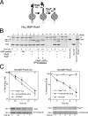
In vitro reproduction of the two-step cleavages of the model substrates by DegS and RseP. A, schematic representations of the His6-MBP-RseA and its cleavage by DegS and RseP. B, demonstration of the two-step substrate cleavages with purified components in detergent. His6-MBP-RseA was incubated with His6-DegS and RseP-HM as indicated. Lanes 15, 16, and 17 show the purified preparations of the indicated proteins individually. Proteins were analyzed by 12.5% SDS-PAGE and CBB staining. Note that the purified preparation of His6-MBP-RseA contained a minute amount of an in vivo generated degradation product (lane 9) with a size similar to the site-1-cleaved intermediate as well as to RseP-HM, and this contaminating fragment was converted to the site-2 cleavage product upon incubation with RseP in lane 14. C, effects of the PDZ mutation on the in vitro cleavage of the model substrates by RseP-HM. His6-MBP-RseA140 (5.8 μg/ml) (left panel) and His6-MBP-RseA (5 μg/ml) (right panel) were incubated with RseP-HM (13 μg/m/and 19 μg/ml for the reaction with the former and the latter substrates, respectively) and RseP (A115V/G214E)-HM (30 μg/m/and 43 μg/ml for the reaction with the former and the latter substrates, respectively). Samples were withdrawn at the indicated time points and analyzed by SDS-PAGE and CBB staining. For B and C, 140 and Full indicate His6-MBP-RseA140 and His6-MBP-RseA, respectively, whereas site-1 and site-2 indicate the DegS and the RseP cleavage products of the model substrates, respectively.

Similar articles

Cited by

References

    1. Raivio, T. L. (2005) Mol. Microbiol. 561119 -1128 - PubMed
    1. Rowley, G., Spector, M., Kormanec, J., and Roberts, M. (2006) Nat. Rev. Microbiol. 4 383-394 - PubMed
    1. Hayden, J. D., and Ades, S. E. (2008) PLoS ONE 3e1573 - PMC - PubMed
    1. Missiakas, D., Mayer, M. P., Lemaire, M., Georgopoulos, C., and Raina, S. (1997) Mol. Microbiol. 24 355-371 - PubMed
    1. De Las Peñas, A., Connolly, L., and Gross, C. A. (1997) Mol. Microbiol. 24 373-385 - PubMed

Publication types

MeSH terms

LinkOut - more resources