Antibody to aquaporin-4 in the long-term course of neuromyelitis optica
- PMID: 18945724
- PMCID: PMC2577801
- DOI: 10.1093/brain/awn240
Antibody to aquaporin-4 in the long-term course of neuromyelitis optica
Abstract
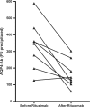
Neuromyelitis optica (NMO) is a severe inflammatory CNS disorder of putative autoimmune aetiology, which predominantly affects the spinal cord and optic nerves. Recently, a highly specific serum reactivity to CNS microvessels, subpia and Virchow-Robin spaces was described in patients with NMO [called NMO-IgG (NMO-immunoglobulin G)]. Subsequently, aquaporin-4 (AQP4), the most abundant water channel in the CNS, was identified as its target antigen. Strong support for a pathogenic role of the antibody would come from studies demonstrating a correlation between AQP4-Ab (AQP4-antibody) titres and the clinical course of disease. In this study, we determined AQP4-Ab serum levels in 96 samples from eight NMO-IgG positive patients (median follow-up 62 months) in a newly developed fluorescence-based immunoprecipitation assay employing recombinant human AQP4. We found that AQP4-Ab serum levels correlate with clinical disease activity, with relapses being preceded by an up to 3-fold increase in AQP4-Ab titres, which was not paralleled by a rise in other serum autoantibodies in one patient. Moreover, AQP4-Ab titres were found to correlate with CD19 cell counts during therapy with rituximab. Treatment with immunosuppressants such as rituximab, azathioprine and cyclophosphamide resulted in a marked reduction in antibody levels and relapse rates. Our results demonstrate a strong relationship between AQP4-Abs and clinical state, and support the hypothesis that these antibodies are involved in the pathogenesis of NMO.
Figures


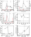
 = CD19 cell counts;
  = clinical relapse;
  = intravenous methylprednisolone;
  = prednisolone;
  = azathioprine;
  = dexamethasone;
  = rituximab;
  = cyclophosphamide;
  = mitoxantrone;
  = plasma exchange; eod = every other day.
 = CD19 cell counts;
  = clinical relapse;
  = intravenous methylprednisolone;
  = prednisolone;
  = azathioprine;
  = dexamethasone;
  = rituximab;
  = cyclophosphamide;
  = mitoxantrone;
  = plasma exchange; eod = every other day.
 = AChR-Ab;
  = TG-Ab;
  = TPO-Ab;
  = clinical relapse;
  = intravenous methylprednisolone;
  = prednisolone;
  = azathioprine;
  = cyclophosphamide;
  = mitoxantrone.
References
-
- Cree BA, Lamb S, Morgan K, Chen A, Waubant E, Genain C. An open label study of the effects of rituximab in neuromyelitis optica. Neurology. 2005;64:1270–2. - PubMed
-
- de Seze J, Stojkovic T, Ferriby D, Gauvrit JY, Montagne C, Mounier-Vehier F, et al. Devic's neuromyelitis optica: clinical, laboratory, MRI and outcome profile. J Neurol Sci. 2002;197:57–61. - PubMed
-
- Hinson SR, Pittock SJ, Lucchinetti CF, Roemer SF, Fryer JP, Kryzer TJ, et al. Pathogenic potential of IgG binding to water channel extracellular domain in neuromyelitis optica. Neurology. 2007;69:2221–31. - PubMed
-
- Jarius S, Franciotta D, Bergamaschi R, Wright H, Littleton E, Palace J, et al. NMO-IgG in the diagnosis of neuromyelitis optica. Neurology. 2007;68:1076–7. - PubMed
-
- Jarius S, Paul F, Franciotta D, Waters P, Zipp F, Hohlfeld R, et al. Mechanisms of disease: aquaporin-4 antibodies in neuromyelitis optica. Nat Clin Pract Neurol. 2008;4:202–14. - PubMed
Publication types
MeSH terms
Substances
LinkOut - more resources
Full Text Sources
Other Literature Sources

