. 2009 Jan;83(1):283-94.
doi: 10.1128/JVI.01865-08. Epub 2008 Oct 22.

Insertional gene activation by lentiviral and gammaretroviral vectors

Affiliations

Insertional gene activation by lentiviral and gammaretroviral vectors

Marieke Bokhoven et al. J Virol. 2009 Jan.

Abstract

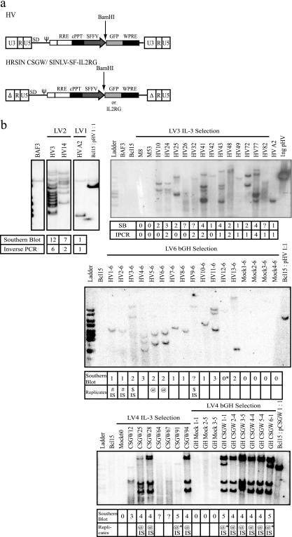
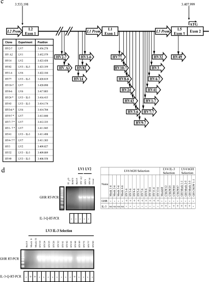
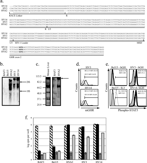
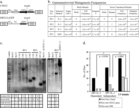
Gammaretroviral and lentiviral vectors are promising tools for gene therapy, but they can be oncogenic. The development of safer vectors depends on a quantitative assay for insertional mutagenesis. Here we report a rapid, inexpensive, and reproducible assay which uses a murine cell line to measure the frequency of interleukin-3 (IL-3)-independent mutants. Lentiviral and gammaretroviral vectors cause insertional mutagenesis at similar frequencies; however, they use different mechanisms. Human immunodeficiency virus (HIV)-based vectors generate mutants by insertion only into the growth hormone receptor (Ghr) locus. The HIV enhancer/promoter is active in the absence of the HIV Tat protein in this locus, and an HIV/Ghr spliced transcript expresses GHR and cells respond to GH. Deletion of the enhancer/promoter in a self-inactivating HIV-based vector prevents this mechanism of insertional mutagenesis. In contrast, gammaretroviral vectors insert into other loci, including IL-3 and genes identified as common insertion sites in the Retroviral Tagged Cancer Gene Database (RTCGD).


Figures

FIG. 1.
FIG. 1.
Characterization of lentiviral-vector insertional mutants. (a) HIV-1 derived lentiviral vectors used in insertional mutagenesis experiments shown as proviral genomes. The HV vector has WT HIV-1 LTRs and an internal SFFV LTR promoter driving GFP expression. The SIN lentiviral vectors HRSIN-CSGW and SINLV SFFV IL2RG (SINLV-SF-IL2RG) lack 400 bp of the U3 region of the HIV-1 LTRs; an internal SFFV promoter drives GFP or IL2RG expression. The location of the unique BamHI restriction site used in Southern blotting is also shown. (b) Southern blots (SB) of lentiviral insertional mutants from experiments LV1, LV2, LV3 (IL-3 selection), LV6, and LV4. Genomic DNA was digested with BamHI, and blots were probed with GFP. Phosphorscreen images are shown of all blots except that of LV3 (IL-3 selection), which was exposed to Kodak BioMax film. The first row of the table below each blot shows estimated vector copy numbers from the Southern blot. In experiments LV1, LV2, and LV3 (IL-3 selection), mutants were independent, as defined by different (or additional) bands on Southern blots, combined with integration site cloning. The number of integration sites cloned by inverse PCR from these mutants is shown in the table below the blots. Replicate clones were obtained in experiments LV4 and LV6; the final number of mutants shown in Table 1 does not include replicate clones. The replicates are identified by common symbols. IPCR, number of insertion site loci identified in the mutant by integration site PCR; IS, integration sites shared by these clones were verified by site-specific PCR; ?, vector copy number was not determined; *, mutant HV12-6 from LV6 was eliminated from further analysis: though it shows a faint band on Southern blot, no integrated vector copies were detected by qPCR. Ladder, 1-kbp DNA marker (Fermentas). (c) Locations of insertions into the Ghr allele on mouse chromosome 15. The 19 HV vector insertions that we mapped occurred in the same transcriptional orientation as Ghr and were in the 125-kb region upstream of exon 2, the first coding exon of Ghr. *, Ghr insertion was recovered by multiplex PCR spanning the region between L1 exon 1 and L5 promoter; all other Ghr insertions were obtained by inverse PCR. (d) RT-PCR for the Ghr transcript. A forward primer in exon 4 and a reverse primer in exon 8a were used to amplify a 552-bp section of the Ghr transcript. Representative gels are shown for mutants from experiments LV1, LV2, and LV3 (IL-3 selection). qRT-PCR to determine IL-3 transcript levels was also performed. A positive result (+) indicates >103 IL-3 copies per 109 18S rRNA copies; a negative result (−) indicates <101 IL-3 copies per 109 18S rRNA copies. The table summarizes the expression of these two transcripts by the LV6 and LV4 experiment mutants. ND, not done. Ladder, 100-bp DNA marker (Fermentas).
FIG. 1.
FIG. 1.
Characterization of lentiviral-vector insertional mutants. (a) HIV-1 derived lentiviral vectors used in insertional mutagenesis experiments shown as proviral genomes. The HV vector has WT HIV-1 LTRs and an internal SFFV LTR promoter driving GFP expression. The SIN lentiviral vectors HRSIN-CSGW and SINLV SFFV IL2RG (SINLV-SF-IL2RG) lack 400 bp of the U3 region of the HIV-1 LTRs; an internal SFFV promoter drives GFP or IL2RG expression. The location of the unique BamHI restriction site used in Southern blotting is also shown. (b) Southern blots (SB) of lentiviral insertional mutants from experiments LV1, LV2, LV3 (IL-3 selection), LV6, and LV4. Genomic DNA was digested with BamHI, and blots were probed with GFP. Phosphorscreen images are shown of all blots except that of LV3 (IL-3 selection), which was exposed to Kodak BioMax film. The first row of the table below each blot shows estimated vector copy numbers from the Southern blot. In experiments LV1, LV2, and LV3 (IL-3 selection), mutants were independent, as defined by different (or additional) bands on Southern blots, combined with integration site cloning. The number of integration sites cloned by inverse PCR from these mutants is shown in the table below the blots. Replicate clones were obtained in experiments LV4 and LV6; the final number of mutants shown in Table 1 does not include replicate clones. The replicates are identified by common symbols. IPCR, number of insertion site loci identified in the mutant by integration site PCR; IS, integration sites shared by these clones were verified by site-specific PCR; ?, vector copy number was not determined; *, mutant HV12-6 from LV6 was eliminated from further analysis: though it shows a faint band on Southern blot, no integrated vector copies were detected by qPCR. Ladder, 1-kbp DNA marker (Fermentas). (c) Locations of insertions into the Ghr allele on mouse chromosome 15. The 19 HV vector insertions that we mapped occurred in the same transcriptional orientation as Ghr and were in the 125-kb region upstream of exon 2, the first coding exon of Ghr. *, Ghr insertion was recovered by multiplex PCR spanning the region between L1 exon 1 and L5 promoter; all other Ghr insertions were obtained by inverse PCR. (d) RT-PCR for the Ghr transcript. A forward primer in exon 4 and a reverse primer in exon 8a were used to amplify a 552-bp section of the Ghr transcript. Representative gels are shown for mutants from experiments LV1, LV2, and LV3 (IL-3 selection). qRT-PCR to determine IL-3 transcript levels was also performed. A positive result (+) indicates >103 IL-3 copies per 109 18S rRNA copies; a negative result (−) indicates <101 IL-3 copies per 109 18S rRNA copies. The table summarizes the expression of these two transcripts by the LV6 and LV4 experiment mutants. ND, not done. Ladder, 100-bp DNA marker (Fermentas).
FIG. 2.
FIG. 2.
Structure of the Ghr transcript, GHR protein expression, and results of functional studies. (a) 5′ RACE was performed on mutants HV A2, HV3, and HV14. A 590-bp product was cloned and sequenced. An alignment of the first 414 bp of the 5′RACE product is shown here. The RACE linker cassette is 46 bp; the Ghr transcript starts at the HIV-1 R region. It contains 289 bp of HV vector sequence from before it splices from the HIV-1 major splice donor to the splice acceptor of Ghr exon 2. The ATG start codon for Ghr is shown in bold. (b) In mutants HV A2, HV3, and HV14, a 5-kb transcript was detected by Northern blotting using a probe that detects the intracellular domain of Ghr. Also shown are the sizes of mouse 28S (4.7 kb) and 18S (1.7 kb) rRNA. (c) A 103-kDa GHR protein was detected in mutant HV A2 and mouse liver, but not in the parental BAF3 cell. A smaller, ∼70kDa protein, the secreted GH binding protein, is also seen. Immunoblot analysis of total cell lysate was performed using an antibody that recognizes the mouse GHR extracellular domain, common to GHR and GH binding protein. Sizes in kDa are shown on the left. (d) Mutants HV3 and HV14 (black peak) express GHR on the cell surface in FACS, unlike the parental Bcl15 cell (solid gray peak), demonstrated by surface staining using an antibody that recognizes the mouse GHR (mGHR) extracellular domain. (e) Parental cells and mutants were stimulated with PBS-0.2% BSA (solid gray peak) or 400 ng/ml human GH (black peak) for FACS. Intracellular staining for phosphorylated STAT5 was performed. Stimulation with 100 ng/ml IL-3 (dashed black peak) also results in STAT5 phosphorylation and acted as a positive control. Selection method (bGH or IL-3) is indicated. MFI, mean fluorescence intensity. (f) Parental cells and mutants were cultured for 48 h in DMEM, 10% FCS, 10% WEHI-3B-cell-conditioned medium (hatched bars); DMEM, 10% FCS (solid black bars); serum-free medium (solid dark-gray bars); or serum-free medium supplemented with 1 μg/ml bGH (solid light-gray bars). Cell proliferation was measured by [3H]thymidine uptake. Error bars indicate standard errors of the means calculated from the results of two independent experiments.
FIG. 3.
FIG. 3.
Characterization of gammaretroviral-vector insertional mutants. (a) The gammaretroviral vectors CNCG and MFG.S eGFP are shown. The locations of the unique HindIII and BamHI restriction sites used in Southern blotting of CNCG and MFG.S eGFP insertional mutants, respectively, are also shown. (b) Gammaretroviral-mutagenesis frequencies. The CNCG vector was used in experiments RV1 and RV3; MFG.S eGFP was used in RV2. IL-3-independent mutants were selected either according to the IL-3 selection protocol (IL-3; RV1 to RV3) or in serum supplemented with 1 μg/ml bGH (bGH; RV3 only). The number of flasks (IL-3 selection) or wells (bGH selection) in which IL-3-independent clones grew out was scored after 7 to 10 days and 4 to 6 weeks, respectively (fourth and seventh columns). Observations from these four experiments using gammaretroviral vectors were pooled. Statistically significant differences between the numbers of wells (P = 0.0286) and mutants (P = 0.0286) in the mock- and vector-transduced groups were seen with a Mann-Whitney U test. The cell frequency was calculated by dividing the number of independent mutants by the total number of target cells in each experiment. The integrant frequency was calculated by dividing the number of independent mutants by the total number of integrants in each experiment. (c) Southern blots of gammaretroviral insertional mutants from experiments RV1 to RV3. Genomic DNA was digested with HindIII (RV1 and RV3) or BamHI (RV2). Blots were probed with the cDNA for enhanced GFP (eGFP). The tables below the blots show estimated vector copy numbers and the number of integration sites in each mutant that were cloned by inverse PCR. In addition, RT-PCR for the Ghr transcript and qRT-PCR for the IL-3 transcript were performed as described in the legend for Fig. 1d. Mutant C94 was eliminated from the analysis as no integrated vector copies were demonstrated by qPCR. (d) Comparison of genes targeted by vector integration in gammaretroviral-vector-transduced IL-3-independent mutants (γRV IL-3 independent); gammaretroviral-vector-transduced IL-3-dependent clones, cloned by limiting dilution (γRV IL-3 dependent); and WT LTR lentiviral-vector-transduced IL-3-independent mutants (LV mutants). Sites near CIS genes or the IL-3 gene are significantly more frequent targets of gammaretroviral-vector integration in RV experiment mutants than in RV experiment clones (two-tailed P value, 0.0186; Mann-Whitney U test). The Ghr gene is preferentially targeted by WT LTR lentiviral-vector integration but not gammaretroviral-vector integration in IL-3-independent mutants (two-tailed P value, 0.0061; Mann-Whitney U test).

Similar articles

Cited by

References

    1. Aiuti, A., S. Slavin, M. Aker, F. Ficara, S. Deola, A. Mortellaro, S. Morecki, G. Andolfi, A. Tabucchi, F. Carlucci, E. Marinello, F. Cattaneo, S. Vai, P. Servida, R. Miniero, M. G. Roncarolo, and C. Bordignon. 2002. Correction of ADA-SCID by stem cell gene therapy combined with nonmyeloablative conditioning. Science 2962410-2413. - PubMed
    1. Akagi, K., T. Suzuki, R. M. Stephens, N. A. Jenkins, and N. G. Copeland. 2004. RTCGD: retroviral tagged cancer gene database. Nucleic Acids Res. 32D523-D527. - PMC - PubMed
    1. Baixeras, E., S. Jeay, P. A. Kelly, and M. C. Postel-Vinay. 2001. The proliferative and antiapoptotic actions of growth hormone and insulin-like growth factor-1 are mediated through distinct signaling pathways in the Pro-B Ba/F3 cell line. Endocrinology 1422968-2977. - PubMed
    1. Blatt, C., D. Aberdam, R. Schwartz, and L. Sachs. 1988. DNA rearrangement of a homeobox gene in myeloid leukaemic cells. EMBO J. 74283-4290. - PMC - PubMed
    1. Carteau, S., C. Hoffmann, and F. Bushman. 1998. Chromosome structure and human immunodeficiency virus type 1 cDNA integration: centromeric alphoid repeats are a disfavored target. J. Virol. 724005-4014. - PMC - PubMed

Publication types