. 2008 Dec;14(12):2618-33.
doi: 10.1261/rna.1168408. Epub 2008 Oct 22.

Suppressors of the cdc-25.1(gf)-associated intestinal hyperplasia reveal important maternal roles for prp-8 and a subset of splicing factors in C. elegans

Affiliations

Suppressors of the cdc-25.1(gf)-associated intestinal hyperplasia reveal important maternal roles for prp-8 and a subset of splicing factors in C. elegans

Michaël Hebeisen et al. RNA. 2008 Dec.

Abstract

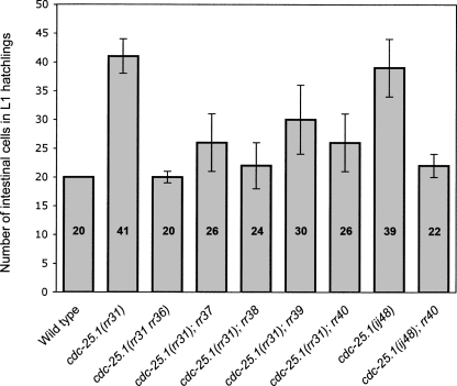
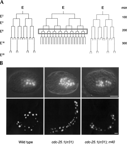
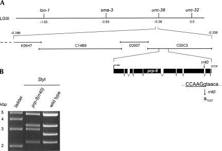
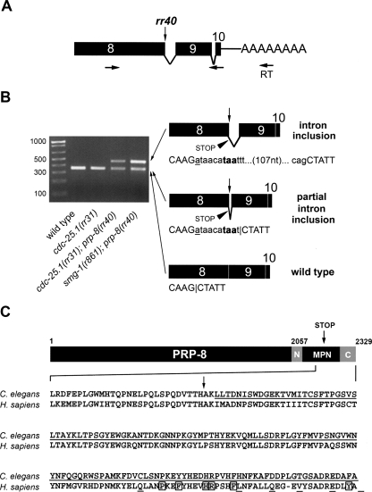
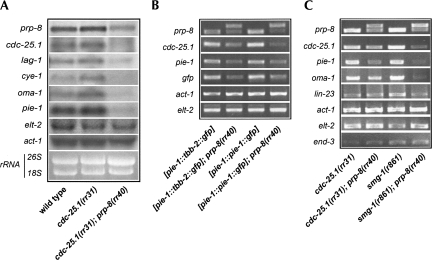
The maternal contribution of gene products enables embryos to initiate their developmental program in the absence of zygotic gene expression. In Caenorhabditis elegans, maternal CDC-25.1 levels are tightly regulated to promote early cell divisions, while stabilization of this phosphatase by gain-of-function mutations gives rise to intestinal-specific hyperplasia. To identify regulators of CDC-25.1 levels and/or function, we performed a modifier screen of the cdc-25.1(gf)-dependent hyperplasia. One of the isolated suppressor mutants possesses a donor splice site mutation in prp-8, a key splicing factor of the U5-specific snRNP. prp-8(rr40) produces aberrant prp-8 splice variants that generate C-terminal truncations at the expense of wild-type prp-8. Levels of maternal transcripts are reduced, including cdc-25.1, while zygotic transcripts appear unperturbed, suggesting a germ-line-specific role for this splicing factor in regulating the splicing, and consequently, the steady-state levels of maternal transcripts. Using a novel feeding RNAi strategy we found that only a subset of splicing factors suppress cdc-25.1(gf), suggesting that they too may play specific roles in germ-line spliceosome function. In humans, mutations in the corresponding hPrp8 C-terminal domain result in retinitis pigmentosa, a retinal-specific disorder. Intriguingly, despite affecting the general splicing apparatus, both human and C. elegans show tissue-specific defects resulting from mutations in this key splicing component. Our findings suggest that in addition to its important regulatory function in the C. elegans germ line, prp-8(rr40) may provide further insight into the etiology of this splicing-associated human disorder.


Figures

FIGURE 1.
FIGURE 1.
Isolation of five viable suppressors of the cdc-25.1(rr31)-associated intestinal hyperplasia. The average number of intestinal cells in L1 hatchings (sample size n > 50) is indicated for each genotype at 20°C. All animals express the intestinal-specific elt-2∷gfp marker that enables visualization and quantification of intestinal cells (Fukushige et al. 1999). Each of the suppressors (rr36rr40) reduces the cdc-25.1(rr31)-associated intestinal hyperplasia specifically during embryogenesis, with no post-embryonic effect. rr36 is the strongest suppressor and corresponds to an intragenic mutation in the cdc-25.1 gene (Hebeisen and Roy 2008). cdc-25.1(ij48) corresponds to a second gain-of-function allele of the phosphatase that produces intestinal-specific hyperplasia similar to cdc-25.1(rr31) (Clucas et al. 2002). ij48 and rr31 both affect residues of the highly conserved DSG(X)4S destruction motif. Error bars represent standard deviation.
FIGURE 2.
FIGURE 2.
The rr40 gene product is required from L3 to adulthood to produce and maintain a functional germ line. Reciprocal temperature-shift assays for cdc-25.1(rr31); rr40 suppressors demonstrate two major requirements for the rr40 gene product. Upshifts from 15°C (permissive) to 25°C (restrictive) are represented by black lines; downshifts from 25°C to 15°C by gray lines. (A) A first temperature-sensitive period is detected between the larval L3 and L4 stages, where rr40 is required to establish a functional gamete-producing germ line. (B) rr40 is also required continuously from the L3 stage until adulthood to maintain a normal brood size and to avoid the rapid and penetrant embryonic and early larval lethality detected in fertile adults that have been shifted at the restrictive temperature.
FIGURE 3.
FIGURE 3.
rr40 specifically suppresses the cdc-25.1(rr31)-associated supernumerary intestinal cell divisions. (A) A representative lineage map of the embryonic intestinal (E) lineage of cdc-25.1(rr31) mutants is shown alongside those of wild-type (left) and cdc-25.1(rr31); rr40 suppressors (right). E represents the founder endodermal cell specified at the embryonic 7-cell stage and the number of E descendants in wild type is indicated on the left. All animals express integrated end-3gfp and elt-2gfp transgenes, which together mark the intestinal nuclei from the embryonic E2 stage onward. The gray box highlights the supernumerary round of division that occurs during the E8 stage in cdc-25.1(rr31) and causes intestinal hyperplasia. Time scale is indicated on the right in minutes after fertilization. (B) Representative elt-2gfp expressing 300-min-old embryos (top) and L1 hatchlings (bottom) demonstrating the rr40-mediated suppression (right) of the cdc-25.1(rr31)-associated extra cell divisions (middle). Thirty-two percent of the cdc-25.1(rr31); rr40 suppressors were phenotypically indistinguishable from wild-type animals at 20°C (left). Scale bars, 10 μm.
FIGURE 4.
FIGURE 4.
Genetic mapping of rr40 indicates that it corresponds to an allele of the highly conserved U5-specific snRNP component prp-8. (A) Meiotic mapping in cdc-25.1(rr31) was used to place rr40 to the left of linkage group III between the visible markers unc-36 and sma-3. Feeding RNAi against the 26 genes present in this interval revealed that only C50C3.6(RNAi) phenocopied the rr40 suppression. Independent sequence analysis indicated that rr40 disrupts the second-last intron donor splice site of the prp-8 gene, converting the highly conserved GT donor sequence to AT and creating a StyI restriction polymorphism (underlined sequence). (B) StyI-digested PCR amplification products of wild-type and prp-8(rr40) genomic DNA confirm the G7337A mutation generated by rr40.
FIGURE 5.
FIGURE 5.
Differentially spliced prp-8 mRNA species are detected in prp-8(rr40) mutants. Total mRNA was isolated from wild-type, cdc-25.1(rr31), cdc-25.1(rr31); prp-8(rr40), and smg-1(r861); prp-8(rr40) mutants and reverse transcribed (RT) with polydT primers in a controlled, semiquantitative manner (Materials and Methods). (A) Primers used for PCR amplification were located in exon 8 (sense) and exon 10 (antisense) of prp-8. The rr40 mutation is indicated by a vertical arrow. (B) Species of cDNA of varying sizes were excised from 2% agarose gels and subjected to sequence analysis (left). The sequences indicated that animals mutant for prp-8(rr40) possess wild-type and at least two aberrant forms of prp-8 mRNA, while only the wild-type prp-8 sequence was detected in both wild-type and cdc-25.1(rr31) animals. Schematic representations of the three mRNA species obtained from the prp-8(rr40) mutants are depicted on the right. Mature mRNA containing full or partial intron inclusions encode premature stop codons (arrowhead) followed by a frameshift. Uppercase letters correspond to exon sequence, lowercase letters represent intron 8. The rr40 G7337A substitution (vertical arrow) is underlined in the sequences. The nucleotides highlighted in bold represent the first stop codon encoded in the aberrant prp-8 mRNAs and the horizontal bars indicate the detected splice sites. The intron between exons 9 and 10 was correctly spliced in all mRNA species. Note that compromising the NMD pathway increases the level of aberrant prp-8(rr40) mRNA. (C) The translation of both aberrant prp-8(rr40) mRNAs is predicted to result in a truncated PRP-8(rr40) protein that lacks the C-terminal-most 138 amino acids (underlined C. elegans sequence after the STOP [vertical arrow]). The crystal structure of the peptide encompassing the N-terminal extension (N), the MPN domain and the C-terminal extension (C) was resolved for C. elegans PRP-8 (Zhang et al. 2007). The rr40-mediated truncation removes half of the PRP-8 MPN interacting domain and the entire C-terminal extension. The corresponding, highly conserved human hPrp8 sequence is represented underneath, with all hPrp8 mutations associated with human autosomal dominant retinitis pigmentosa RP13 (boxed and underlined residues represent substitution and frameshift mutations, respectively).
FIGURE 6.
FIGURE 6.
prp-8(rr40) reduces the levels of prp-8 and other maternal mRNA transcripts, without affecting zygotic transcripts. (A) Quantitative Northern analysis of the indicated mRNA performed on wild-type, cdc-25.1(rr31), and cdc-25.1(rr31); prp-8(rr40) animals. Wild-type and cdc-25.1(rr31) control animals show little variation in their mRNA levels, while prp-8 levels (top) and the levels of five other maternally provided transcripts (cdc-25.1, lag-1, cye-1, oma-1, pie-1) are substantially reduced in cdc-25.1(rr31); prp-8(rr40) suppressors compared with the rRNA loading controls and the zygotically transcribed actin (act-1) and endodermal-specific GATA factor (elt-2) genes. (B) Representative semiquantitative RT–PCRs performed on AZ244 and JH1327 animals (first and third column, respectively; see Materials and Methods for complete genotypes). These transgenic animals possess different full-length genomic, intron-containing GFP constructs exclusively in their germ line (pie-1 promoter). Both the endogenous and transgenic maternal mRNA levels are reduced in a prp-8(rr40) mutant background (second and last column, respectively), while the zygotic act-1 and elt-2 messages seem unaffected. (C) The prp-8(rr40)-associated reduction in maternal mRNA levels (cdc-25.1, pie-1, and oma-1) does not depend on the NMD of unspliced pre-mRNAs, as single RT–PCR amplicons corresponding to correctly spliced mRNAs are produced in the smg-1(r861); prp-8(rr40) double mutant, with the exception of prp-8(rr40). Every primer set encompasses one or multiple genomic introns. Representative semiquantitative RT–PCR products indicate that compromise of the NMD pathway modestly decreases the levels of the maternal mRNAs, while the levels of zygotically expressed transcripts (act-1, elt-2, end-3, and lin-23) are more stable. Note that lin-23 is contributed both maternally and zygotically.

Similar articles

Cited by

References

    1. Alphey L., Jimenez J., White-Cooper H., Dawson I., Nurse P., Glover D.M. twine, a cdc25 homolog that functions in the male and female germ line of Drosophila . Cell. 1992;69:977–988. - PubMed
    1. Aroian R.V., Levy A.D., Koga M., Ohshima Y., Kramer J.M., Sternberg P.W. Splicing in Caenorhabditis elegans does not require an AG at the 3′ splice acceptor site. Mol. Cell. Biol. 1993;13:626–637. - PMC - PubMed
    1. Ashcroft N., Golden A. CDC-25.1 regulates germ line proliferation in Caenorhabditis elegans . Genesis. 2002;33:1–7. - PubMed
    1. Ashcroft N.R., Kosinski M.E., Wickramasinghe D., Donovan P.J., Golden A. The four cdc25 genes from the nematode Caenorhabditis elegans . Gene. 1998;214:59–66. - PubMed
    1. Bao Z., Zhao Z., Boyle T.J., Murray J.I., Waterston R.H. Control of cell cycle timing during C. elegans embryogenesis. Dev. Biol. 2008;318:65–72. - PMC - PubMed

Publication types

MeSH terms