Review
. 2009 Jan;75(1):1-12.
doi: 10.1124/mol.108.051938. Epub 2008 Oct 22.

Topology of class A G protein-coupled receptors: insights gained from crystal structures of rhodopsins, adrenergic and adenosine receptors

Affiliations
Review

Topology of class A G protein-coupled receptors: insights gained from crystal structures of rhodopsins, adrenergic and adenosine receptors

Debarshi Mustafi et al. Mol Pharmacol. 2009 Jan.

Abstract

Biological membranes are densely packed with membrane proteins that occupy approximately half of their volume. In almost all cases, membrane proteins in the native state lack the higher-order symmetry required for their direct study by diffraction methods. Despite many technical difficulties, numerous crystal structures of detergent solubilized membrane proteins have been determined that illustrate their internal organization. Among such proteins, class A G protein-coupled receptors have become amenable to crystallization and high resolution X-ray diffraction analyses. The derived structures of native and engineered receptors not only provide insights into their molecular arrangements but also furnish a framework for designing and testing potential models of transformation from inactive to active receptor signaling states and for initiating rational drug design.


Figures

Fig. 1.
Fig. 1.
Structural transformations of rhodopsin during photolysis. Alignment of the first three photointermediates, bovine rhodopsin colored by helix: helix I (blue), helix II (cyan), helix III (violet), helix IV (red), helix V (orange), helix VI (yellow), helix VII (green), and helix 8 (magenta); bovine bathorhodopsin (magenta); and bovine lumirhodopsin (green) reveals that the most pronounced structural changes occur in the chromophore region (dashed rectangle). The chromophore from each of the three structures is shown on the right to highlight the changes as photolysis proceeds. Retinal is colored red for ground state rhodopsin, magenta for bathorhodopsin, and green for lumirhodopsin. From the ground state to bathorhodopsin, the C11 = C12 bond adopts a trans configuration after illumination. From bathorhodopsin to lumirhodopsin, a conformational change in the β-ionone ring of the retinal is apparent. Changes in MII and opsin are more pronounced, with opsin showing slight side chain shifts of helices surrounding the chromophore binding site that allow retinal entry and exit as illustrated in Fig. 4.
Fig. 2.
Fig. 2.
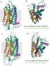
Comparison of rhodopsin with the photoactivated state resembling the MII structure and squid rhodopsin. A, side view. Molecules are colored as the bovine photoactivated structure (green ribbon) and bovine rhodopsin by helix I (blue), helix II (cyan), helix III (violet), helix IV (red), helix V (orange), helix VI (yellow), helix VII (green), and helix 8 (magenta). These structures revealed smaller conformational changes than predicted from EPR studies. B, cytoplasmic view. Although the photoactivated structure resembling MII is structurally similar to that of the ground-state rhodopsin, portions of the C-II and C-III loops are disordered in the photoactive state. The C-III loop does not even follow the path of the loop found in the ground-state crystals. This difference may reflect the importance of these loops' dynamics in transducin activation. C, side view of squid rhodopsin is shown as a green ribbon overlying bovine rhodopsin colored by helix. D, these two structures show immense structural homology, with the most noticeable difference being the larger C-III loop in squid rhodopsin as a result of an extra sequence in that region. The difference in the C-III loop, involved in the binding of transducin, suggests that this may be an important structural motif for specifying different modes of coupling with Gq-type G proteins in invertebrate rhodopsins.
Fig. 3.
Fig. 3.
Comparison of rhodopsin and opsin structures. A, side view. Molecules colored as bovine opsin II (green ribbon) and bovine rhodopsin by helix: helix I (blue), helix II (cyan), helix III (violet), helix IV (red), helix V (orange), helix VI (yellow), helix VII (green), and helix 8 (magenta). Despite overall helical plasticity, the structural overlay illustrates slight shifts in the side chains that could explain retinal entry/exit (see Fig. 4). B, cytoplasmic view. The opsin and rhodopsin structures show similar tracings of the cytoplasmic loops, but slight differences may indicate the importance of these loop movements in activation or relaxation back to the ground state.
Fig. 4.
Fig. 4.
Structural comparison between opsin and rhodopsin illustrating a pathway for retinal exchange. The opsin structure shows two different openings to the retinal-binding pocket (i.e., one between the extracellular ends of helice V and VI and the other between helices I and VII. These openings suggest different chromophore entrance and exit routes. Both bovine opsin and bovine rhodopsin are colored by helix: helix I (blue), helix II (cyan), helix III (violet), helix IV (red), helix V (orange), helix VI (yellow), helix VII (green), and helix 8 (magenta), whereas retinal is depicted as red sticks. A, use of the online software Caver shows a tunneling path of retinal through an entry site between helix V and helix VI of opsin, with three key Phe residues lining the path (Phe208, Phe212, and Phe273). B, this retinal entry path is blocked in rhodopsin as a result of shifts in helices V and VI that cause repositioning of these three key Phe residues. C, a distinct pathway for retinal exit from opsin is formed between helices I and VII and is illustrated as a clear hole in the solvent accessible surface. D, similar to the retinal entry pathway, the exit pathway is closed off in rhodopsin, most notably by a shift in Phe293, as evidenced by the closed solvent accessible surface.
Fig. 5.
Fig. 5.
Comparison of β1-adrenergic receptor with rhodopsin, β2-adrenergic receptor, and A2A adenosine receptor structures. The turkey β1-adrenergic receptor was mutated to facilitate its crystallization. The mutated receptor evidenced enhanced thermostability and preferentially existed in an antagonist-binding state. Stretches of amino acid sequence were deleted from the N-terminal region (i.e., the loop connecting helices V and VI and the C-terminal region). A, the β1-adrenergic receptor is colored by helix: helix I (blue), helix II (cyan), helix III (violet), helix IV (red), helix V (orange), helix VI (yellow), helix VII (green), and helix VIII (magenta). Thermostabilizing mutations (R68S, M90V, Y227A, A282L (not resolved in the shown structure), F327A, and F338M) and those that either increased functional expression (C116L) or eliminated a palmitoylation site (C358A) are shown as balls/sticks. B, structure of the β1-adrenergic receptor (colored by helix) is shown with bovine rhodopsin (green ribbon) to illustrate key structural differences. Despite some differences in the transmembrane helices, the main distinction is in the organization of the cytoplasmic (C-) loops. The C-II loop in the β1-adrenergic receptor structure forms a short α-helix, whereas this loop is more extended in rhodopsin. In addition, rhodopsin has a native C-III loop that is absent from the β1-adrenergic receptor structure as a result of deletions needed for crystallization. C, the structure of the β1-adrenergic receptor (colored by helix) is displayed with the human β2-adrenergic receptor (green ribbon) to show the key structural differences. In this case, the C-II loops of both the β1- and β2-adrenergic receptors are similar, but the α-helical conformation of the β1-adrenergic receptor cannot be accommodated within the two crystallized structures of the β2-adrenergic receptor because of lattice contacts with adjacent molecules. D, the structure of the β1-adrenergic receptor (colored by helix) is displayed with the human A2A adenosine receptor (green ribbon) to show key structural differences. The main difference between these two structures lies in the distinct folding of the extracellular loops, with the adenosine receptor adopting a more open surface for ligand binding as a result of this folding.
Fig. 6.
Fig. 6.
Structural comparison of the β2-adrenergic and A2A adenosine fusion receptors with rhodopsin. Shown are structures of the human β2-adrenergic receptor fused to T4 lysozyme (A) and in complex with Fab (B) and the human A2A adenosine receptor fused to T4 lysozyme (C). Bovine rhodopsin is colored by helix: helix I (blue), helix II (cyan), helix III (violet), helix IV (red), helix V (orange), helix VI (yellow), helix VII (green), and helix 8 (magenta), whereas each of the two fusion β2-adrenergic receptors are displayed as green ribbons. There are differences in the transmembrane helices between the β2-adrenergic receptor and rhodopsin, with the most pronounced disparity occurring in helix I because the Pro residue kink is absent in the β2-adrenergic receptor structures. In addition, the β2-adrenergic receptor structures show minimal inter-receptor contacts illustrating that protein engineering may have affected the crystal packing and disrupted any possible dimeric interfaces of these structures. The overall helical differences between the adenosine receptor and rhodopsin are minimal, but the adenosine receptor has a distinct arrangement of the extracellular loops for a more open structure of the ligand binding pocket that is shifted closer to helices VI and VII.

References

    1. Becker OM, Shacham S, Marantz Y, and Noiman S (2003) Modeling the 3D structure of GPCRs: advances and application to drug discovery. Curr Opin Drug Discov Devel 6 353-361. - PubMed
    1. Buczylko J, Saari JC, Crouch RK, and Palczewski K (1996) Mechanisms of opsin activation. J Biol Chem 271 20621-20630. - PubMed
    1. Chabre M, Cone R, and Saibil H (2003) Biophysics: is rhodopsin dimeric in native retinal rods? Nature 426 30-31. - PubMed
    1. Cherezov V, Rosenbaum DM, Hanson MA, Rasmussen SG, Thian FS, Kobilka TS, Choi HJ, Kuhn P, Weis WI, Kobilka BK, et al. (2007) High-resolution crystal structure of an engineered human beta(2)-adrenergic G protein-coupled receptor. Science 318 1258-1265. - PMC - PubMed
    1. Civelli O (2005) GPCR deorphanizations: the novel, the known and the unexpected transmitters. Trends Pharmacol Sci 26 15-19. - PubMed

Publication types

LinkOut - more resources