. 2009 Feb;23(2):523-33.
doi: 10.1096/fj.08-113191. Epub 2008 Oct 22.

Circadian clock and output genes are rhythmically expressed in extratesticular ducts and accessory organs of mice

Affiliations

Circadian clock and output genes are rhythmically expressed in extratesticular ducts and accessory organs of mice

Piotr Bebas et al. FASEB J. 2009 Feb.

Abstract

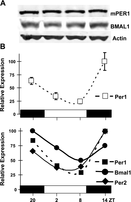
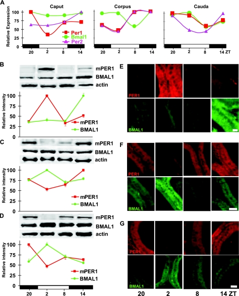
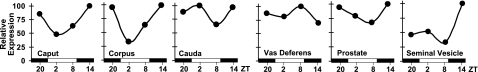
Circadian clocks regulate multiple rhythms in mammalian tissues. In most organs core clock gene expression is oscillatory, with negative components Per and Cry peaking in antiphase to Bmal1. A notable exception is the testis, where clock genes seem nonrhythmic. Earlier mammalian studies, however, did not examine clock expression patterns in accessory ductal tissue required for sperm maturation and transport. Previous studies in insects demonstrated control of sperm maturation in vas deferens by a local circadian system. Sperm ducts express clock genes and display circadian pH changes controlled by vacuolar-type H(+)-ATPase and carbonic anhydrase (CA-II). It is unknown whether sperm-processing rhythms are conserved beyond insects. To address this question in mice housed in a light-dark environment, we examined temporal patterns of mPer1 and Bmal1 gene expression and protein abundance in epididymis, vas deferens, seminal vesicles, and prostate. Results demonstrate variable tissue-specific patterns of expression of the two genes, with variations in levels of clock proteins and their nucleo-cytoplasmic cycling observed among examined tissues. Strikingly, mPer1 and Bmal1 mRNA and proteins oscillate in antiphase in the prostate, with similar peak-trough patterns as observed in the suprachiasmatic nuclei, the brain's central clock. Genes encoding CA and a V-ATPase subunit, which are rhythmically expressed in sperm ducts of moths, are also rhythmic in some segments of murine sperm ducts. Our data suggest that some sperm duct segments may contain peripheral circadian systems whereas others may express clock genes in a pleiotropic manner.


Figures

Figure 1.
Figure 1.
Core clock gene expression, but not protein abundance, is rhythmic in homogenized mouse testis. A) Representative Western blot demonstrating levels of mPER1, BMAL1, and β-actin in testis homogenates, collected at 6 h intervals, of mice entrained to 12:12 LD. B) Top panel: mean ± se relative expression levels of mPer1 transcript (open squares), derived from testes of individual mice (n=4) at each time point, as determined by real-time qPCR. Expression levels are determined by the ΔΔCt method; each time point is normalized to reference gene Ppia and compared to ZT20. Highest relative expression level is set to 100. Horizontal bars represent day (open) and night (filled), with ZT indicated below. Bottom panel: relative expression values of mPer1 (filled squares), mPer2 (filled diamonds), and Bmal1 (filled circles) at ZT20, ZT2, ZT8, and ZT14, derived from pooled testis RNA samples. Data shown are efficiency-corrected, normalized, relative gene-expression values calculated from the mean of reaction triplicates; the highest relative expression level is set to 100. Of note is the high similarity between the means of individual mPer1 expression levels in the top panel compared to mPer1 expression levels derived from pooled testis homogenates in the bottom panel.
Figure 2.
Figure 2.
Daily changes in clock gene expression and protein abundance in epididymis. Male mice entrained to 12:12 LD were sacrificed every 6 h at ZT20, ZT2, ZT8, ZT14; for every type of analysis in each time point, organs from 4–5 individuals were collected. A) Graphs represent expression levels, determined by real-time qPCR, of 3 genes: mPer1 (red line and squares), mPer2 (purple line and triangles), and Bmal1 (green line and circles) in epididymal caput, corpus, and cauda, respectively. Efficiency-corrected, normalized, relative gene-expression values for the mean of reaction triplicates are shown at each time point. Horizontal bars represent day (open) and night (filled), with ZT indicated below. BD) Representative Western blots of mPER1, BMAL1, and β-actin in caput (B), corpus (C), and cauda (D). Graphs below blots represent densitometric analysis of protein bands. Clock protein/β-actin ratios were calculated and normalized to a percentage of darkest band intensity; values are averages ± sem from 3 Western blots. E–G) Representative immunohistochemical staining for mPER1 and BMAL1 in epithelia of epididymal tubules of caput (E), corpus (F), and cauda (G). A minimum of 20 images was analyzed per time point in each mouse tissue; average staining intensity is shown in Supplemental Fig. 2. Scale bars = 20 μm.
Figure 3.
Figure 3.
Variable temporal expression of clock genes and proteins in vas deferens, prostate, and seminal vesicles. Clock gene and protein expression profiles are shown analogous to Fig. 2 (compare for marking and description details), with all organs dissected from the same mice as above. A) Daily changes in the levels of mPer1, mPer2, Bmal1 mRNAs in the tissues of whole vas deferens, prostate, and seminal vesicles, respectively, based on real-time qPCR analysis. B–D) Western blots of mPER1, BMAL1, and β-actin (control) in the whole vas deferens (B), prostate (C), and seminal vesicles (D), with corresponding densitometric analysis of protein bands staining intensity shown below each Western blot. E–G) Immunohistochemical identification of mPER1 and BMAL1 in epithelia of vas deferens (E), dorso-lateral prostate (F), and seminal vesicles (G). Scale bars = 20 μm. For densitometrical analysis, see Supplemental Fig. 2.
Figure 4.
Figure 4.
Carbonic anhydrase II gene and protein expression patterns in specific reproductive ducts and accessory glands. Car2 gene expression in (from top to bottom) caput, corpus, and cauda epididymis, vas deferens, prostate, and seminal vesicles, as revealed by real-time qPCR analysis (left panels) and corresponding immunohistochemical identification of CA-II protein (right panels). Real-time qPCR data shown are efficiency corrected, normalized, relative gene-expression values for the mean of reaction triplicates. Horizontal bars represent day (open) and night (filled), with ZT indicated below. For immunohistochemistry, CA-II protein (green) is costained with DNA-intercalating dye Hoechst 33258 (blue). Scale bars = 20 μm. For quantitative analysis of CA-II positive cells for each time point and organ, see Supplemental Fig. 3.
Figure 5.
Figure 5.
Expression patterns of V-ATPase subunit B2 isoform in reproductive ducts and accessory glands. Real-time qPCR results show gene expression profiles for the B2 subunit of vacuolar-type proton pump V-ATPase (Atp6v1b2) across a 24-h period in 3 parts of epididymis (caput, corpus, cauda), vas deferens, prostate, and seminal vesicles. Data are efficiency corrected, normalized, relative gene-expression values for the mean of reaction triplicates. Total RNAs were obtained from the same individuals sacrificed for investigating clock gene and Car2 expression (see Figs. 234). Horizontal bars represent day (open) and night (filled), with ZT indicated below.

References

    1. Davidson A J, Yamazaki S, Menaker M. SCN: ringmaster of the circadian circus or conductor of the circadian orchestra? Novartis Found Symp. 2003;253:110–121. discussion 121–125, 281–284. - PubMed
    1. Boden M J, Kennaway D J. Circadian rhythms and reproduction. Reproduction. 2006;132:379–392. - PubMed
    1. Alvarez J D, Hansen A, Ord T, Bebas P, Chappell P E, Giebultowicz J M, Williams C, Moss S, Sehgal A. The circadian clock protein BMAL1 is necessary for fertility and proper testosterone production in mice. J Biol Rhythms. 2008;23:26–36. - PMC - PubMed
    1. Alvarez J D, Chen D, Storer E, Sehgal A. Non-cyclic and developmental stage-specific expression of circadian clock proteins during murine spermatogenesis. Biol Reprod. 2003;69:81–91. - PubMed
    1. Bittman E L, Doherty L, Huang L, Paroskie A. Period gene expression in mouse endocrine tissues. Am J Physiol. 2003;285:R561–R569. - PubMed

Publication types

LinkOut - more resources