. 2008 Oct 22;28(43):10885-92.
doi: 10.1523/JNEUROSCI.3015-08.2008.

Swiss Cheese, a protein involved in progressive neurodegeneration, acts as a noncanonical regulatory subunit for PKA-C3

Affiliations

Swiss Cheese, a protein involved in progressive neurodegeneration, acts as a noncanonical regulatory subunit for PKA-C3

Alexandre Bettencourt da Cruz et al. J Neurosci. .

Abstract

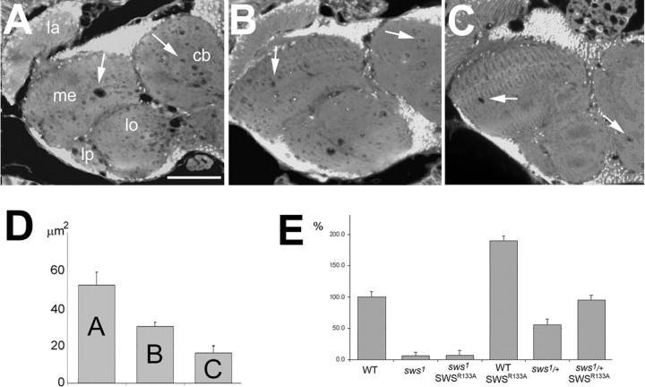
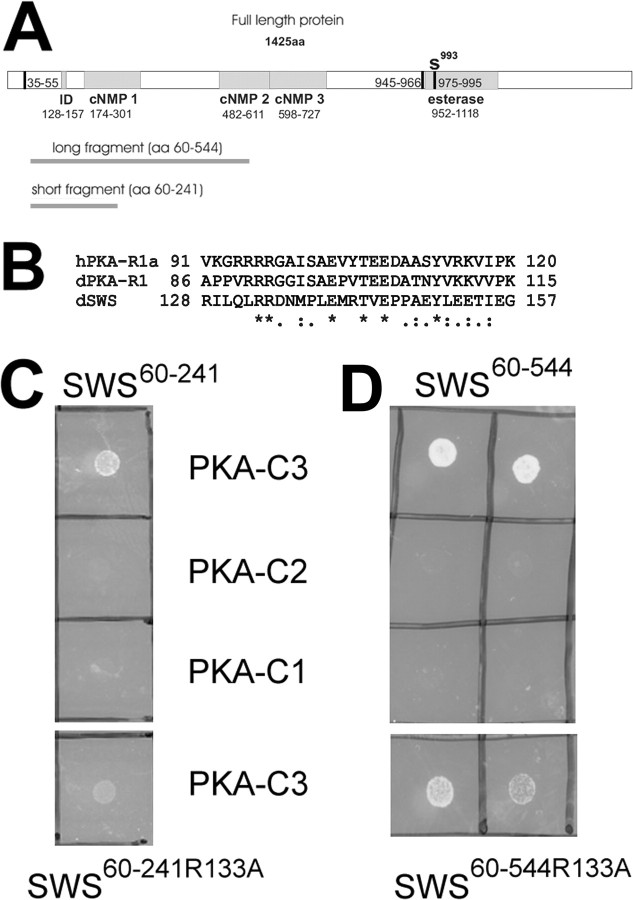
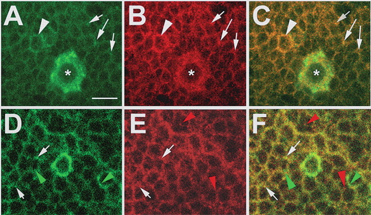
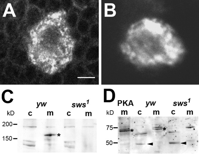
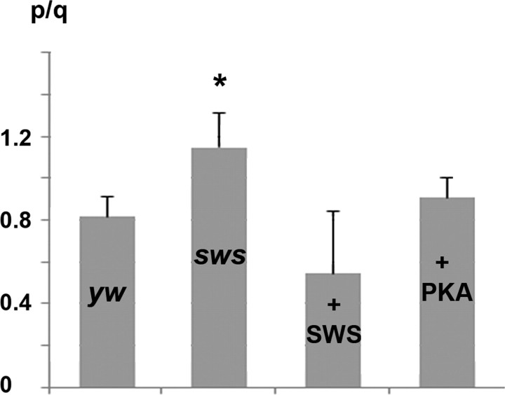
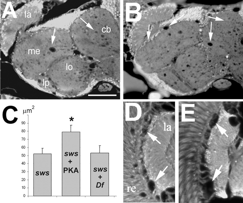
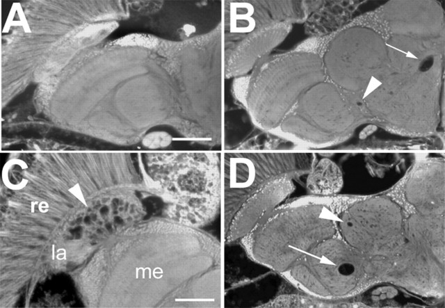
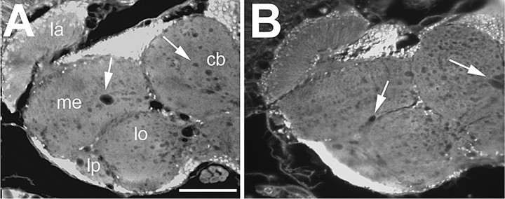
The Drosophila Swiss Cheese (SWS) protein and its vertebrate ortholog Neuropathy Target Esterase (NTE) are required for neuronal survival and glial integrity. In humans, NTE is the target of organophosphorous compounds which cause a paralyzing axonal degeneration and recently mutations in NTE have been shown to cause a Hereditary Spastic Paraplegia called NTE-related Motor-Neuron Disorder. SWS and NTE are concentrated in the endoplasmic reticulum and both have been shown to have an esterase function against an artificial substrate. However, the functional mechanisms and the pathways in which SWS/NTE are involved in are still widely unknown. Here, we show that SWS interacts specifically with the C3 catalytic subunit of cAMP activated protein kinase (PKA-C3), which together with orthologs in mouse (Pkare) and human (PrKX) forms a novel class of catalytic subunits of unknown function. This interaction requires a domain of SWS which shows homology to regulatory subunits of PKA and, like conventional regulatory subunits, the binding of SWS to the PKA-C3 inhibits its function. Consistent with this result, expression of additional PKA-C3 induces degeneration and enhances the neurodegenerative phenotype in sws mutants. We also show that the complex formation with the membrane-bound SWS tethers PKA-C3 to membranes. We therefore propose a model in which SWS acts as a noncanonical subunit for PKA-C3, whereby the complex formation regulates the localization and kinase activity of PKA-C3, and that disruption of this regulation can induce neurodegeneration.


Figures

Figure 1.
Figure 1.
Expression of SWSR133A in the nervous system of sws1 mutant flies does only partially rescue the degenerative phenotype and affects esterase activity. A, A sws1 fly shows the characteristic vacuolization in the neuropil (arrows). B, Expressing SWSR133A pan-neuronally in these flies leads to a significant reduction in vacuole formation. C, Expressing wild-type SWS under the same conditions almost completely reverts the mutant phenotype. D, Mean area of vacuoles in μm2 in the three genotypes shown in A, B, and C. Number of measured flies was as follows: n = 24 for Appl-GAL4, sws1 (A), n = 28 for Appl-GAL4, sws1; UAS-SWSR133A (B), and n = 34 for Appl-GAL4, sws1; UAS-SWS (C). SEMs are indicated. All sections are horizontal paraffin sections through the heads of 14-d-old flies. E, Esterase activity in fly head homogenates revealed that expressing the SWSR133A construct pan-neuronally did not restore the esterase function in sws1. In contrast, this construct showed esterase function in a wild-type background and heterozygous sws1/+ flies, approximately doubling the activity. All values are expressed relative to wild type (100%). SEMs are indicated. Scale bar: (in A) A–C, 50 μm. la, Lamina; me, medulla; lo, lobula; lb, lobula plate; cb, central brain.
Figure 2.
Figure 2.
SWS binds specifically to PKA-C3. A, The different fragments used in the yeast two-hybrid experiments are shown as dark gray lines below a schematic of the SWS protein. The predicted transmembrane (TM) domains are shown as vertical black bars and the interaction domain (ID), cyclic nucleotide binding sites (cNMP1–3), and esterase domain are indicated as gray boxes. The active site serine which is localized in the putative third transmembrane domain within the esterase domain is indicated by an S. B, Sequence comparison of the interaction domain of SWS with the human and Drosophila R1 regulatory subunits. Identical amino acids are indicated by asterisks, highly conserved amino acids by colons, and conserved amino acids by dots. C, Using the smaller SWS (SWS60–241) fragment, we obtained colonies at the restrictive temperature when cotransfected with PKA-C3 (first row) but not after cotransfection with PKA-C2 or PKA-C1 (second and third row). Using the same fragment with a mutation in the conserved arginine133 still produced colonies after cotransfection with PKA-C3 at the restrictive temperature but these colonies grew less well (fourth row). D, The same results were obtained in cotransfection studies using the larger SWS60–544 fragment.
Figure 3.
Figure 3.
SWS and PKA-C3 colocalize in neurons. A–C, Brain whole-mounts stained with anti-PKA-C3 (green) and anti-SWS (red) show expression in most or all neurons with stronger expression of both proteins in some neurons (arrowheads), including a few large neurons, which highly express PKA-C3 (asterisks). In addition, both proteins can be colocalized to the same vesicles (arrows). D–F, In some vesicles PKA-C3 (green) colocalizes with the ER marker GRP78 (red), although both proteins can also be found separately (green arrowheads and red arrowheads). Thickness of the optical sections was 0.1 μm. Scale bar, 5 μm.
Figure 4.
Figure 4.
PKA-C3 is mislocalized in the absence of SWS. A, B, Whereas PKA-C3 can be found in a mostly punctuate pattern in yw control flies, its pattern is less distinct in a sws1 mutant fly (B). C, In Western blots, the SWS protein (asterisks), which contains several transmembrane domains, is exclusively found in membrane fractions (m) from yw control flies, while it is missing in sws1 mutants. D, PKA-C3 is detected as a 66 kDa band in membrane fractions from yw control flies and, in increased amount in Appl-GAL4/UAS-PKA-C3 (mPKA) flies, whereas it is not detectable in membrane fractions from sws1 mutant flies. Cytosolic fractions (c) reveal an increase in a 50 kDa band in sws1, which could be a degradation product of PKA-C3. Scale bar: (in A) A, B, 2 μm.
Figure 5.
Figure 5.
PKA activity in fly head homogenates. In the presence of cAMP (0.4 mm), PKA activity is increased by ∼30% in sws1 extracts compared with yw control flies, whereas expressing additional SWS in wild type reduces PKA activity (although not quite statistically significant; p = 0.054). Expression of additional PKA-C3 via Appl-GAL4 did not increase the overall PKA activity compared with yw controls. n = 16 for yw, n = 21 for sws1, n = 13 for elav-GAL4/UAS-SWS, and n = 15 for Appl-GAL4/UAS-PKA/C3. p/q, quotient of the luminosity value of phosphorylated to unphosphorylated kemptide peptide. Values were normalized to ng protein in the lysates. SEMs are indicated. *p < 0.01.
Figure 6.
Figure 6.
Overexpression of PKA-C3 enhances the neurodegenerative phenotype of sws. A, B, Compared with the sws1 mutant fly (A), flies expressing PKA-C3 via the APPL-GAL4 driver (B) showed a significant increase in vacuolization. C, This is confirmed by measuring the mean area of vacuoles in μm2 in sws1 (n = 24) and sws1; UAS-PKA-C3 flies (n = 33). sws1 flies carrying one copy of the deficiency Df(2)brm11 showed no significant difference in the mean area of vacuoles in the central brain (n = 22). Sections in A and B are horizontal paraffin sections through the heads of 14-d-old flies. D, Vacuoles (arrows) in the lamina cortex of a 7-d-old sws1 fly heterozygous for Df(2)brm11. In this region, Df(2)brm11 significantly reduces the area of vacuoles compared with sws1 without the deficiency (E) [n = 17 for sws1, n = 13 for sws1; Df(2)brm11]. Scale bar: (in A), A, B, 50 μm. la, Lamina; me, medulla; lo, lobula; lb, lobula plate; cb, central brain; re, retina.
Figure 7.
Figure 7.
Neuronal expression of PKA-C3 induces vacuolization. A, A 1-d-old male fly hemizygous for Appl-GAL4 and heterozygous for UAS-PKA-C3 shows no degeneration, whereas a 30-d-old one (B) shows some, sometimes quite large vacuoles (arrowhead and arrow). C, In ∼10% of these aged flies, the lamina shows severe vacuolization (arrowhead). D, Removing one copy of sws in 30-d-old females heterozygous for Appl-GAL4 and UAS-PKA-C3 also results in the formation of vacuoles similar to the ones seen in males, whereas these heterozygous females do not show vacuoles without removing one copy of sws (data not shown). All sections are horizontal paraffin head sections. Scale bars: (in A) A, B, D, 50 μm; C, 25 μm. re, Retina, la, lamina.
Figure 8.
Figure 8.
Expression of a catalytically inactive SWS construct results in a partial rescue. A, sws1. B, Pan-neuronal expression of SWSS985D, which has no kinase activity (Mühlig-Versen et al., 2005), but an intact PKA-C3 interaction domain in the nervous system of sws1 mutant flies can partially rescue the degenerative phenotype. Mean area of vacuoles (in μm2) in Appl-GAL4, sws1 flies (n = 24), and Appl-GAL4, sws1; UAS-SWSS985D (n = 38). Sections are horizontal paraffin sections through the heads of 14-d-old flies. SEMs are indicated. Scale bar, 50 μm. la, Lamina; me, medulla; lo, lobula; lb, lobula plate; cb, central brain.

References

    1. Akassoglou K, Malester B, Xu J, Tessarollo L, Rosenbluth J, Chao MV. Brain-specific deletion of neuropathy target esterase/swisscheese results in neurodegeneration. Proc Natl Acad Sci U S A. 2004;101:5075–5080. - PMC - PubMed
    1. Bettencourt da Cruz A, Schwärzel M, Schulze S, Niyyati M, Heisenberg M, Kretzschmar D. Disruption of the MAP1B-related protein FUTSCH leads to changes in the neuronal cytoskeleton, axonal transport defects, and progressive neurodegeneration in Drosophila. Mol Biol Cell. 2005;16:2433–2442. - PMC - PubMed
    1. Blaschke RJ, Monaghan AP, Bock D, Rappold GA. A novel murine PKA-related protein kinase involved in neuronal differentiation. Genomics. 2000;64:187–194. - PubMed
    1. Bradford MM. A rapid and sensitive method for the quantitation of microgram quantities of protein utilizing the principle of protein-dye binding. Anal Biochem. 1976;72:248–254. - PubMed
    1. Brand AH, Perrimon N. Targeted gene expression as a means of altering cell fates and generating dominant phenotypes. Development. 1993;118:401–415. - PubMed

Publication types

MeSH terms