. 2008 Oct 22;28(43):10893-904.
doi: 10.1523/JNEUROSCI.3299-08.2008.

Schizophrenia-related neural and behavioral phenotypes in transgenic mice expressing truncated Disc1

Affiliations

Schizophrenia-related neural and behavioral phenotypes in transgenic mice expressing truncated Disc1

Sanbing Shen et al. J Neurosci. .

Abstract

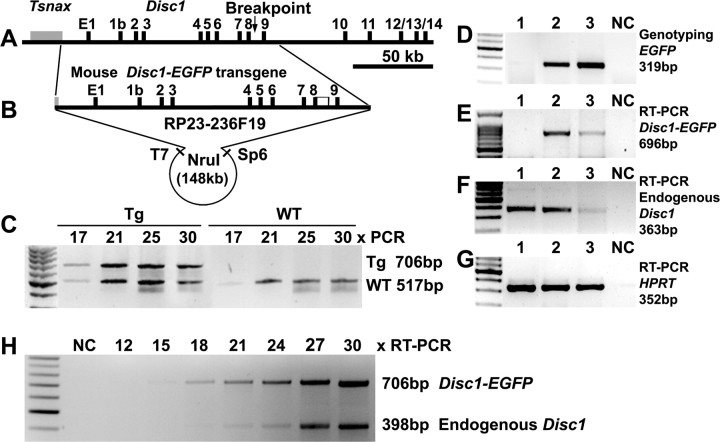
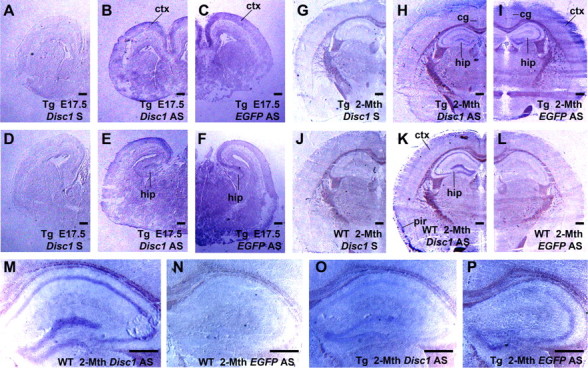
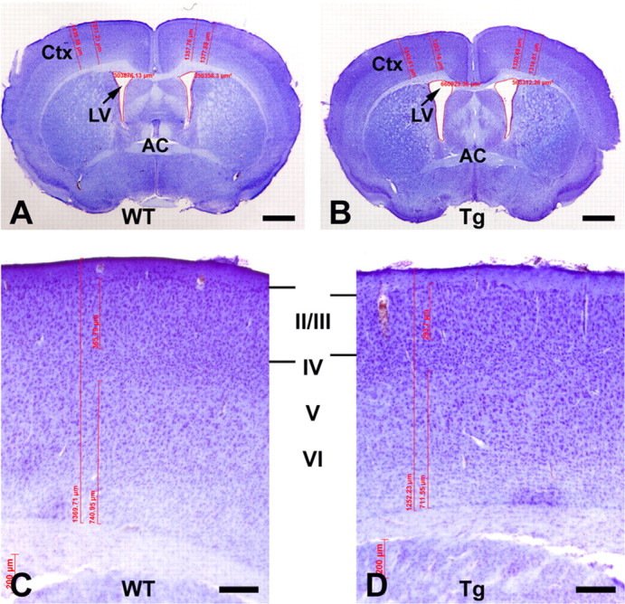
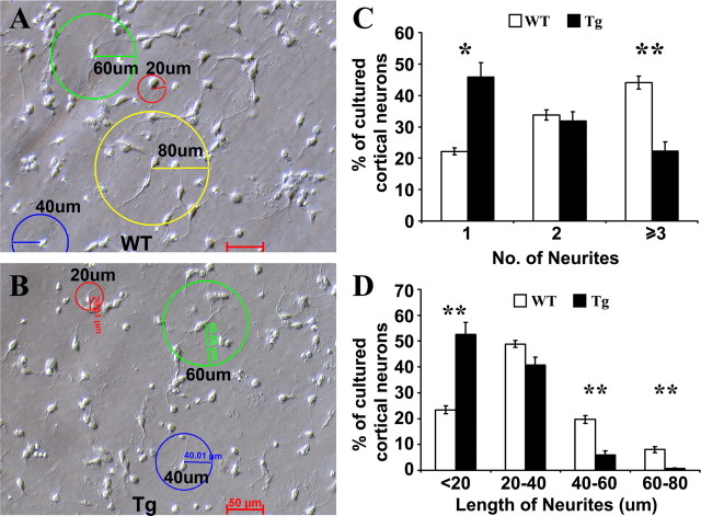
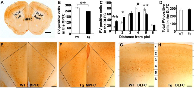
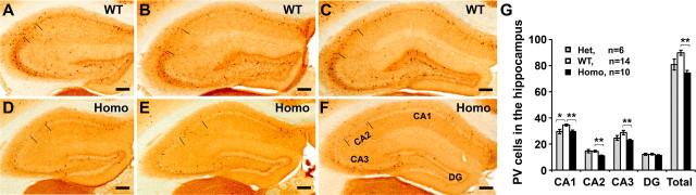
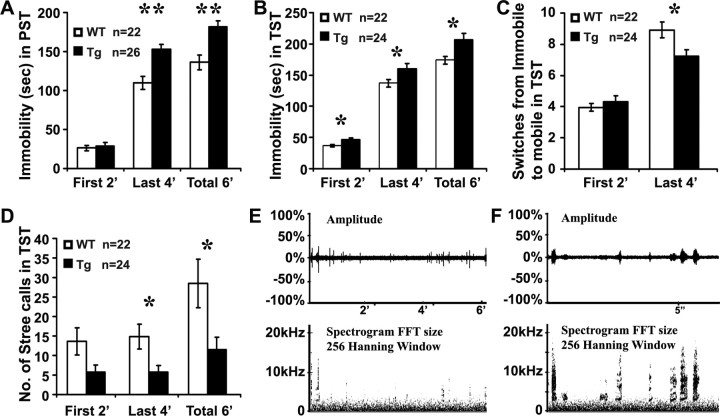
Disrupted-in-Schizophrenia-1 (DISC1), identified by positional cloning of a balanced translocation (1;11) with the breakpoint in intron 8 of a large Scottish pedigree, is associated with a range of neuropsychiatric disorders including schizophrenia. To model this mutation in mice, we have generated Disc1(tr) transgenic mice expressing 2 copies of truncated Disc1 encoding the first 8 exons using a bacterial artificial chromosome (BAC). With this partial simulation of the human situation, we have discovered a range of phenotypes including a series of novel features not previously reported. Disc1(tr) transgenic mice display enlarged lateral ventricles, reduced cerebral cortex, partial agenesis of the corpus callosum, and thinning of layers II/III with reduced neural proliferation at midneurogenesis. Parvalbumin GABAergic neurons are reduced in the hippocampus and medial prefrontal cortex, and displaced in the dorsolateral frontal cortex. In culture, transgenic neurons grow fewer and shorter neurites. Behaviorally, transgenic mice exhibit increased immobility and reduced vocalization in depression-related tests, and impairment in conditioning of latent inhibition. These abnormalities in Disc1(tr) transgenic mice are consistent with findings in severe schizophrenia.


Figures

Figure 1.
Figure 1.
The truncated mouse Disc1 transgene and expression. A, Genomic organization of the mouse Disc1 locus, with an arrow corresponding to the breakpoint in the Scottish family. B, A BAC clone RP23–236F19 containing ∼148 kb mouse Disc1 genomic DNA, starting from the 3′UTR of Tsnax, and ending with 16.7 kb of Disc1 intron 9. The BAC was fused to an EGFP at the end of exon 8 followed by a PolyA. The NruI fragment (148,730 bp) was purified for microinjection. C, M19 heterozygous transgenics contained 2 copies of the transgene, as similar intensity of PCR products were obtained from the truncated and endogenous Disc1 genes at 17–30 cycles. D, Two E17.5 embryos (lanes 1–2) and a transgenic mother (lane 3) were genotyped by PCR with EGFP primers. E–G, RT-PCR with primers for the transgene (E), the endogenous Disc1 (F), and a housekeeping HPRT (G), showing comparable levels of the truncated (E) and full-length (F) Disc1 mRNA. H, Relative expression of endogenous Disc1 and Disc1tr-EGFP demonstrated by RT-PCR at 12, 15, 18, 21, 24, 27, and 30 cycles. The average ratio of the RT-PCR products 706 bp:398 bp at 18–30 cycles was 1.09 ± 0.19, showing comparable abundance of endogenous and truncated Disc1 mRNA (Fig. 1H). NC, Negative control.
Figure 2.
Figure 2.
Comparable temporal and spatial patterns of expression of truncated and full-length Disc1. In situ hybridization was performed using DIG-labeled sense (S) and antisense (AS) RNA probes on E17.5 (A–F) and 2-month-old brain sections (G–P). Whereas the Disc1 AS probe hybridized to the endogenous full-length Disc1 transcripts, EGFP AS probe detected the Disc1tr-EGFP messages only. Both the endogenous and transgenic transcripts (purple) were detected at the regions of cerebral cortex (ctx), including cingulate (cg) and piriform (pir) cortex, CA1, CA2, CA3, and dentate gyrus of the hippocampus (hip). Scale bars, 500 μm.
Figure 3.
Figure 3.
Enlarged lateral ventricles (LV) and reduced cerebral cortex (Ctx) in Disc1tr transgenic (Tg) mice. WT (A) and Tg (B) brain sections were Nissl stained, imaged at the level where the anterior commissure (AC) crossed the midline, and quantified with AxioVision Rel. 4.5. The LV was significantly (p < 0.05) enlarged in Tg brains (0.819 ± 0.079 mm2, n = 11) compared with WT littermates (0.568 ± 0.043 mm2, n = 15). C, D, Magnified view of the cerebral cortex from WT and Tg representatives showing changes in layers II–III. Statistical analyses detected moderate but significant (p < 0.05) reduction in Tg cerebral cortex (1353.6 ± 19.9 μm, n = 11) compared with WT littermates (1409.2 ± 10.0 μm, n = 15). Cortical layers II/III was thinned (p < 0.01) from 347.2 ± 4.5 μm in WT to 302.7 ± 3.4 μm in Tg mice. Scale bars: A, B, 1 mm; C, D, 200 μm.
Figure 4.
Figure 4.
Reduced neurogenesis in Disc1tr transgenic (Tg) embryos. A–D, A pulse of BrdU was injected into four E15.5 pregnant females and newborn brains were processed with an anti-BrdU antibody. For each brain, four images were taken from the cerebral cortex at the left and right sides of two consecutive sections with the largest lateral ventricles. Images were arbitrarily divided into 5 layers as shown, and BrdU-positive cells were quantified from each area (400 μm wide × 150 μm high). A and B were from two WT littermates, C and D from two Tg newborns. E, Statistical analyses revealed significant reduction of BrdU-positive cells in the arbitrarily assigned layer 1 of Tg mice (140.4 ± 4.9, n = 13), compared with their WT littermates (158.6 ± 5.3, n = 8). F, The total number of BrdU-incorporated cells was also significantly reduced in Tg mice compared with that in WT littermates. Scale bars, 200 μm. *p < 0.05.
Figure 5.
Figure 5.
Partial agenesis of the corpus callosum (CC) in 2-month-old transgenic brains. Coronal sections of WT (A, C) and Tg (B, D) brains were Nissl-stained. A, B, Images represented average thickness of rostral CC in WT (A) and Tg (B) brains where the AC crossed the midline, showing significant reduction of the CC in 11 Tg brains compared with 15 WT. At the SCO level (arrowed in C and D), a thick layer of the CC crossed the midline in all 15 WT mice (C), whereas Tg CC failed to cross the midline in 9 of 11 cases (D). Scale bars: A, B, 200 μm; C, D, 1 mm.
Figure 6.
Figure 6.
Fewer and shorter neurites in cultured transgenic (Tg) neurons. WT (A) and Tg (B) newborn cortex were dissociated and neurons were cultured for 26 h in vitro. Images (6–8 fields/mouse) were randomly taken under a 20× objective lens. Neurons (1817 WT and 845 Tg) were quantified for the number (C) and length of neurites with 20 (red), 40 (blue) 60 (green), and 80 μm (yellow) rings (D), respectively. The data were presented as mean ± SEM. *p < 0.05, **p < 0.01. Scale bars, 50 μm.
Figure 7.
Figure 7.
Parvalbumin (PV) interneurons in the prefrontal cortex of WT and Tg brains. A, A brain section stained with anti-PV, showing areas of MPFC and DLFC where magnified images (E–H) were taken for counting PV cells. B, Statistical analyses of PV cells in the MPFC of WT and Tg mice as illustrated in E (WT) and F (Tg). C, PV-positive cells in 6 arbitrarily assigned layers (1386 μm wide × 267 μm high) of the DLFC as illustrated in G and H. D, Total number of PV cells in the DLFC showing no difference between WT and Tg mice. Scale bars: A, 1 mm; E–H, 200 μm.
Figure 8.
Figure 8.
PV interneurons are reduced in the hippocampus of Disc1tr transgenic mice. A–F, Brain sections from 14 WT (A–C), 6 heterozygous (data not shown), and 10 homozygous (D–F) transgenic mice were stained with anti-PV. PV-positive cells at the CA1, CA2, CA3, and dentate gyrus (DG) were quantified from 6 comparable images of each mouse brain as shown. G, Statistical analyses of the mean PV cells in each area of the hippocampus, showing significant reduction of the PV interneurons in the CA1 of the heterozygotes, and in the CA1–CA2–CA3 of the homozygotes. Scale bars: 200 μm. *p < 0.05, **p < 0.01.
Figure 9.
Figure 9.
Disc1tr transgenic mice are defective in conditioning of latent inhibition. A, The horizontal activity (mean ± SEM) in numbers of beam breaks per second (bb/s) during the different phases of conditioning. B, Pooled activity (bb/s) during the 5 × 10 s tone period. C, Pooled activity (bb/s) during the 5 × 2 s shock period. D, Total number of beam breaks (mean ± SEM) during the 120 s retention test on the following day. Note that only the npe-WT group showed considerable “freezing” during the tone (B), shock (C), or retention (D) period. *p < 0.05. npe, Non-preexposed; pe, preexposed to tone.
Figure 10.
Figure 10.
Increased immobility (A–C) and reduced vocalization (D–F) of Disc1tr transgenic mice in depression tests. Disc1tr transgenic (Tg) mice and WT littermates were tested individually in 6 min PST (A) or TST (B–F). Transgenic mice showed increased immobility in PST (A) and TST (B), with a reduced number of switches from immobile to mobile status in the last 4 min of the TST (C). D, Disc1tr transgenic mice made significantly fewer stress calls. E, An example of vocalizations (squeaks) recorded by a bat detector showing amplitude and frequency (kHz) of calls during the 6 min TST. F, Magnified view of the calls. *p < 0.05, **p < 0.01.

Similar articles

Cited by

References

    1. Assadi AH, Zhang G, Beffert U, McNeil RS, Renfro AL, Niu S, Quattrocchi CC, Antalffy BA, Sheldon M, Armstrong DD, Wynshaw-Boris A, Herz J, D'Arcangelo G, Clark GD. Interaction of reelin signaling and Lis1 in brain development. Nat Genet. 2003;35:270–276. - PubMed
    1. Benes FM, Lim B, Matzilevich D, Walsh JP, Subburaju S, Minns M. Regulation of the GABA cell phenotype in hippocampus of schizophrenics and bipolars. Proc Natl Acad Sci U S A. 2007;104:10164–10169. - PMC - PubMed
    1. Blackwood DH, Fordyce A, Walker MT, St Clair DM, Porteous DJ, Muir WJ. Schizophrenia and affective disorders—cosegregation with a translocation at chromosome 1q42 that directly disrupts brain-expressed genes: clinical and P300 findings in a family. Am J Hum Genet. 2001;69:428–433. - PMC - PubMed
    1. Braff DL, Geyer MA, Swerdlow NR. Human studies of prepulse inhibition of startle: normal subjects, patient groups, and pharmacological studies. Psychopharmacology. 2001;156:234–258. - PubMed
    1. Brandon NJ, Handford EJ, Schurov I, Rain JC, Pelling M, Duran-Jimeniz B, Camargo LM, Oliver KR, Beher D, Shearman MS, Whiting PJ. Disrupted in Schizophrenia 1 and Nudel form a neurodevelopmentally regulated protein complex: implications for schizophrenia and other major neurological disorders. Mol Cell Neurosci. 2004;25:42–55. - PubMed

Publication types

MeSH terms