. 2008 Oct 22;28(43):10972-82.
doi: 10.1523/JNEUROSCI.1521-08.2008.

Lesions of the medial striatum in monkeys produce perseverative impairments during reversal learning similar to those produced by lesions of the orbitofrontal cortex

Affiliations

Lesions of the medial striatum in monkeys produce perseverative impairments during reversal learning similar to those produced by lesions of the orbitofrontal cortex

Hannah F Clarke et al. J Neurosci. .

Abstract

The ability to switch responding between two visual stimuli based on their changing relationship with reward is dependent on the orbitofrontal cortex (OFC). OFC lesions in humans, monkeys, and rats disrupt performance on a common test of this ability, the visual serial discrimination reversal task. This finding is of particular significance to our understanding of psychiatric disorders such as obsessive-compulsive disorder (OCD) and schizophrenia, in which behavioral inflexibility is a prominent symptom. Although OFC dysfunction can occur in these disorders, there is considerable evidence for more widespread dysfunction within frontostriatal and frontoamygdalar circuitry. Because the contribution of these subcortical structures to behavioral flexibility is poorly understood, the present study compared the effects of excitotoxic lesions of the medial striatum (MS), amygdala, and OFC in the marmoset monkey on performance of the serial reversal task. All monkeys were able to learn a novel stimulus-reward association but, compared with both control and amygdala-lesioned monkeys, those with MS or OFC lesions showed a perseverative impairment in their ability to reverse this association. However, whereas both MS and OFC groups showed insensitivity to negative feedback, only OFC-lesioned monkeys showed insensitivity to positive feedback. These findings suggest that, for different reasons, both the MS and OFC support behavioral flexibility after changes in reward contingencies, and are consistent with the hypothesis that striatal and OFC dysfunction can contribute to pathological perseveration.


Figures
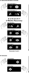
Figure 1.
Figure 1.
A, B, Stimulus exemplars used for the various stages of the reversal and extinction paradigms. The rewarded and unrewarded stimuli on each discrimination is indicated by the “+” and “−,” respectively. The real stimuli were multicolored.
Figure 2.
Figure 2.
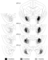
Schematic diagram of a series of coronal sections throughout the striatum of the marmoset monkey illustrating the extent of the lesion. The three shades of gray indicate regions that were lesioned in all three monkeys, any two monkeys, and one monkey.
Figure 3.
Figure 3.
Low-power (A, B) and high-power (C) photomicrographs of cresyl violet-stained coronal sections through a representative region of the striatum in a sham-operated control (A) and a striatal-lesioned marmoset (B). Dotted lines in A indicate the extent of an intact caudate nucleus, and in C indicate the boundary between the lesioned area on the left and intact neurons on the right after a medial striatal lesion. Comparison of the caudate nucleus in A and B clearly shows the absence of the medial caudate in B and the corresponding enlargement of the lateral ventricle. Caud, Intact caudate nucleus; Caud, spared region of the caudate nucleus after a medial striatal lesion; NA, nucleus accumbens; IC, internal capsule; P, putamen; LV, lateral ventricle. The asterisks mark the same position in B and C.
Figure 4.
Figure 4.
Orbitofrontal cortex. Schematic coronal sections taken through the frontal lobe of the marmoset indicating the extent of the orbitofrontal lesion. The coronal sections depicted in the inset show the distribution of granular (G), dysgranular (dG), and agranular (aG) cortices within the marmoset frontal lobe. The dotted areas of the inset represent the area targeted for the orbitofrontal lesion. Shading is as described in Figure 2.
Figure 5.
Figure 5.
A schematic diagram of a series of coronal sections through the anterior temporal lobe of the marmoset illustrating the extent of the amygdala lesion. The individual nuclei of the amygdala can be seen in the single hemisphere to the left of the figure. L, Lateral nucleus; B, basal nucleus; Bmg, basal nucleus magnocellular subdivision; Bpc, basal nucleus parvocellular subdivision; Bi, basal intermediate nucleus; AA, anterior amygdala; AB, accessory basal nucleus; ABmg, magnocellular subdivision of the AB; ABpc, parvocellular subdivision of the AB; C, central nucleus; Co, cortical region of the amygdala; M, medial nucleus; PL, paralaminar nucleus; Hipp, hippocampus. Shading is as described in Figure 2.
Figure 6.
Figure 6.
Serial discrimination reversal performance (square-root transformed). A, Total errors to criterion. B, Perseverative errors. Analysis by repeated-measures ANOVA revealed an error type × group interaction (p = 0.001) attributable to increased perseverative responding in the MS and OFC groups. C, Error type data collapsed across reversals. Amygdala, Amygdala-lesioned monkeys; MS, medial striatal-lesioned monkeys; OFC, orbitofrontal cortex-lesioned monkeys. *REGWQ post hoc test, p < 0.05. Error bars represent SEM.
Figure 7.
Figure 7.
The mean probabilities of monkeys shifting their responding to the other stimulus after making either an incorrect choice (and therefore not receiving reward) (A) or a correct choice and receiving reward (B). Black line/asterisk indicates which groups differ from one another at p = 0.05 using the REGWQ post hoc homogenous subset test. Gray dotted line/asterisk indicates significance at p < 0.05 when the MS- and OFC-lesioned monkeys were directly compared according to our a priori hypothesis. Error bars represent SEM.
Figure 8.
Figure 8.
A, Mean responses made in extinction (square-root transformed) for all monkeys, including individual performances. *p < 0.05 when comparing amygdala-lesioned monkeys to controls only. B, Total numbers of perseverative errors in reversal learning compared with the total number of responses made in extinction, for individual monkeys. Error bars represent SEM.

References

    1. Alexander GE, Crutcher MD, DeLong MR. Basal ganglia-thalamocortical circuits-parallel substrates for motor, oculomotor, prefrontal and limbic functions. Prog Brain Res. 1990;85:119–146. - PubMed
    1. Annett LE, McGregor A, Robbins TW. The effects of ibotenic acid lesions of the nucleus accumbens on spatial learning and extinction in the rat. Behav Brain Res. 1989;31:231–242. - PubMed
    1. Block AE, Dhanji H, Thompson-Tardif SF, Floresco SB. Thalamic-prefrontal cortical-ventral striatal circuitry mediates dissociable components of strategy set shifting. Cereb Cortex. 2007;17:1625–1636. - PubMed
    1. Burns LH, Everitt BJ, Robbins TW. Effects of excitotoxic lesions of the basolateral amygdala on conditional discrimination learning with primary and conditioned reinforcement. Behav Brain Res. 1999;100:123–133. - PubMed
    1. Butter CM. Perseveration in extinction and in discrimination reversal tasks following selective frontal ablations in Macaca mulatta. Physiol Behav. 1969;4:163–171.

Publication types

Substances