. 2009 Mar 19;113(12):2732-41.
doi: 10.1182/blood-2008-05-158642. Epub 2008 Oct 22.

Capture and transfer of HIV-1 particles by mature dendritic cells converges with the exosome-dissemination pathway

Affiliations

Capture and transfer of HIV-1 particles by mature dendritic cells converges with the exosome-dissemination pathway

Nuria Izquierdo-Useros et al. Blood. .

Abstract

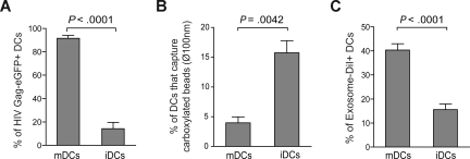
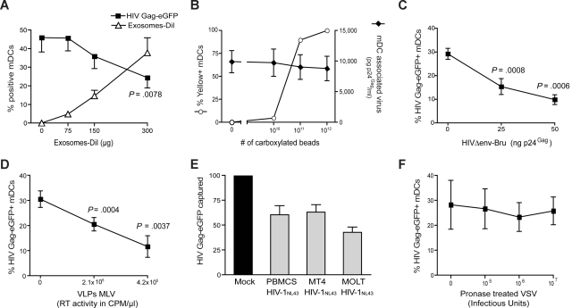
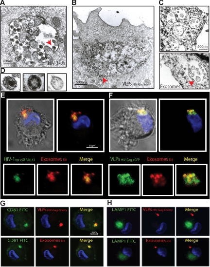
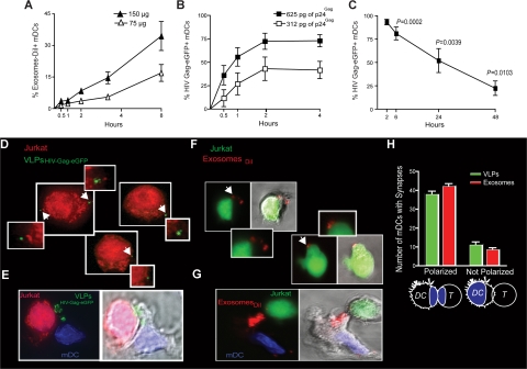
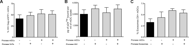
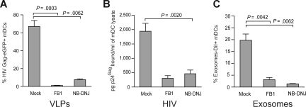
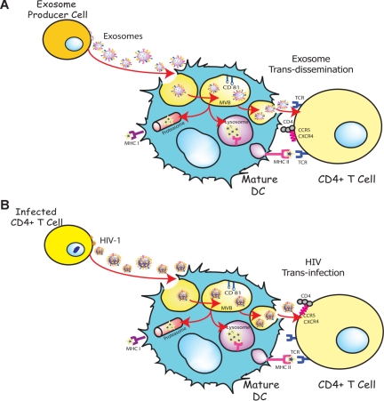
Exosomes are secreted cellular vesicles that can be internalized by dendritic cells (DCs), contributing to antigen-specific naive CD4(+) T-cell activation. Here, we demonstrate that human immunodeficiency virus type 1 (HIV-1) can exploit this exosome antigen-dissemination pathway intrinsic to mature DCs (mDCs) for mediating trans-infection of T lymphocytes. Capture of HIV-1, HIV-1 Gag-enhanced green fluorescent protein (eGFP) viral-like particles (VLPs), and exosomes by DCs was up-regulated upon maturation, resulting in localization within a CD81(+) compartment. Uptake of VLPs or exosomes could be inhibited by a challenge with either particle, suggesting that the expression of common determinant(s) on VLP or exosome surface is necessary for internalization by mDCs. Capture by mDCs was insensitive to proteolysis but blocked when virus, VLPs, or exosomes were produced from cells treated with sphingolipid biosynthesis inhibitors that modulate the lipid composition of the budding particles. Finally, VLPs and exosomes captured by mDCs were transmitted to T lymphocytes in an envelope glycoprotein-independent manner, underscoring a new potential viral dissemination pathway.


Figures

Figure 1
Figure 1
Maturation of DCs enhances VLPHIV-Gag-eGFP and ExosomeDiI capture. (A) Comparative capture of VLPHIV-Gag-eGFP by DCs. A total of 105 DCs were pulsed for 4 hours at 37°C with 2500 pg p24Gag in 0.1 mL, washed with PBS, and fixed to analyze the percentage of eGFP-positive cells by FACS. Data show mean values and SEM from 5 independent experiments, including cells from 6 donors. mDCs captured significantly greater amounts of VLPs compared with iDCs (P < .0001, paired t test). (B) Comparative capture of carboxylated yellow fluorescent beads by DCs. A total of 5 × 10⁵ iDCs and mDCs were incubated at 4°C and 37°C for 2 hours with approximately 1.8 × 10¹⁰ beads. Cells were washed, fixed, and analyzed by FACS. Graph displays the percentage of DCs that captured beads at 37°C, after subtracting binding percentages at 4°C. Data show mean values and SEM from 4 independent experiments, including cells from 7 donors. iDCs significantly captured greater amounts of beads compared with mDCs (P = .0042, paired t test). (C) Comparative capture of Jurkat-derived ExosomesDiI by DCs. A total of 105 DCs were pulsed for 8 hours at 37°C with 150 μg exosomes, washed with PBS, and fixed to analyze the percentage of DiI-positive cells by FACS. Data show mean values and SEM from 4 independent experiments, including cells from 11 donors. mDCs capture significantly greater amounts of exosomes compared with iDCs (P < .0001, paired t test).
Figure 2
Figure 2
Competition experiments suggest that different particles derived from cholesterol-enriched domains use the same entry pathway into mDCs. (A) Capture of VLPHIV-Gag-eGFP by mDCs previously exposed to increasing amounts of Jurkat-derived ExosomesDiI. Cells were preincubated for 30 minutes with increasing amounts of ExosomesDiI and then pulsed with 625 pg of VLPHIV-Gag-eGFP p24Gag for 1 hour at 37°C, washed with PBS, fixed, and analyzed by FACS to determine the percentage of eGFP- and DiI-positive cells. mDCs captured fewer VLPHIV-Gag-eGFP in the presence of increasing amounts of ExosomesDiI (P = .0078, paired t test). (B) Capture of HIVΔenv-NL43 by mDCs previously exposed to increasing amounts of yellow carboxylated 100-nm beads. A total of 5 × 10⁵ mDCs were preincubated for 30 minutes with the beads and then pulsed for 1 hour at 37°C with 130 ng HIVΔenv-NL43 p24Gag in 0.5 mL and extensively washed with PBS. Each sample was then divided and either fixed for analysis by FACS for bead capture or lysed with 0.5% Triton (at a final concentration of 5 × 10⁵ cells per milliliter) to measure p24Gag content in the cell lysate by an ELISA. Results represent the percentage of yellow positive mDCs (○) and the amount of pg of p24Gag bound per mL of cell lysate (♦). (C,D) Capture of VLPHIV-Gag-eGFP by mDCs previously exposed to increasing amounts of HIVΔenv-Bru (C) and VLPMLV-Gag (D). Cells were preincubated for 30 minutes with increasing amounts of HIVΔenv-Bru or VLPMLV-Gag and then pulsed with 625 pg of VLPHIV-Gag-eGFP p24Gag for 1 hour at 37°C, washed with PBS, and fixed to analyze the percentage of eGFP-positive cells by FACS. mDCs capture less VLPHIV-Gag-eGFP in the presence of increasing concentrations of particles derived from cholesterol-enriched membrane microdomains (P values on the graphs, paired t test). (E) The data represent the relative VLPHIV-Gag-eGFP capture by mDCs that had been preincubated with 200 ng of p24Gag of HIVNL43 obtained from either MT4, MOLT, or PHA-stimulated PBMCs and normalized to the level of VLPHIV-Gag-eGFP capture by mock-treated mDCs (set at 100%). mDCs captured less VLPHIV-Gag-eGFP in the presence of these different viral stocks. (F) Capture of VLPHIV-Gag-eGFP by mDCs that had been preincubated with increasing amounts of pronase-treated VSV particles. Cells were preincubated for 30 minutes in the presence of pronase-treated VSV particles and then pulsed with the 625 pg of VLPHIV-Gag-eGFP p24Gag for 1 hour at 37°C, washed with PBS, and fixed to analyze the percentage of eGFP-positive cells by FACS. mDCs captured similar amounts of VLPHIV-Gag-eGFP in the presence of pronase-treated VSV particles. Panels A through F show mean values and SEM from 3 independent experiments, including cells from at least 4 different donors.
Figure 3
Figure 3
mDCs retain HIV-1, VLPs and exosomes in the same CD81+ intracellular compartment. (A-C) Electron micrographs of mDCs sections exposed in parallel to HIVNL43, VLPHIV-Gag-eGFP, and ExosomesDiI, showing similar large vesicles containing these particles. Arrows indicate captured particles magnified in panel D, where comparative micrographs show, from left to right: HIVNL43, VLPHIV-Gag-eGFP, and a Jurkat-derived exosome. (E) Confocal images of a section of mDC exposed to HIVvpr-eGFP/NL43 and ExosomesDiI for 4 hours and stained with DAPI. Top images show the mDC, where the red and green fluorescence that merged with DAPI either with or without the bright-field cellular shape are presented. Bottom images show magnification of vesicles containing these particles where individual green and red fluorescence and the combination of both are depicted. (F) Confocal images of a section of a mDC exposed to VLPHIV-Gag-eGFP and ExosomesDiI as described in panel E. (G) Confocal images of a section of a mDC exposed to red fluorescent VLPHIV-Gag-Cherry (top) or ExosomesDiI (bottom) in parallel for 4 hours, fixed, and permeabilized to stain with DAPI and FITC-CD81. Images shown, from left to right, depict individual green and red fluorescence channels and the combination of both merged with DAPI. (H) Confocal images obtained as in panel G, except that cells were stained with DAPI and FITC-LAMP-1. mDCs retain VLPHIV-Gag-eGFP and ExosomesDiI in a CD81+ LAMP1 compartment.
Figure 4
Figure 4
VLP and exosome uptake in mDCs is a dose-dependent mechanism that increases over time, allowing efficient transfer to target T cells. (A) Time course of mDCs (n = 4) exposed to 2 different concentrations of ExosomesDiI and fixed at each of the indicated time points and analyzed by FACS. Exosome capture by mDCs increases over time in a dose dependent manner. (B) Time course of mDCs (n = 4) exposed to 2 different concentrations of VLPHIV-Gag-eGFP and fixed at each of the indicated time points and analyzed by FACS. VLPHIV-Gag-eGFP capture by mDCs increases over time in a dose-dependent manner. (C) Fate of VLPHIV-Gag-eGFP captured by mDCs and followed by flow cytometry for 2 days. Graph shows the percentage of Gag-eGFP-positive cells measured by FACS at the indicated time points. P values on the graph reveal that, at 48 hours after pulse with VLPHIV-Gag-eGFP, a significant percentage of mDCs still retained VLPs (one sample t test). Data (mean and SEM from 3 independent experiments) include cells from 4 different donors. (D) Orange cell tracker dye-labeled Jurkat T cells were analyzed by deconvolution microscopy after 4 hours of coculture with mDCs previously pulsed with VLPHIV-Gag-eGFP and extensively washed before coculture. The cells shown in the panels are projections of stack images obtained by merging the red and green fluorescence. Arrows indicate Gag-eGFP dots associated to Jurkat T cells, magnified in the nearby marked boxes (E). Viral synapse could also be observed in these cocultures, where mDCs pulsed with VLPHIV-Gag-eGFP were stained with DAPI. Images shown, from left to right, depict the red and green fluorescence channels merged with DAPI, the bright-field cellular shape and the combination of both. (F) Jurkat T cells labeled with a green cell tracker dye were analyzed by confocal microscopy after 4 hours of coculture with mDCs previously pulsed with ExosomesDiI and extensively washed. Images were obtained by merging the red and green fluorescence. Arrows indicate DiI dots associated with Jurkat T cells, magnified in the nearby marked boxes. Bright-field cellular shape merged with the red and green fluorescence is also shown. (G) Exosome polarization to the site of DC-T cell–contact, where mDCs pulsed with ExosomesDiI were stained with DAPI. Images shown, from left to right, depict the red and green fluorescence channels merged with DAPI, the bright-field cellular shape and the combination of both. (H) Quantification of mDCs forming synapses like those shown in panels E and G. Polarization of particles toward the synapse was considered when VLPsHIV-Gag-eGFP (green) or ExosomesDiI (red) were found within one-third of the cell proximal to the contact zone (as represented in the illustration by the blue colored area). Mean values and SEM of 50 synapses from 2 donors counted by 3 distinct observers.
Figure 5
Figure 5
VLPs, HIV-1 and exosomes enter mDCs through a mechanism resistant to proteolysis. mDC capture of (A) VLPHIV-Gag-eGFP, (B) HIVNL43, or (C) ExosomesDiI. Pronase-treated or -untreated mDCs were pulsed for 15 minutes at 37°C with pronase or mock-treated VLPHIV-Gag-eGFP, HIVNL43, and ExosomesDiI. Protease pretreatments of either the cells or the particles were insufficient to prevent the capture of VLPHIV-Gag-eGFP, ExosomesDiI or HIVNL43 by mDCs. (C-E) Mean values and SEM from 6 independent experiments, including cells from at least 4 different donors.
Figure 6
Figure 6
VLP, HIV-1, and exosome capture can be inhibited when particles are produced from ceramide-deficient cells. (A) A total of 105 mDCs were exposed to 2500 pg of p24Gag VLPHIV-Gag-eGFP produced from either FB1, NB-DNJ, or mock-treated HEK-293T cells, fixed, and analyzed by FACS to measure the percentage of eGFP-positive cells. mDCs captured significantly greater amounts of VLPHIV-Gag-eGFP produced from mock treated HEK-293T cells (P = .0003, paired t test). Mean values and SEM from 3 independent experiments, including cells from 5 donors are plotted. (B) mDCs were exposed to 10 ng of HIV-1Lai or HIV-1NL43 p24Gag produced from either FB1, NB-DNJ, or mock-treated HEK-293T cells, washed thoroughly to remove unbound particles, lysed, and assayed to measure the cell-associated p24Gag content by an ELISA. mDCs captured greater amounts of HIV-1 produced from mock-treated HEK-293T cells. Mean values and SEM from 2 independent experiments and cells from 3 donors are plotted. (C) mDCs (105) were exposed to 250 μg of ExosomesDiI produced from either FB1, NB-DNJ, or mock-treated Jurkat cells, fixed, and analyzed by FACS to measure the percentage of DiI-positive cells. mDCs significantly captured greater amounts of ExosomesDiI produced from mock-treated Jurkat cells (P = .0042, paired t test). Mean values and SEM from 3 independent experiments and cells from 6 donors are plotted.
Figure 7
Figure 7
HIV can exploit a preexisting exosome trans-dissemination pathway intrinsic to mature DCs, allowing the final trans-infection of CD4+ T cells. (A) Exosomes can transfer antigens from infected, tumoral, or antigen-presenting cells to mDCs, increasing the number of DCs bearing a particular antigen and amplifying the initiation of primary adaptive immune responses through the MHC II pathway, cross-presentation, or the release of intact exosomes, a mechanism described here as trans-dissemination. (B) HIV gains access into mDCs by hijacking this exosome trans-dissemination pathway, thus allowing for the final trans-infection of CD4+ T cells.

References

    1. Cameron PU, Freudenthal PS, Barker JM, Gezelter S, Inaba K, Steinman RM. Dendritic cells exposed to human immunodeficiency virus type-1 transmit a vigorous cytopathic infection to CD4+ T cells. Science. 1992;257:383–387. - PubMed
    1. Granelli-Piperno A, Delgado E, Finkel V, Paxton W, Steinman RM. Immature dendritic cells selectively replicate macrophagetropic (M-tropic) human immunodeficiency virus type 1, while mature cells efficiently transmit both M- and T-tropic virus to T cells. J Virol. 1998;72:2733–2737. - PMC - PubMed
    1. Geijtenbeek TB, Kwon DS, Torensma R, et al. DC-SIGN, a dendritic cell-specific HIV-1-binding protein that enhances trans-infection of T cells. Cell. 2000;100:587–597. - PubMed
    1. McDonald D, Wu L, Bohks SM, Kewal Ramani VN, Unutmaz D, Hope TJ. Recruitment of HIV and its receptors to dendritic cell-T cell junctions. Science. 2003;300:1295–1297. - PubMed
    1. Sanders RW, de Jong EC, Baldwin CE, Schuitemaker JH, Kapsenberg ML, Berkhout B. Differential transmission of human immunodeficiency virus type 1 by distinct subsets of effector dendritic cells. J Virol. 2002;76:7812–7821. - PMC - PubMed

Publication types

MeSH terms