. 2009 Jan 29;113(5):1129-38.
doi: 10.1182/blood-2008-07-169821. Epub 2008 Oct 22.

Placenta growth factor induces 5-lipoxygenase-activating protein to increase leukotriene formation in sickle cell disease

Affiliations

Placenta growth factor induces 5-lipoxygenase-activating protein to increase leukotriene formation in sickle cell disease

Nitin Patel et al. Blood. .

Abstract

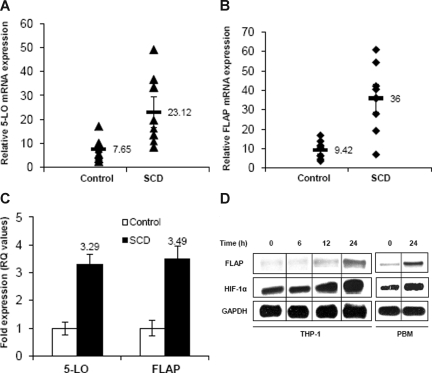
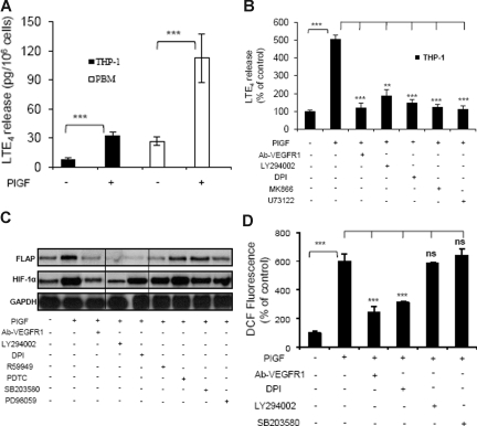
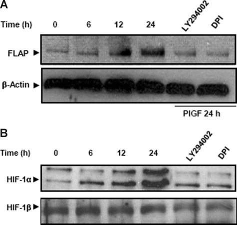
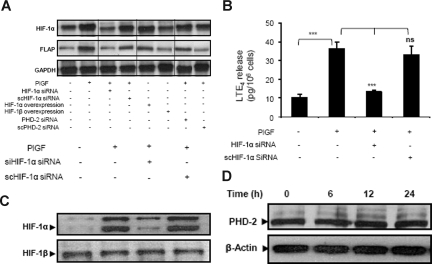
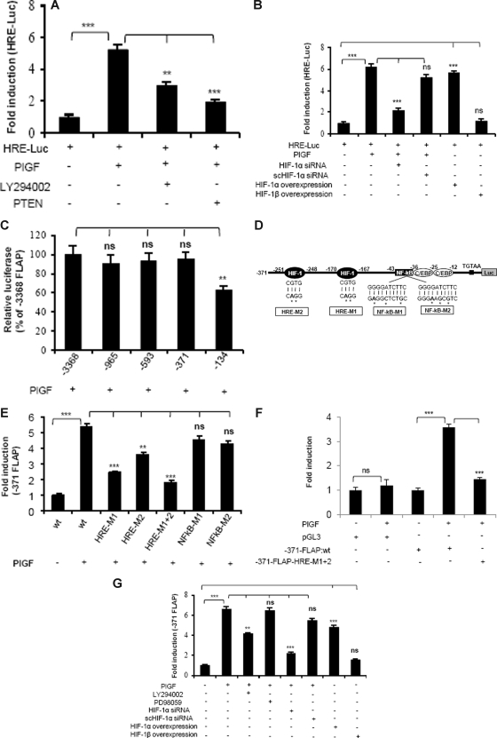
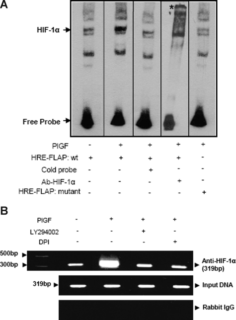
Individuals with sickle cell disease (SCD) have increased inflammation, a high incidence of airway hyperreactivity (AH), and increased circulating leukotrienes (LT). We show that expression of 5-lipoxygenase and 5-lipoxygenase activating protein (FLAP), key catalytic molecules in the LT pathway, were significantly increased in peripheral blood mononuclear cells (MNCs) in patients with SCD, compared with healthy controls. Placenta growth factor (PlGF), elaborated from erythroid cells, activated MNC and THP-1 monocytic cells to induce LT production. PlGF-mediated increased FLAP mRNA expression occurred via activation of phosphoinositide-3 (PI-3) kinase, nicotinamide adenine dinucleotide phosphate (NADPH) oxidase, and hypoxia inducible factor-1alpha (HIF-1alpha). HIF-1alpha small interfering RNA (siRNA) reduced PlGF-induced FLAP expression. FLAP promoter-driven luciferase constructs demonstrated that PlGF-mediated luciferase induction was abrogated upon mutation of HIF-1alpha response element (HRE), but not the nuclear factor-kappaB (NF-kappaB) site in the FLAP promoter; a finding confirmed by chromatin immunoprecipitation (ChIP) analysis. PlGF also increased HIF-1alpha binding to the HRE in the FLAP promoter. Therefore, it is likely that the intrinsically elevated levels of PlGF in SCD subjects contribute to increased LT, which in turn, mediate both inflammation and AH. Herein, we identify a mechanism of increased LT in SCD and show HIF-1alpha as a hypoxia-independent target of PlGF. These studies provide new avenues to ameliorate these complications.


Figures

Figure 1
Figure 1
5-LO and FLAP mRNA expression in MNC, PBM, and THP-1. (A,B) qRT-PCR analysis of mRNA in MNC isolated from SCD subjects at steady state (n = 9) and healthy controls (n = 9). Each data point represents ΔCt values of 5-LO and FLAP expression from individual subjects. Mean values are represented as black bars. Error bars represent SEM. (C) ΔΔCt (RQ) values show fold expression of 5-LO and FLAP mRNA in SCD compared with healthy controls. (D) RPA analysis of FLAP, HIF-1α, and GAPDH in PlGF-treated THP-1 cells and PBM for the indicated time periods. Data are representative of 3 independent experiments. Where indicated, the vertical lines show repositioned gel lanes.
Figure 2
Figure 2
PlGF-induced LTE4 release, FLAP mRNA expression, and ROS formation. (A) PBM or THP-1 cells were treated with PlGF (250 ng/mL) for 24hours and (B) THP-1 cells were pretreated for 30 minutes with Ab-VEGFR1 (2 μg/mL), LY294002 (10 μM), DPI (10 μM), MK866 (10 μM), and U73122 (10 μM), followed by treatment with PlGF for 24 hours. The supernatants were collected and assayed for LTE4 release by ELISA. (C) RPA analysis of total RNA isolated from THP-1 cells pretreated for 30 minutes with Ab-VEGFR1, LY294002, DPI, R59949 (10 μM), PDTC (10 μM), SB203580 (10 μM), and PD98059 (10 μM) before PlGF treatment for 24 hours. (D) THP-1 cells were loaded with DCFH-DA dye for 30 minutes, incubated with indicated inhibitors for an additional 30 minutes and treated with PlGF for 4 hours. The cells were lysed, and the fluorescence was measured. Data are expressed as means plus or minus SEM of 3 independent experiments (***P < .001; **P < .01; ns, P > .05). Where indicated, the vertical lines show repositioned gel lanes.
Figure 3
Figure 3
PlGF increases FLAP and HIF-1α protein in THP-1 cells. THP-1 cells were pretreated for 30 minutes with LY294002 and DPI followed by PlGF treatment for indicated time periods. (A) Cytosolic proteins were subjected to western blot analysis using antibody to FLAP. The same membrane was reprobed with β-actin antibody to normalize protein loading. (B) Nuclear extracts were subjected to western blot analysis using antibody to HIF-1α. The same membrane was reprobed with HIF-1β antibody to normalize the protein loading. Proteins were visualized by enhanced chemiluminescence corresponding to their expected molecular weights: FLAP (18 kDa), β-actin (42 kDa), HIF-1α (120 kDa), and HIF-1β (95 kDa). Data are representative of 3 independent experiments.
Figure 4
Figure 4
PlGF-mediated FLAP and LTE4 expression involves HIF-1α. THP-1 cells were transfected with indicated siRNA constructs or expression plasmids, followed by PlGF treatment for 24 hours. (A) RPA, (B) LTE4 release, and (C) Western blot analysis of nuclear extract using HIF-1α antibody. (D) Western blot analysis of cytosolic extracts from THP-1 cells treated with PlGF for indicated time period (6-24 hours) using PHD-2 antibody. Data are representative of 3 independent experiments. Data are expressed as means plus or minus SEM of 3 independent experiments (***P < .001; **P < .01; ns, P > .05). Where indicated, the vertical lines show repositioned gel lanes.
Figure 5
Figure 5
PlGF augments HRE-Luc and FLAP-Luc promoter via activation of PI-3 kinase and HIF-1α. THP-1 cells cotransfected with HRE-Luc and β-galactosidase plasmid were (A) either pretreated with LY294002 or cotransfected with PTEN and (B) cotransfected with indicated plasmids before treatment with PlGF for 24 hours. (C) Deletion analysis of FLAP promoter. THP-1 cells were cotransfected with indicated deletion construct and β-galactosidase plasmid, followed by PlGF treatment for 24 hours. (D) Schematics of FLAP promoter (−371 bp) indicating the location of HIF-1α, NF-κB, and C/EBP binding sites. (E,F) PlGF augments minimal FLAP promoter activity through HREs but not NF-κB in THP-1 (E) and PBM (F). THP-1 cells or PBM were cotransfected with indicated promoter constructs and β-galactosidase plasmid, followed by PlGF treatment for 24 hours. (G) THP-1 cells were treated with either indicated pharmacologic inhibitors or transfected with siRNA or HIF expression plasmids. These cells were then cotransfected with −371-FLAP-Luc and β-galactosidase plasmid, followed by PlGF treatment for 24 hours. Luciferase and β-galactosidase activities were measured as described in “Transient transfection.” The luciferase activity was normalized to that of the promoterless pGL3 basic vector. Data are expressed as mean plus or minus SEM of 3 independent experiments (***P < .001; **P < .01; ns, P > .05).
Figure 6
Figure 6
PlGF augments HIF-1α binding to FLAP promoter in vitro (EMSA) and in vivo (ChIP). (A) Nuclear extracts from THP-1 cells (10 μg) were incubated with a biotinylated double-stranded oligonucleotide corresponding to the region (−179 to −159 bp) of the FLAP promoter containing the proximal HRE located at −170 to −167 bp. Where indicated, 50-fold excess of unlabeled wild-type probe (lane 3) or antibody to HIF-1α (lane 4) was added. EMSA analysis was also performed with a probe containing a mutation of the HRE (−170 bp to −167 bp, lane 5). * denotes supershifted band. Data are representative of 2 independent experiments. (B) THP-1 cells were pretreated with indicated pharmacologic inhibitors before PlGF stimulation for 4 hours. The soluble chromatin was isolated and immunoprecipitated with either HIF-1α antibody (top panel) or control rabbit IgG (bottom panel). The primers used to amplify the products flanking HIF-1α binding sites in the FLAP promoter are indicated in Table 1. The middle panel represents the amplification of input DNA before immunoprecipitation. Data are representative of 2 independent experiments. Where indicated, the vertical lines show repositioned gel lanes.

Similar articles

Cited by

References

    1. Solovey A, Gui L, Ramakrishnan S, Steinberg MH, Hebbel RP. Sickle cell anemia as a possible state of enhanced anti-apoptotic tone: survival effect of vascular endothelial growth factor on circulating and unanchored endothelial cells. Blood. 1999;93:3824–3830. - PubMed
    1. Belcher JD, Marker PH, Weber JP, Hebbel RP, Vercellotti GM. Activated monocytes in sickle cell disease: potential role in the activation of vascular endothelium and vaso-occlusion. Blood. 2000;96:2451–2459. - PubMed
    1. Kaul DK, Liu XD, Choong S, et al. Anti-inflammatory therapy ameliorates leukocyte adhesion and microvascular flow abnormalities in transgenic sickle mice. Am J Physiol Heart Circ Physiol. 2004;287:H293–H301. - PubMed
    1. Croizat H, Nagel RL. Circulating cytokines response and the level of erythropoiesis in sickle cell anemia. Am J Hematol. 1999;60:105–115. - PubMed
    1. Miller ST, Sleeper LA, Pegelow CH, et al. Prediction of adverse outcomes in children with sickle cell disease. N Engl J Med. 2000;342:83–89. - PubMed

Publication types

MeSH terms