. 2008 Oct 28;105(43):16677-82.
doi: 10.1073/pnas.0808597105. Epub 2008 Oct 22.

FADD and caspase-8 control the outcome of autophagic signaling in proliferating T cells

Affiliations

FADD and caspase-8 control the outcome of autophagic signaling in proliferating T cells

Bryan D Bell et al. Proc Natl Acad Sci U S A. .

Abstract

Fas-associated death domain protein (FADD) and caspase-8 (casp8) are vital intermediaries in apoptotic signaling induced by tumor necrosis factor family ligands. Paradoxically, lymphocytes lacking FADD or casp8 fail to undergo normal clonal expansion following antigen receptor cross-linking and succumb to caspase-independent cell death upon activation. Here we show that T cells lacking FADD or casp8 activity are subject to hyperactive autophagic signaling and subvert a cellular survival mechanism into a potent death process. T cell autophagy, enhanced by mitogenic signaling, recruits casp8 through interaction with FADD:Atg5-Atg12 complexes. Inhibition of autophagic signaling with 3-methyladenine, dominant-negative Vps34, or Atg7 shRNA rescued T cells expressing a dominant-negative FADD protein. The necroptosis inhibitor Nec-1, which blocks receptor interacting protein kinase 1 (RIP kinase 1), also completely rescued T cells lacking FADD or casp8 activity. Thus, while autophagy is necessary for rapid T cell proliferation, our findings suggest that FADD and casp8 form a feedback loop to limit autophagy and prevent this salvage pathway from inducing RIPK1-dependent necroptotic cell death. Thus, linkage of FADD and casp8 to autophagic signaling intermediates is essential for rapid T cell clonal expansion and may normally serve to promote caspase-dependent apoptosis under hyperautophagic conditions, thereby averting necrosis and inflammation in vivo.


Conflict of interest statement

The authors declare no conflict of interest.

Figures
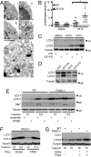
Fig. 1.
Fig. 1.
Increased autophagy in cells lacking FADD function. (A) Naïve (I and II), SIINFEKL activated (III–V), OT1 (I and III), and OT1-FD (II, IV, and V) cells were analyzed by TEM; representative micrographs presented (Scale bar: 0.5 μm). (B) Number of autophagosomes per cell section (*P value: 0.005). (C and D) Immunoblot showing extent of LC3 processing in naïve and activated (C), or 3-day activated (D) OT1, OTII, OT1-FD, and OTII-FD cells [Molecular weight (arrow heads) in kDa]. (E) Immunoblot of WT and FAD−/− MEFs infected with either scrambled or Atg7 shRNA with 10% or 0.5% serum. (F) Immunoblot of FADD−/− MEFs infected with MIT-empty or MIT-FADD, followed by growth with 10% or 0.5% serum. (G) Immunoblot of WT and FADD−/− MEFs grown with 10% serum, with or without E64d and pepA for the last 4 h.
Fig. 2.
Fig. 2.
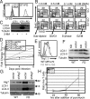
Inhibition of autophagic signaling rescues FADDdd T cells. (A) Activated CFSE-labeled CD8+ T cell proliferation for 3 days, ±0.8 mM 3-methyladenine. (B) Three-day activated T cells pulsed with BrdU for last hour of culture. (C) Immunoblot of activated T cells at 3 days, ±3-MA. (D–F) Activated T cells infected with Vps34-KD or empty retrovirus. (D) Fold change in Thy1.1 expression compared to 2 days postinfection. (E and F) Thy1.1 expression and immunoblot of Thy1.1+ CD8+ cells at 3 days postinfection. (G and H) Immunoblot and live cell recovery of WT and FADDdd (FD) T cells infected with shAtg7 followed by growth in the presence of 10 μg/ml puromycin. (H) Expressed as a ratio of live shAtg7 cells over live empty-vector control cells (All error bars: ±SD).
Fig. 3.
Fig. 3.
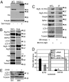
FADD, casp8, and RIPK1 form a complex with Atg5-Atg12/Atg16L. (A) Immunoblot to show expression level of TAP-FADD construct in MEFs. (B) FADD−/− MEFs grown in 10% serum were infected with Mit-TAP-HA-FADD for 24 h, followed by cell lysis and TAP purification. Whole cell lysate (WCL) and post-TAP-FADD purification immunoblot shown. Anti-HA antibody detects TAP-HA-FADD (**) and TAP-HA-FADD post-TEV cleavage (*). (C) WT MEFs grown in 10% serum were infected with Mit-empty or Mit-HA-Atg5 retroviruses, followed by cell lysis. Immunoblot of WCL and anti-HA immunoprecipitated fractions. (D) IETDase (casp8) and DEVDase (casp3) activities in naïve, 36-h activated, or 36-h activated + last 6 h with anti-FAS plate-bound antibody WT and FADDdd T cells, ±3-methyladenine. Activities expressed as ratios of activated versus naïve cells (All error bars: ±SD). (E) Immunoblot of naïve and 2-day activated OT1 T cells plus or minus 50 μM zVAD-FMK.
Fig. 4.
Fig. 4.
Nec-1 restores OT1-FD T cell proliferation and survival through inhibition of RIPK1 signaling. (A) Three-day activated CD8+ T cells grown in increasing doses of Nec-1 and pulsed with BrdU for last hr of culture. (B) CFSE-labeled CD8+ T cell proliferation, ±10 μM Nec-1. (C) AnnexinV staining on CD8+ T cells from B. (D) Live CD8+ cell recovery from cells in B (triplicate cultures: ±SD; P values: *, 0.017; **, 0.0025; ***, 0.005). (E) Immunoblot of 3-day activated CD8+ T cells, ±10 μM Nec-1.
Fig. 5.
Fig. 5.
Nec-1 restores cell proliferation and survival in T cells lacking casp8 through inhibition of RIPK1 signaling. (A) Immunoblot of 3-day activated CD8+ T cells ±10 μM Nec-1. (B) Live CD8+ T cell recovery from 3-day activated WT, casp8+/−, and casp8−/− cells (triplicate cultures: ±SD). (C) CFSE-labeled CD8+ T cell proliferation, ±10 μM Nec-1. (D) Three-day activated CD8+ T cells ±Nec-1 and pulsed with BrdU for last h of culture. (E) Live cell recovery of WT and casp8−/− (C8) T cells infected with shAtg7 or scrambled control followed by growth in the presence of 10 μg/ml puromycin. Expressed as a ratio of live T cells infected with shAtg7 vs. a scrambled hairpin (All error bars: ±SD). (F) Proposed mechanism of FADD, casp8, and RIPK1 negative feedback on autophagic signaling.

References

    1. Kischkel F, et al. Cytotoxicity-dependent APO-1 Fas/CD95-associated proteins form a death-inducing signaling complex DISC with the receptor. EMBO J. 1995;14:5579–5588. - PMC - PubMed
    1. Walsh CM, Luhrs KA, Arechiga AF. The “fuzzy logic” of the death-inducing signaling complex in lymphocytes. J Clin Immunol. 2003;23:333–353. - PubMed
    1. Varfolomeev EE, et al. Targeted disruption of the mouse Caspase 8 gene ablates cell death induction by the TNF receptors, Fas/Apo1, and DR3 and is lethal prenatally. Immunity. 1998;9:267–276. - PubMed
    1. Zhang J, Cado D, Chen A, Kabra N, Winoto A. Fas-mediated apoptosis and activation-induced T cell proliferation are defective in mice lacking FADD/Mort1. Nature. 1998;392:296–300. - PubMed
    1. Yeh WC, et al. FADD: essential for embryo development and signaling from some, but not all, inducers of apoptosis. Science. 1998;279:1954–1958. - PubMed

Publication types

Substances