. 2008 Oct 28;105(43):16549-54.
doi: 10.1073/pnas.0801795105. Epub 2008 Oct 22.

Side-chain recognition and gating in the ribosome exit tunnel

Affiliations

Side-chain recognition and gating in the ribosome exit tunnel

Paula M Petrone et al. Proc Natl Acad Sci U S A. .

Abstract

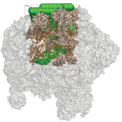
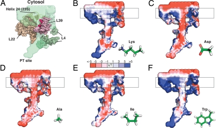
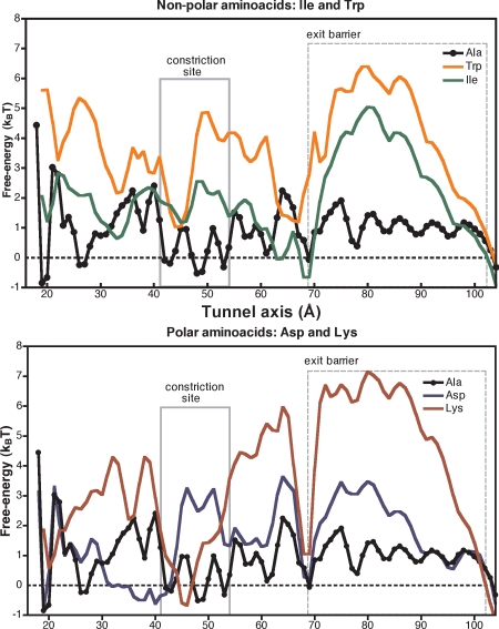
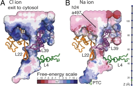
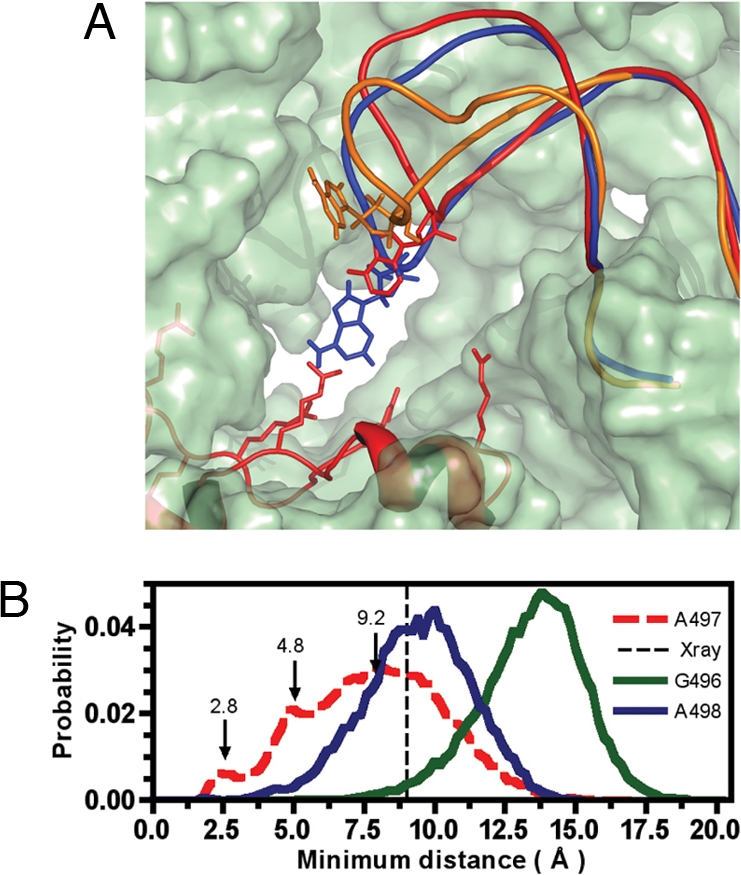
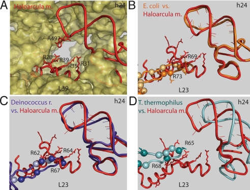
The ribosome is a large complex catalyst responsible for the synthesis of new proteins, an essential function for life. New proteins emerge from the ribosome through an exit tunnel as nascent polypeptide chains. Recent findings indicate that tunnel interactions with the nascent polypeptide chain might be relevant for the regulation of translation. However, the specific ribosomal structural features that mediate this process are unknown. Performing molecular dynamics simulations, we are studying the interactions between components of the ribosome exit tunnel and different chemical probes (specifically different amino acid side chains or monovalent inorganic ions). Our free-energy maps describe the physicochemical environment of the tunnel, revealing binding crevices and free-energy barriers for single amino acids and ions. Our simulations indicate that transport out of the tunnel could be different for diverse amino acid species. In addition, our results predict a notable protein-RNA interaction between a flexible 23S rRNA tetraloop (gate) and ribosomal protein L39 (latch) that could potentially obstruct the tunnel's exit. By relating our simulation data to earlier biochemical studies, we propose that ribosomal features at the exit of the tunnel can play a role in the regulation of nascent chain exit and ion flux. Moreover, our free-energy maps may provide a context for interpreting sequence-dependent nascent chain phenomenology.


Conflict of interest statement

The authors declare no conflict of interest.

Figures

Fig. 1.
Fig. 1.
Large-subunit Haloarcula marismortui (PDB ID code 1S72) and in-box simulated model. Tan, modeled rRNA (tubes) and protein (spheres); green, solvent-accessible volume.
Fig. 2.
Fig. 2.
Free-energy maps of the chemical environment inside the ribosome tunnel simulated with molecular dynamics. (A) Inner-tunnel volume (green) and proteins near the ribosome tunnel. The gate A497 in helix 24 is highlighted in spheres. In dark-pink spheres, arginines of L39. (B–F) Free-energy profiles for single amino acid side chains in the tunnel. Each graph is a transverse cut of the tunnel; the exit to cytosol is at the top and peptidyl transferase center at the bottom. Free energies are measured in kBT with reference to each probe's solvated state outside the tunnel. Red-colored areas are regions of favorable residence; white denotes neutral areas. High free-energy areas are represented in increasing tones of blue (error analysis is in Fig. S13). Boxed in gray, location of free-energy barrier predicted by our simulations.
Fig. 3.
Fig. 3.
Free-energy profiles along the longitudinal axis of the ribosomal tunnel. The scale corresponds to the structural elements as shown in Fig. S14. Flexible parts of simulated model located in the range 20–105 Å. (Upper) Hydrophilic side chains Asp and Lys. (Lower) Hydrophobic side chains Trp and Ile. Boxed in dashes, free-energy barrier predicted by our simulations. The uncertainty to the free-energy profiles is plotted in Fig. S15.
Fig. 4.
Fig. 4.
Transversal cut of the free-energy profile for Cl and Na+ ions in the ribosome exit tunnel. Free energies are measured in kBT with reference to each ion's solvated state outside the tunnel.
Fig. 5.
Fig. 5.
Conformational change of the tip of rRNA helix24 (gate) relative to arginines in L39 (latch) predicted with MD simulations. (A) Exterior view of the ribosome tunnel. Shown are A497 in sticks and x-ray structure in red. In blue, A497 is 2.7 Å away from the closest arginine. In orange, A497 is 16.7 Å away from the closest arginine. (B) Histogram of minimum distance between the rRNA tetraloop and L39. Each curve shows the interactions of rRNA bases G496, A497 and A498, with arginines (R20, R31, and R39) in L39. Two close-contact peaks are shown at 2.8 Å and 4.8 Å for A497 and the maximum at 7.8 Å. In dashes, the x-ray minimum distance of 9.04 Å (details in SI Text Discussion).
Fig. 6.
Fig. 6.
Structural alignment of different ribosome x-ray structures to H. marismortui (in red color, PDB ID code 1S72). Exterior view of the exit of the ribosomal tunnel that shows rRNA helix 24 from 23S and proteins L23 and L39. Positive residues are in spheres, and arginines are in dark spheres and sticks. (A) H. marismortui [PDB ID code 1S72 (15)]. (B) E. coli [PDB ID code 2AW4 (14)], in orange. (C) D. radiodurans [PDB ID code 1NKW (23)], in blue. (D) T. thermophilus [PDB ID code 1VSA (24)], in cyan.

References

    1. Nakatogawa H, Ito K. The ribosomal exit tunnel functions as a discriminating gate. Cell. 2002;108:629–636. - PubMed
    1. Nakatogawa H, Ito K. Intraribosomal regulation of expression and fate of proteins. ChemBioChem. 2004;5:48–51. - PubMed
    1. Muto H, Nakatogawa H, Ito K. Genetically encoded but nonpolypeptide prolyl-tRNA functions in the A site for SecM-mediated ribosomal stall. Mol Cell. 2006;22:545–552. - PubMed
    1. Mankin AS. Nascent peptide in the “birth canal” of the ribosome. Trends Biochem Sci. 2006;31:11–13. - PubMed
    1. Dresios J, Derkatch IL, Liebman SW, Synetos D. Yeast ribosomal protein L24 affects the kinetics of protein synthesis and ribosomal protein L39 improves translational accuracy, while mutants lacking both remain viable. Biochemistry. 2000;39:7236–7244. - PubMed

Publication types

LinkOut - more resources